

March 2008 - Mycological Society of America
March 2008 - Mycological Society of America
March 2008 - Mycological Society of America

You also want an ePaper? Increase the reach of your titles
YUMPU automatically turns print PDFs into web optimized ePapers that Google loves.
Newsletter <strong>of</strong> the <strong>Mycological</strong> <strong>Society</strong> <strong>of</strong> <strong>America</strong><br />
— In This Issue —<br />
Feature Article<br />
The Chytrid Epidemic Revisited<br />
MSA Business<br />
From the President’s Corner<br />
MSA Secretary’s Email Express<br />
Inoculum Editor’s Note<br />
MSA Abstracts 2007<br />
<strong>Mycological</strong> News<br />
Northern Thailand and the Mushroom Research Centre<br />
Did Fusarium or Warm Temperatures Kill New York Bats?<br />
Smoky Mountains Treetop Exploration Airs on Wild Chronicles<br />
Announcing the <strong>2008</strong> Annual <strong>Mycological</strong> <strong>Society</strong> <strong>of</strong> <strong>America</strong><br />
Meeting<br />
MNRC <strong>2008</strong> Award for Best Student Poster<br />
Great Lakes-St. Lawrence Spring Workshop in Mycology<br />
John W. Rippon Fellowship in Medical Mycology<br />
Middle Atlantic States Mycology Conference (MASMC <strong>2008</strong>)<br />
Introduction to Food- and Air-Borne Fungi<br />
Fleshy Fungi <strong>of</strong> the Highlands Plateau<br />
<strong>2008</strong> Seminars at the Humboldt Institute<br />
APS Centennial Celebration <strong>2008</strong><br />
China-Japan Pan-Asia Pacific Mycology Forum <strong>2008</strong><br />
International Congress on Systematics and Ecology<br />
<strong>of</strong> Myxomycetes<br />
Saccardo’s Sylloge Fungorum Now Available Online<br />
Seeking Air-Dried Collections <strong>of</strong> Red Nectria-Like Fungi<br />
Fungal Conservation in Canada and the USA<br />
Mycologist’s Bookshelf<br />
Mycology, Vols. 1 and 2, DVD-ROM<br />
Exploitation <strong>of</strong> Fungi: Symposium <strong>of</strong> the British<br />
<strong>Mycological</strong> <strong>Society</strong><br />
Compendium <strong>of</strong> Brassica Diseases<br />
Compendium <strong>of</strong> Rose Diseases and Pests<br />
Field Guide to North <strong>America</strong>n Truffles<br />
Annotated List <strong>of</strong> Polypores for the Iberian Peninsula<br />
and Balearic Islands<br />
Pioneering Women in Plant Pathology<br />
Recently Received Books<br />
Previously Listed Books<br />
Take a Break<br />
MycoLotus 1 Crossword<br />
Cookery Corner<br />
<strong>Mycological</strong> Jobs<br />
Faculty Position in Molecular Plant Pathology at Ohio State<br />
Ph.D. or Postdoctoral Fellow in Food and Bioproduct Sciences<br />
M.S. or Ph.D. Graduate Assistantship in Marine Mycology<br />
Research<br />
<strong>Mycological</strong> Classifieds<br />
Mushrooms in Their Natural Habitats (Vols. I and II), 1949<br />
Mold testing and Identification Services<br />
Mycology On-Line<br />
Calendar <strong>of</strong> Events<br />
Sustaining Members<br />
— Important Dates —<br />
April 15, <strong>2008</strong><br />
Deadline for Submission to Inoculum 59(3)<br />
July 28–August 5, <strong>2008</strong><br />
China-Japan and Pan Asia Pacific Mycology Forum<br />
Changchun, China<br />
August 9–14, <strong>2008</strong><br />
MSA Meeting<br />
State College, Pennsylvania, U.S.<br />
September 4–7, <strong>2008</strong><br />
NAMA Annual Foray in memory <strong>of</strong> Dr. Orson K. Miller<br />
McCall, Idaho<br />
Editor — Jinx Campbell<br />
Dept. <strong>of</strong> Coastal Sciences, Gulf Coast Research Lab<br />
University <strong>of</strong> Southern Mississippi<br />
703 East Beach Drive, Ocean Springs, MS 39564<br />
Telephone: (228) 818-8878, Fax: (228) 872-4264<br />
Email: jinx.campbell@usm.edu<br />
The Chytrid Epidemic Revisited<br />
Supplement to<br />
Mycologia<br />
Vol. 59(2)<br />
<strong>March</strong> <strong>2008</strong><br />
by Frank H. Gleason and Deborah J. Macarthur<br />
Spring has finally come to Big Pond after a long harsh<br />
winter. The snow is beginning to melt in the catchment<br />
bringing nutrients into the pond. The ice on the pond has<br />
melted so that sunlight can penetrate into the water. The<br />
temperature is rising on the surface. Many species <strong>of</strong> phytoplankton<br />
are beginning to resume growth. The surface <strong>of</strong><br />
the pond is suddenly dusted with pine and sweet gum<br />
pollen, which provide substrates for many microorganisms.<br />
Mosquito and midge larvae abound feeding on<br />
plankton. Tadpoles are beginning to swim along the edge.<br />
Birds are diving in to look for fish. However, unnoticed by<br />
the casual observer, resistant sporangia <strong>of</strong> chytrids, which<br />
have over-wintered attached to substrates throughout the<br />
water column, are beginning to release zoospores. Soon<br />
many different substrates will become colonized by<br />
chytrid zoospores, and the chytrid epidemic is about to<br />
Continued on following page<br />
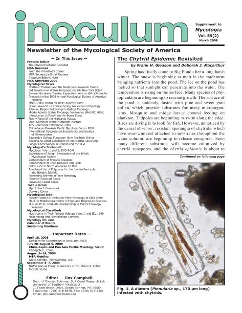
Fig. 1. A diatom (Pinnularia sp., 170 µm long)<br />
infected with chytrids.
egin. This kind <strong>of</strong> phenomenon occurs in many<br />
aquatic ecosystems throughout the world.<br />
The chytrid epidemic appears suddenly as long as<br />
substrates are available (Sparrow, 1960). The life<br />
cycle <strong>of</strong> most chytrids is completed rapidly, and many<br />
zoospores are released from each sporangium. Therefore,<br />
a large increase in population numbers is possible<br />
within a very short period <strong>of</strong> time. For this reason<br />
all chytrids are considered to be ruderal in their life<br />
histories (sensu Dix and Webster, 1995, p.7).<br />
Nearly fifty years ago Frederick Sparrow (1960,<br />
pp. 104-114) defined the chytrid epidemic to include<br />
host-parasite interactions involving chytrids (Phyla<br />
Blastocladiomycota and Chytridiomycota). Sparrow’s<br />
examples considered only phytoplankton and fungi as<br />
hosts but zooplankton and larger aquatic animals need<br />
to be included as well. According to Sparrow, beginning<br />
in early spring, population densities <strong>of</strong> parasitic<br />
chytrids rapidly increase for a short period, then decline<br />
due to the loss <strong>of</strong> available substrates and finally produce<br />
resting spores. We would expect to find the same<br />
pattern <strong>of</strong> growth in both parasitic and saprophytic<br />
chytrids in aquatic ecosystems.<br />
Donald Barr, Hilda Canter, John Couch, John Karling,<br />
Joyce Longcore, Frederick Sparrow, Howard<br />
Whisler, Guy Willoughby and many other mycologists<br />
have spent their lives recording the presence <strong>of</strong> parasitic<br />
chytrids on aquatic plants and animals and saprophytic<br />
chytrids colonizing nonliving substrates in<br />
aquatic environments. Sparrow (1960, pp. 1073-1104)<br />
provides a long list <strong>of</strong> substrates reported for the<br />
growth <strong>of</strong> chytrids. Studies on chytrid parasites and<br />
saprophytes in freshwater lakes, particularly in northern<br />
Michigan, the Netherlands and the English Lake<br />
District, have continued for many years. Dix and Webster<br />
(1995, pp. 227-231) briefly summarized the roles<br />
<strong>of</strong> chytrid parasites and saprophytes in freshwater<br />
ecosystems. Yet, the ecological importance <strong>of</strong> this<br />
group <strong>of</strong> fungi and its roles in food web dynamics remain<br />
unappreciated and are not very well understood.<br />
Furthermore, chytrids sometimes have been either totally<br />
ignored or wrongly classified in studies on the biodiversity<br />
<strong>of</strong> fresh water ecosystems (Kagami et al.,<br />
2007; Lefèvre et al., 2007).<br />
Recently, there has been renewed interest in the<br />
ecology <strong>of</strong> chytrids in aquatic environments. Ibelings<br />
et al. (2003) and Kagami et al. (2007) have<br />
again examined the large number <strong>of</strong> species <strong>of</strong> phytoplankton<br />
affected by chytrid parasites. The role <strong>of</strong><br />
Batrachochytrium dendrobatidis in reduction in size<br />
<strong>of</strong> amphibian populations and in extinctions <strong>of</strong><br />
species is currently under investigation (Fisher &<br />
Garner 2007). Chytridiomycosis is an emerging in-<br />
2 Inoculum 59(2), <strong>March</strong> <strong>2008</strong><br />
fectious disease <strong>of</strong> amphibians world-wide. Kagami<br />
et al. (2007) have suggested that chytrid zoospores<br />
are an excellent food source for zooplankton and facilitate<br />
the transfer <strong>of</strong> energy to higher trophic levels<br />
in food webs. Chytrid zoospores are rich in<br />
stored lipids and glycogen. Studies on DNA extracted<br />
from water samples from a mountain lake in<br />
France have revealed a significant number <strong>of</strong> ribosomal<br />
DNA sequences belonging to known and unknown<br />
clades <strong>of</strong> chytrids (Lefèvre et al., 2007).<br />
Chytrid biodiversity is apparently surprisingly high<br />
in many freshwater lakes. Research on the ecology<br />
<strong>of</strong> chytrids in freshwater lakes in France is being<br />
continued by T. Sime-Ngando and his colleagues<br />
and students at Laboratoire Microorganismes:<br />
Génome et Environnement, Université Blaise Pascal,<br />
63177 AUBIERRE cedex.<br />
Evidence is emerging that the interactions between<br />
the chytrid parasite and its host may involve<br />
strong phenotypic selection pressures. A recent<br />
study by D. J. Macarthur (unpublished data), funded<br />
by the NSW Environmental Trust, involved the inoculation<br />
<strong>of</strong> cultures <strong>of</strong> the bloom forming cyanobacteria<br />
Anabaena circinalis and Microcysis aeruginosa<br />
with diatoms infected with chytrids (Fig. 1) and with<br />
pure cultures <strong>of</strong> chytrids in the Rhizophydiales from<br />
our culture collection. One <strong>of</strong> these isolates, SPP,<br />
adapted to the new host environments by parasitising<br />
both species <strong>of</strong> cyanobacteria. SPP was isolated into<br />
pure culture from a phytoplankton sample containing<br />
infected diatoms (by E. Lefèvre) and was tentatively<br />
identified as Rhizophydium sp. These results tend to<br />
support the assertion by Kagami et al. (2007) that the<br />
host range <strong>of</strong> chytrid parasites, <strong>of</strong>ten thought to be<br />
host-specific, could be altered by environmental<br />
stress. More interesting, however, was an apparent<br />
enhancement <strong>of</strong> growth and survival <strong>of</strong> A. circinalis<br />
cultures. Clearly, in this case, there are benefits to<br />
both the chytrid parasite and the cyanobacteria host.<br />
However, more data is required in order to unravel<br />
the complicated interactions between the chytrid parasite<br />
and its host.<br />
The growth and survival <strong>of</strong> chytrids are quite<br />
sensitive to physical factors, such as moisture, temperature,<br />
salinity and dissolved oxygen (Gleason et<br />
al., 2007) and possibly toxic chemicals. With global<br />
warming and environmental deterioration on the increase,<br />
it is high time that more mycologists begin<br />
to undertake intensive studies on the ecology <strong>of</strong> the<br />
chytrids in aquatic ecosystems. The survival <strong>of</strong><br />
many aquatic ecosystems in their present state could<br />
even depend on the activities <strong>of</strong> chytrids along with<br />
other groups <strong>of</strong> microorganisms.
Literature Cited<br />
Dix NJ, Webster J, 1995. Fungal ecology. Chapman &<br />
Hall, London.<br />
Fisher MC, Garner WJ, 2007. The relationship between<br />
the emergence <strong>of</strong> Batrachochytrium dendrobatidis,<br />
the international trade <strong>of</strong> amphibians<br />
and introduced amphibian species. Fungal Biology<br />
Reviews 21: 2-9.<br />
Gleason FH, Mozley-Standridge SE, Porter D, Boyle<br />
DG, Hyatt A, 2007. Preservation <strong>of</strong> Chytridiomycota<br />
in culture collections. <strong>Mycological</strong> Research<br />
111: 129-136.<br />
Ibelings BW, de Bruin A, Kagami M, Rijkeboer M,<br />
Brehm M, van Donk E, 2004. Host parasite interactions<br />
between freshwater phytoplankton and<br />
chytrid fungi (Chytridiomycota). Journal <strong>of</strong> Phycology<br />
40: 437-453.<br />
From the President’s Corner . . .<br />
Preparations are well underway for<br />
the <strong>2008</strong> Annual Meetings in August at<br />
Penn State University. David Geiser<br />
has been on top <strong>of</strong> preparations for several<br />
years now so the foray sites have<br />
been identified, the lunches have been<br />
ordered, and all we need now are some<br />
<strong>of</strong> those big ol’ mushrooms that<br />
abound in the forests <strong>of</strong> central Pennsylvania<br />
to get us <strong>of</strong>f to a great start. Jo<br />
Taylor and her committee have been<br />
hard at work on organizing the program<br />
and have selected three gangbuster<br />
symposia emphasizing everything<br />
from discussions on techniques<br />
for measuring fungal diversity to recent<br />
advances in live-microscopy tools<br />
to take a closer look at the hyphal apex. It’s not too<br />
early to start selecting some treasures and novelties<br />
for the annual social and auction. Last year we made<br />
over $10,000 at LSU and we surely want to reach<br />
that mark again. Let Endowment Chair Betsy<br />
Arnold know so she can start making a list.<br />
Our goal to increase the amounts in the mentor<br />
travel funds is doing well, but again we ask for your<br />
support <strong>of</strong> your favorite mentor so we can get all the<br />
funds over $10,000 and be able to assist young mycologists<br />
in attending our annual meetings. Our<br />
mentor research funds did get a tremendous boost<br />
from long-time member and all-around good guy<br />
Tom Volk who donated $10,000 in honor <strong>of</strong> med-<br />
MSA BUSINESS<br />
Don Hemmes, President<br />
Kagami M, de Bruin A, Ibelings BW, van Donk E,<br />
2007. Parasitic chytrids: their effects on phytoplankton<br />
communities and food-web dynamics.<br />
Hydrobiologia 578: 113-129.<br />
Lefèvre E, Bardot C, Noel C, Carrias J-F, Viscogliosi E,<br />
Amblard C, Sime-Ngando T, 2007. Unveiling fungal<br />
zo<strong>of</strong>lagellates as members <strong>of</strong> freshwater picoeukaryotes:<br />
evidence from a molecular diversity<br />
study in a deep meromictic lake.<br />
Environmental Microbiology 9: 61-71.<br />
Sparrow FK, 1960. Aquatic Phycomycetes. 2nd ed,<br />
The University <strong>of</strong> Michigan Press, Ann Arbor, MI.<br />
Frank H. Gleason and Deborah J. Macarthur<br />
are members <strong>of</strong> the School <strong>of</strong> Biological<br />
Sciences at the University <strong>of</strong> Sydney in<br />
Australia. For more information, send an<br />
email to frankjanet@ozemail.com.au.<br />
ical mycologist John Rippon. We have<br />
an article detailing Dr. Rippon’s career<br />
in this issue <strong>of</strong> Inoculum. Tom added<br />
another $500 on top <strong>of</strong> that to make<br />
sure the funds were awarded this year.<br />
I hope you all saw the criteria for the<br />
award that was sent in an email blast to<br />
the membership and on the webpage.<br />
With a symposium in cell biology and<br />
research awards in medical mycology,<br />
we hope to get more mycologists in<br />
these fields back in the fold and participating<br />
in the annual meetings.<br />
The council will hold its annual<br />
mid-year meetings at University <strong>of</strong><br />
California at Berkeley in late <strong>March</strong>. I<br />
just couldn’t talk anyone into the trip<br />
to Hawai’i. Thanks go to John Taylor for making<br />
local arrangements. If any <strong>of</strong> you have issues you<br />
would like to have brought up at the mid-year meetings,<br />
please email me or secretary Cathie Aime as<br />
soon as possible and we’ll make sure they get on the<br />
agenda.<br />
And while I’m at it, don’t you all think the Inoculum<br />
issues are just terrific! Kudos go to Jinx<br />
Campbell for a job well done. If you appreciate her<br />
efforts as much as I do, drop her an email and give<br />
her a pat on the back.<br />
—Don Hemmes<br />
President<br />
Inoculum 59(2), <strong>March</strong> <strong>2008</strong> 3
MSA Secretary’s Email Express<br />
MSA Council has completed two email<br />
polls since my last report, approving the following:<br />
• MSA Full Council poll <strong>2008</strong>-01: Council<br />
approved the establishment <strong>of</strong> a new<br />
Graduate Research Award in the name<br />
<strong>of</strong> John W. Rippon for research in Medical<br />
Mycology. The Award was established<br />
by a generous donation from<br />
Tom Volk and will be presented for the<br />
first time this year at the <strong>2008</strong> Annual<br />
MSA meeting at Penn State.<br />
• MSA Exec Council poll <strong>2008</strong>-02: MSA<br />
council approved the allocation <strong>of</strong> an<br />
additional $1500 to supplement the<br />
symposium budget for the <strong>2008</strong> Annual<br />
Meeting at Penn State. This will allow<br />
Program Chair Jo Taylor to provide<br />
support for three top-notch symposia<br />
this year.<br />
New Members: It is my pleasure to extend a warm welcome<br />
to the following new (or returning) members. New<br />
memberships will be formally approved by the <strong>Society</strong> at the<br />
Annual Business Meeting at Penn State in <strong>2008</strong>.<br />
• Canada: Kevin J Beiler, Laura Biggs<br />
• China: Meichun Xiang<br />
• Colombia: Maria Camila Pizano<br />
• Gambia: Felix C. Onyemachi<br />
• Germany: Martin Strohmeyer<br />
• Panama: Meike Piepenbring<br />
• United States: Aaron Brown, Catharine Mattison Catranis,<br />
Sharron L. Crane, Georgia Davis, Stephen L.<br />
Franke, Nancy Fisher Gregory, Jonatan Hernandez-roa,<br />
Inoculum Editor’s Note<br />
You will notice we have some great new changes to Inoculum.<br />
From this issue there is an expanded list <strong>of</strong> contents.<br />
This list is hyperlinked so you can click on the article you<br />
want to read and jump straight to it. No more scrolling<br />
through looking for a specific page! Also all web sites are<br />
hyperlinked so you can click on them direct from Inoculum<br />
and be taken through cyberspace to the webpage directly.<br />
Additionally, email addresses have been hyperlinked so one<br />
click on those will open up a new mail message window with<br />
the recipient’s address.<br />
If you have any comments or suggestions to improve Inoculum<br />
or some content you wish to see added, please let me<br />
know. If you would like to submit an article or have any<br />
news or classifieds etc for Inoculum, please send your materials<br />
to me at jinx.campbell@usm.edu. Do not send materials<br />
to Kay Rose at Allen Press. All submissions should be sent<br />
as attachments, preferably in Word. If you submit pictures,<br />
these need to be sent as separate JPGS or GIFFS, not em-<br />
4 Inoculum 59(2), <strong>March</strong> <strong>2008</strong><br />
Cathie Aime<br />
Cedar Nelson Hesse, David R Huff, Joe D.<br />
McFarland, Brienne Jean Meyer, Antonis<br />
Rokas, Noah Rosenzweig, Sergio Manuel<br />
Salcedo, Paul Hamilton Scott, Brian Seitzman,<br />
Anna R Simonin, Jennifer Talbot, Andrew<br />
David Thaler<br />
Emeritus candidates: I have received<br />
two applications for emeritus status by long<br />
standing members Jack Fell (Key Biscayne,<br />
FL) and F Brent Reeves, Jr. (Fort Collins<br />
CO). Emeritus status is conferred upon retired<br />
or retiring members who have at least<br />
15 years good standing with the <strong>Society</strong>.<br />
REMINDER: MSA Directory Update: Is<br />
your information up-to-date in the MSA directory?<br />
The <strong>Society</strong> is relying more and<br />
more on email to bring you the latest MSA<br />
news, awards announcements and other<br />
timely information, and our newsletter. To ensure that you<br />
receive <strong>Society</strong> blast emails and the Inoculum as soon as it<br />
comes out, and so that your colleagues can keep in touch,<br />
please check the accuracy <strong>of</strong> your email address and contact<br />
information in the online directory. This can be accessed via<br />
our web site at www.msafungi.org. If you need assistance<br />
with updating your membership information, or help with<br />
your membership log-in ID and password, please contact our<br />
Association Manager at Allen Press, the always-helpful Kay<br />
Rose at krose@allenpress.com.<br />
—Cathie Aime<br />
MSA Secretary<br />
maime@agcenter.lsu.edu<br />
bedded in the word document. Please do not send your submission<br />
in the body <strong>of</strong> the email as copy <strong>of</strong>ten has to be<br />
cleaned <strong>of</strong> extraneous symbols, brackets, etc and reformatted<br />
for use. My concern is that on reformatting and cleaning up,<br />
typographical errors may occur that could alter the context <strong>of</strong><br />
your submission.<br />
Inoculum is published in odd numbered months (January,<br />
<strong>March</strong>, May, July, September, November). The deadline<br />
for submitting is the 15 th <strong>of</strong> even numbered months: February,<br />
April, June, August, October, except December, which<br />
is the 10 th .<br />
If you would like to review a book or CD, please contact<br />
Amy Rossman at Amy.Rossman@ars.usda.gov. She<br />
will send it to you, you write the review, and then you can<br />
keep the book. Titles for review can be found in the Mycologist’s<br />
Bookshelf section.<br />
—Jinx Campbell<br />
Inoculum Editor<br />
jinx.campbell@usm.edu
MSA ABSTRACTS 2007<br />
Abstracts from 2007 MSA Meeting at LSU, Baton Rouge, Louisiana<br />
Acevedo, Carmen T. University <strong>of</strong> Puerto Rico, Rio Piedras Campus,<br />
Department <strong>of</strong> Biological Sciences, P.O. Box 23323, San Juan, PR<br />
00931-3323. ctacevedo@uprrp.edu. Fungi diversity in Puerto Rican<br />
mangroves and algae and their potential as bioremediation agents.<br />
Mangroves and algae are suitable substrates for marine fungi. Red and<br />
brown algae and red mangrove Rhizophora mangle were examined for<br />
the presence <strong>of</strong> endophytic marine fungi and various cultures were<br />
screened for the ability to degrade phenanthrene. Ten species <strong>of</strong> marine<br />
fungi were collected from mangroves, ten species in four genera <strong>of</strong><br />
brown algae, and eighteen species in twelve genera <strong>of</strong> red algae from<br />
beaches in Puerto Rico. Thirty marine manglicolous and endophytic<br />
fungi were used for biotransformation <strong>of</strong> phenanthrene assays. Thirteen<br />
isolates transformed significant amounts <strong>of</strong> phenanthrene in culture,<br />
eleven <strong>of</strong> which were endophytes. Two <strong>of</strong> the metabolites <strong>of</strong> phenanthrene<br />
produced were identified by HPLC/MS as dihydrodiol phenanthrene<br />
and phenanthrol. Surfactants were tested for their ability to solubilize<br />
phenanthrene, and therefore increase the biotransformation <strong>of</strong><br />
phenanthrene. Four different fungi were used with surfactants in two<br />
concentrations. Results indicate that the surfactants examined can either<br />
enhance or inhibit biotransformation depending on the fungus and<br />
concentration. Xylaria biotransformed significant amounts <strong>of</strong> phenanthrene<br />
with and without surfactants. Results suggest that marine fungi<br />
and particularly endophytes are potentially useful for bioremediation in<br />
marine environments. Symposium Presentation<br />
Adams, Gerard C. Department <strong>of</strong> Plant Pathology, Michigan State University,<br />
East Lansing, MI 48824, USA. gadams@msu.edu. A multigene<br />
phylogenetic analysis <strong>of</strong> species <strong>of</strong> Valsa revealing lineages <strong>of</strong><br />
medically important strains. Valsa is a cosmopolitan genus <strong>of</strong> fungi<br />
in the Diaporthales. Species in the genus cause cankers on woody angiosperms<br />
and gymnosperms and occasionally parasitize herbaceous<br />
plants. Identification <strong>of</strong> Valsa species based on morphology has long<br />
been problematical. Species discovery has been ongoing yet description<br />
<strong>of</strong> new species has foundered on the lack <strong>of</strong> distinguishing features.<br />
Members <strong>of</strong> the Diaporthales are recognized as a pr<strong>of</strong>itable resource<br />
for drug discovery. Valsa contains several strains that have<br />
received U.S. Patents because they produce unique compounds with<br />
important medical properties. The compounds include Cytosporacins,<br />
Cytosporin A, B & C, Cytosporone D & E, and Grahamimycin A, A1<br />
& B. Most <strong>of</strong> the strains are not identified. We examine the lineage <strong>of</strong><br />
the patented strains in relation to clades containing well-characterized<br />
species using multigene phylogenetic analysis. The analysis is based on<br />
partial sequences from DNA <strong>of</strong> the following five nuclear genes: the<br />
complete ITS, the 3’-end <strong>of</strong> the LSU, EF-1 alpha, beta-tubulin, and histone<br />
H3. Poster<br />
Aime, M. Catherine 1 , Henkel, Terry W. 2 * and Ryvarden, Leif. 3<br />
1 USDA-ARS, Systematic Botany and Mycology Lab, Beltsville, MD<br />
20705, USA, 2 Department <strong>of</strong> Biological Sciences, Humboldt State<br />
University, Arcata, CA 95521,USA, 3 Department <strong>of</strong> Botany, University<br />
<strong>of</strong> Oslo, Blindern, N-0316 Oslo, Norway. twh5@humboldt.edu.<br />
Polyporoid fungi <strong>of</strong> Guyana: diversity, new species, and ecological<br />
roles. Seven years <strong>of</strong> field work in a remote region <strong>of</strong> Guyana have uncovered<br />
a diverse assemblage <strong>of</strong> polypores associated with mixed<br />
and/or ectotrophic Dicymbe corymbosa (Caesalpiniaceae)-dominated<br />
rainforests in the Pakaraima Mountains. As a result, the total known<br />
species <strong>of</strong> polypores in Guyana has nearly doubled, from 55 to 91, including<br />
nine new species in seven genera <strong>of</strong> the Polyporales, Hymenochaetales<br />
and Russulales (Amauroderma, Ceriporia, Dichomitus,<br />
Fomitopsis; Coltricia, Coltriciella; Wrightoporia) and new distribution<br />
records for rarely collected species (e.g. Antrodiella dentipora,<br />
Antrodiella luteocontexta, Junghuhnia minuta). We report new species<br />
that expand current generic and family concepts, such as Amauroderma<br />
coltricioides, which is the first known species in the Ganodermataceae<br />
with smooth basidiospores. In addition to describing a variety <strong>of</strong><br />
wood-decay strategies among these polypores, we provide habitat,<br />
morphological and molecular evidence supporting an ectomycorrhizal<br />
nutritional mode for Coltricia spp. and examine the ecological consequences<br />
<strong>of</strong> Phellinus heart rot in D. corymbosa forests. Contributed<br />
Presentation<br />
Alexander, Mark* and Baird, Richard. Plant Pathology, P.O. Box<br />
9655, 206 Dorman Hall, Mississippi State, MS 39762, USA. MAlexander@plantpath.msstate.edu.<br />
Baseline data on pathogenic and ectomycorrhizal<br />
fungi associated with old growth eastern hemlock in<br />
the GSMNP and effects <strong>of</strong> imidacloprid on rhizospheric fungi and<br />
future restoration efforts. At its current rate <strong>of</strong> spread the exotic Hemlock<br />
Wooly Adelgid (HWA) will infest and devastate the entire native<br />
range <strong>of</strong> eastern hemlock within the next 2 decades. Baseline data on<br />
all associated fungal organisms must be obtained before forest habitat<br />
succession occurs. The effects <strong>of</strong> the loss <strong>of</strong> hemlock on associated<br />
soilborne fungal communities and on subsequent hemlock seedling establishment<br />
and regeneration are unknown. Select stands are being preserved<br />
using the systemic insecticide imidacloprid. The current application<br />
method, soil drenching, has an unknown impact on belowground<br />
fungal communities. Two study sites were selected in the Great Smoky<br />
Mountains National Park at two elevations within pure stands <strong>of</strong> mature<br />
hemlock. Twenty mature trees randomly selected at each site were<br />
subjected to one <strong>of</strong> 5 imidacloprid treatments varying rate and frequency<br />
and replicated 4 times at 2 elevations. Effects <strong>of</strong> treatments<br />
were analyzed by collecting a 120cm root sample from each replicate<br />
tree for identification <strong>of</strong> fungi using cultural techniques and molecular<br />
sequence data. Baseline data on fungal population diversity and abundance<br />
were compared among treatments and controls. In addition, all<br />
terrestrial macr<strong>of</strong>ungi within the plots were collected monthly. Ectomycorrhizal<br />
fungi were isolated from fresh sporocarps and cryogenically<br />
stored in a fungal repository. Poster<br />
Alspaugh, Andrew. Dept. <strong>of</strong> Medicine, Duke University School <strong>of</strong><br />
Medicine, Durham, NC, USA. andrew.alspaugh@duke.edu. Transcriptional<br />
pr<strong>of</strong>iling in the human fungal pathogen Cryptococcus<br />
ne<strong>of</strong>ormans. Similar to other microbial pathogens, Cryptococcus ne<strong>of</strong>ormans<br />
must coordinate the expression <strong>of</strong> many genes to adapt to the<br />
environment <strong>of</strong> the infected host. We have previously demonstrated<br />
that the cAMP signal transduction pathway coordinately regulates the<br />
expression <strong>of</strong> genes required for capsule and melanin production.<br />
Using whole genome microarrays, we have begun to explore the transcriptional<br />
network controlled by the cAMP pathway. Several transcription<br />
factors act downstream <strong>of</strong> this conserved pathway, including<br />
the Nrg1 protein. In addition to capsule and melanin production, C.<br />
ne<strong>of</strong>ormans must also control subtle morphogenic events to retain full<br />
virulence. The cytoskeletal changes required for altered morphogenesis<br />
are regulated by Ras signaling pathways. Gene microarray studies have<br />
also demonstrated the role <strong>of</strong> Ras-dependent gene expression in morphogenesis.<br />
Transcriptional pr<strong>of</strong>iling <strong>of</strong> targeted C. ne<strong>of</strong>ormans mutant<br />
strains with altered virulence has begun to demonstrate the complex<br />
ways in which a microbial pathogen develops an adaptive cellular response<br />
to the host environment. Symposium Presentation<br />
Arnold, A. Elizabeth. Department <strong>of</strong> Plant Sciences, University <strong>of</strong> Arizona,<br />
Tucson, AZ 85721, USA. arnold@ag.arizona.edu. Barcoding<br />
endophytic fungi: lessons, limitations, and linkages with multilocus<br />
data sets. Drawing from surveys <strong>of</strong> endophytic fungi from (1) all major<br />
Continued on following page<br />
Inoculum 59(2), <strong>March</strong> <strong>2008</strong> 5
lineages <strong>of</strong> land plants and (2) biogeographic provinces ranging from<br />
the arctic to the tropics, I will explore the degree to which a large-scale,<br />
single-locus (bar-code) data set based on the nuclear ribosomal internal<br />
transcribed spacer (ITS) can further our understanding <strong>of</strong> fungal diversity<br />
and ecology. Specifically, I will (1) address patterns <strong>of</strong> geographical<br />
distributions, taxonomic makeup, host specificity, and diversity <strong>of</strong><br />
endophytic fungi using a data set <strong>of</strong> over 6000 ITS sequences from cultures<br />
and environmental samples; (2) explore empirical approaches for<br />
delimiting meaningful taxonomic units from ITS data alone; (3) highlight<br />
a variety <strong>of</strong> limitations imposed by the single-locus and ITS-specific<br />
approach, and demonstrate that such issues vary in intensity<br />
among clades <strong>of</strong> Ascomycota and among different geographic sites; (4)<br />
discuss the degree to which ITS data are congruent and incongruent<br />
with inferences based on multi-locus datasets; and (5) describe several<br />
new methods for visualizing ITS data in spatial and phylogenetic contexts,<br />
with the goal <strong>of</strong> critically evaluating the biological realism and inferential<br />
strength <strong>of</strong> the bar-code approach for studies <strong>of</strong> highly diverse<br />
fungi. Symposium Presentation<br />
Atkinson, Toni J. 1 *, Orlovich, David, A. 2 and Miller, Andrew N. 11 Section<br />
for Biodiversity, Illinois Natural History Survey, 1816 S. Oak St.,<br />
Champaign, IL 61820, USA, 2 Department <strong>of</strong> Botany, University <strong>of</strong><br />
Otago, P.O. Box 56, Dunedin 9054, New Zealand.<br />
toni@botany.otago.ac.nz. From the Land <strong>of</strong> the Long White Cloud<br />
to the Great Smoky Mountains: New Zealand and Appalachian diversity<br />
among woody decay pyrenomycetes. The New Zealand archipelago,<br />
a temperate, oceanic island group 1600 km south-east <strong>of</strong><br />
Australia, forms the largest landmass in the south Pacific. Island biotas<br />
are usually considered ‘depauperate’ when compared with those <strong>of</strong><br />
continents. New Zealand does have fewer plant and animal taxa than a<br />
continent, but its biota has long been noted for its uniqueness. Recent<br />
research among woody decay pyrenomycetes in New Zealand, while<br />
finding a high level <strong>of</strong> endemism, nonetheless shows that families, genera,<br />
and frequently morphological species are shared with the continental<br />
northern hemisphere. From knowledge to date, we will discuss<br />
the striking morphological and molecular similarities and differences<br />
between New Zealand and Appalachian members <strong>of</strong> the Lasiosphaeriaceae,<br />
Chaetosphaeriaceae, and Helminthosphaeriaceae. Contributed<br />
Presentation<br />
Avis, Peter G.*, Leacock, Patrick and Mueller, Greg M. Department <strong>of</strong><br />
Botany, The Field Museum <strong>of</strong> Natural History, 1400 South Lake Shore<br />
Drive, Chicago IL 60605, USA. pavis@fieldmuseum.org. Scale dependent<br />
responses <strong>of</strong> ectomycorrhizal fungal communities to simulated<br />
nitrogen deposition in oak forests <strong>of</strong> the Chicago region. Nitrogen<br />
deposition can dramatically impact the diversity and species<br />
composition <strong>of</strong> ectomycorrhizal communities, but it is uncertain at<br />
what level <strong>of</strong> added nitrogen or at what spatial scale these responses<br />
occur in temperate deciduous ecosystems. We tested the impact <strong>of</strong> projected<br />
realistic increases in nitrogen deposition levels in the Chicago region<br />
by measuring the response <strong>of</strong> ectomycorrhizal fungi to nitrogen<br />
fertilization at two oak dominated forests. We systematically surveyed<br />
ectomycorrhizal sporocarps in treatment and control plots from 2003-<br />
2006, but did not detect any significant differences in either abundance<br />
or species richness <strong>of</strong> ectomycorrhizal mushrooms. Belowground, we<br />
measured ectomycorrhizal fungi colonizing roots by morphological<br />
and molecular methods including terminal restriction length fragment<br />
length polymorphisms and sequencing. We detected significant differences<br />
between treatment and controls in species richness and composition<br />
at the scale <strong>of</strong> the treatment plots but not at the scale <strong>of</strong> the soil core<br />
or individual roots. Such responses indicate that realistic future increases<br />
<strong>of</strong> nitrogen deposition could impact ectomycorrhizal communities,<br />
especially at larger spatial scales. Contributed Presentation<br />
Baucom, Deana*, Romero, Marie and Creamer, Rebecca. New Mexico<br />
State University, Las Cruces, NM 88003, USA. dbaucom@nmsu.edu.<br />
Morphological and genetic characterization <strong>of</strong><br />
new fungal endophytes <strong>of</strong> locoweed found in six western states.<br />
Toxic locoweeds (Astragalus and Oxytropis spp.) found throughout the<br />
6 Inoculum 59(2), <strong>March</strong> <strong>2008</strong><br />
western USA are accountable for significant losses to grazing animals.<br />
Fungal endophytes <strong>of</strong> locoweed are responsible for production <strong>of</strong> the<br />
toxic alkaloid swainsonine and have been shown to cause symptoms <strong>of</strong><br />
locoweed toxicity outside <strong>of</strong> the plant environment. Fungal endophytes<br />
<strong>of</strong> locoweed have been characterized previously from only a few <strong>of</strong> the<br />
many species <strong>of</strong> Astragalus and Oxytropis. To further expand our understanding<br />
<strong>of</strong> this endophytic fungus, we examined culture morphology<br />
and genetics <strong>of</strong> fungi isolated from nine locoweed species collected<br />
from six states. Although all isolates were typically slow growing in<br />
culture, as indicative <strong>of</strong> the locoweed fungal endophyte, we found<br />
novel morphological characteristics that were not seen in the previously<br />
limited examination <strong>of</strong> locoweed species. Genetic differences were<br />
also observed in nucleic acid sequences <strong>of</strong> the ITS (internal transcribed<br />
spacer) and gpd (glyceraldehyde phosphate dehydrogenase) regions <strong>of</strong><br />
the different isolates. The morphological and genetic differences we<br />
found illustrate the diversity <strong>of</strong> the fungal endophyte and allow us to<br />
distinguish between isolates collected from a number <strong>of</strong> different locoweed<br />
species. Poster<br />
Beard, Charles E. Department <strong>of</strong> Entomology, Soils, and Plant Sciences,<br />
Clemson University, Clemson, SC 29634, USA.<br />
cbrd@clemson.edu. Trichospore shapes <strong>of</strong> the trichomycete fungus<br />
Harpella melusinae. The trichomycete fungus Harpella melusinae is a<br />
common symbiote in the midgut <strong>of</strong> larval black flies. The variation and<br />
wide distribution <strong>of</strong> Harpella melusinae probably represents the existence<br />
<strong>of</strong> a species complex, but limited morphological characters are<br />
available for discriminating possible cryptic species. The asexual<br />
spores (trichospores) <strong>of</strong> the fungus vary from coiled to straight. Straight<br />
and coiled or curved trichospores have not been found on the same thallus.<br />
Straight-spored thalli might represent a species or genotype distinct<br />
from coiled- or curved-spore thalli. We are testing the heritability <strong>of</strong><br />
spore shape by allowing horizontal transmission <strong>of</strong> the fungus from<br />
field-collected larvae to lab-reared trichomycete-free larvae. The<br />
straight spore shape (from Simulium innoxium) carries over to the new<br />
host (Simulium vittatum). Coiled spores are more difficult to collect and<br />
horizontal transmission is less successful, suggesting that the lab-reared<br />
larvae are less competent hosts for the coiled spores from field-collected<br />
larvae (Simulium tuberosum grp.), or that the coiled spores are less<br />
infective in this study. Spore shape might be related to other parameters<br />
such as host physiology. We also demonstrate that horizontal transmission<br />
between host species occurs. Contributed Presentation<br />
Bechara, Mark A. 1 *, Heinemann, Paul 1 , Walker, Paul N. 1 and Romaine,<br />
C. Peter. 21 Department <strong>of</strong> Agricultural and Biological Engineering, 249<br />
Agriculture Engineering Building, The Pennsylvania State University,<br />
University Park, PA 16802, USA, 2 Department <strong>of</strong> Plant Pathology, 211<br />
Buckhout Laboratory, The Pennsylvania State University, University<br />
Park, PA 16802, USA. mab568@psu.edu. The development <strong>of</strong> noncomposted<br />
grain-based substrates for mushroom production. Two<br />
different systems for Agaricus bisporus (button mushroom) production<br />
are proposed as alternatives to the traditional environmentally problematic<br />
mushroom production system that relies on composting <strong>of</strong> plant and<br />
animal organic matter. Each system involves processing grains into suitable<br />
mushroom substrates. The first system proposes the use <strong>of</strong> commercial<br />
grain spawn, the vehicle typically used to inoculate traditional<br />
substrates, supplemented with high protein delayed-release supplements.<br />
In this system, grain spawn producers supply mushroom producers the<br />
entire substrate for mushroom production. The second system consists <strong>of</strong><br />
producing mushrooms on sterilized grains supplemented with oilseeds.<br />
In this system, an aseptic processing system would be located on-site at<br />
the mushroom production facility to sterilize grain substrates. For the second<br />
system, mushroom producers would need to get their inoculum from<br />
grain spawn producers to inoculate the sterilized substrates. The highest<br />
yield <strong>of</strong> mushrooms for the commercial grain spawn substrate supplemented<br />
with delayed-release supplements was 13.7 kg/m 2 , whereas yield<br />
from substrates composed <strong>of</strong> cereal grains and oilseeds was 16.9 kg/m 2 .<br />
A discussion about the advantages and disadvantages <strong>of</strong> each alternative<br />
mushroom production system will be addressed. Poster<br />
Continued on following page
Beiler, Kevin J. 1 *, Durall, Daniel M. 2 and Simard, Suzanne W. 1 1 Department<br />
<strong>of</strong> Forest Sciences, Vancouver, BC V6T 1Z4, Canada, 2 Department<br />
<strong>of</strong> Biology, University <strong>of</strong> British Columbia-Okanagan,<br />
Kelowna, BC V1Y 1V7, Canada. KJBeiler@interchange.ubc.ca. Structure<br />
<strong>of</strong> mycorrhizal networks between Rhizopogon vesiculosus/ R.<br />
vinicolor and Pseudotsuga menziesii trees. We investigated the structure<br />
<strong>of</strong> mycorrhizal networks (MNs) formed between genets <strong>of</strong> Rhizopogon<br />
vinicolor/ R. vesiculosus and multiple cohorts <strong>of</strong> Interior Douglas-fir<br />
(P. menziesii) trees in British Columbia. Structure was<br />
determined based on DNA obtained from tuberculate mycorrhizas sampled<br />
within a 30m x 30m plot, and tree needles obtained from trees inside<br />
and within 10m <strong>of</strong> the plot. Microsatellite regions <strong>of</strong> DNA were<br />
used to distinguish both tree and fungal individuals, and to match the<br />
identities <strong>of</strong> tree roots in mycorrhizas with trees above ground. This data<br />
was used to model MN structure from the phytological perspective with<br />
trees as nodes and R. vinicolor/ R. vesiculosus genets colonizing >1 tree<br />
as links. Based on 210 mycorrhizas collected among 55 trees, we recovered<br />
21 R. vesiculosus genets, 22 R. vinicolor genets, and 77 tree<br />
genotypes, 69 <strong>of</strong> which were linked to other trees through shared fungal<br />
genets. The degree <strong>of</strong> tree-node connectivity ranged from 0 to 30, with<br />
an average <strong>of</strong> 1.4 fungal genets and 5.7 linkages per tree. Thus, there is<br />
a high degree <strong>of</strong> connectivity between Douglas-fir trees and R. vesiculosus/<br />
R. vinicolor genets in this site, with an uneven and clustered degree<br />
distribution. Continuing work will resolve the MN structure by integrating<br />
spatial data with genetic and network analyses. Poster<br />
Berbee, Mary L. Department <strong>of</strong> Botany, University <strong>of</strong> British<br />
Columbia, Vancouver BC, V6T 1Z4, Canada.<br />
berbee@interchange.ubc.ca. What makes a fungus? Fungalspecific<br />
genes and the origin <strong>of</strong> chitinous cell walls. The<br />
Chytridiomycota and Zygomycota include ancient fungal lineages<br />
that may have originated hundreds <strong>of</strong> millions <strong>of</strong> years before<br />
plants invaded land. Complete genomic sequences are now<br />
available for species in both groups. We have been studying<br />
genes that distinguish the fungi from other kingdoms. Among<br />
the fungal specific genes, genes involved in cell wall construction,<br />
notably chitin synthases and chitin deacetylases were diverse<br />
among fungi and divergent compared with their closest<br />
paralogues in other organisms. Both chitin synthases and chitin<br />
deacetylases were more numerous in the basal fungi than in the<br />
Ascomycota, where the genes have been best characterized.<br />
While most filamentous ascomycete species have ~7-8 paralogues<br />
<strong>of</strong> the chitin synthases, the zygomycete Rhizopus<br />
oryzae has 25; the chytrid Batrachochytrium dendrobatidis has<br />
16 and an EST library <strong>of</strong> Blastocladiella emersonii has at least<br />
8 different chitin synthases. Suggesting that diverse chitin synthases<br />
were common to the ancestor <strong>of</strong> almost all fungi, the earliest<br />
duplications <strong>of</strong> the cell wall genes, and establishment <strong>of</strong><br />
fungal specific biosynthetic domains, preceded the divergence<br />
<strong>of</strong> the chytrids from other fungi. Contributed Presentation<br />
Berube, Jean A. 1 * and Stefani, Franck O.P. 21 Canadian Forest Service,<br />
1055 du PEPS, P.O. Box 10380, Quebec City, QC, G1V 4C7, Canada,<br />
2 CRBF, Faculte de foresterie et de geomatique, Universite Laval, Quebec<br />
City, QC, G1K 7P4, Canada. jberube@cfl.forestry.ca. Foliar endophyte<br />
biodiversity <strong>of</strong> cloned needles versus plated needles. We<br />
compared the foliar endophyte biodiversity <strong>of</strong> black spruce (P. mariana)<br />
cloned needles versus the endophytes recorded in Petri plated needles.<br />
Three-years old asymptomatic healthy needles were collected in<br />
Valcartier near Quebec City, surface sterilized and then plated on nutrient<br />
agar or DNA extracted, ITS PCR amplified and cloned. Twentythree<br />
plated needles yielded only three foliar endophyte species and<br />
never more than one endophyte per needle, whereas six cloned needles<br />
yielded 11 OTU’s, with an average <strong>of</strong> 6 OTU’s per cloned needle. The<br />
most common foliar endophyte from plated needles was also found in<br />
cloned needles but the two other rare foliar endophytes from plated<br />
needles were not found in cloned needles. Cloned needles yielded 9<br />
new foliar endophytes, <strong>of</strong> which eight seem to be new fungal species.<br />
In this sampling protocol, one single cloned needle yielded more foliar<br />
endophyte OTU’s than the normal sampling effort <strong>of</strong> plated needles<br />
from a spruce stand. Cloning needles also lead to the discovery <strong>of</strong> 9<br />
new foliar endophytes never recorded before as foliar endophytes using<br />
traditional plating methods. Poster<br />
Binder, Manfred 1 *, Matheny, P. Brandon 1 , Larsson, Karl-Henrik 2 ,<br />
Larsson, Ellen 2 and Hibbett, David S. 11 Clark University, Biology Department,<br />
Lasry Center for Bioscience, 950 Main Street, Worcester,<br />
MA 01610, USA, 2 Goteborg University, Department <strong>of</strong> Plant and Environmental<br />
Sciences, Carl Skottsbergs Gata 22 B, P. O. Box 461,<br />
40530 Goteborg, Sweden. mbinder@clarku.edu. New perspectives on<br />
the early evolution <strong>of</strong> Agaricomycetidae. The Agaricomycetidae is a<br />
terminal clade <strong>of</strong> Basidiomycota that includes the well-known Agaricales<br />
and Boletales, which are dominated by pileate-stipitate forms, and<br />
the more obscure Atheliales, which is a relatively small group <strong>of</strong> resupinate<br />
taxa. We have developed a six-locus nuclear dataset (nuc-ssu,<br />
nuc-lsu, ITS, RPB1, RPB2, tef1), with taxon sampling focused on resupinate<br />
forms that may be related to the Agaricomycetidae. Our analyses<br />
<strong>of</strong> these data corroborate the view that Boletales evolved from athelioid<br />
forms. We have also resolved an additional early-branching clade<br />
within the Agaricomycetidae that is composed primarily <strong>of</strong> resupinate<br />
forms, as well as the pagoda fungus, Podoserpula pusio. This clade,<br />
which we tentatively call the Anomoporiales, is the sister group <strong>of</strong> the<br />
Agaricales. Thus, our results suggest that the greatest radiation <strong>of</strong><br />
pileate-stipitate mushrooms resulted from the elaboration <strong>of</strong> resupinate<br />
ancestors. Contributed Presentation<br />
Blackwell, Meredith 1 *, Suh, Sung-Oui 1 and Nguyen, Nhu H. 21 Department<br />
<strong>of</strong> Biological Sciences, Louisiana State University, Baton Rouge,<br />
LA 70803, USA, 2 Department <strong>of</strong> Plant and Microbial Biology, University<br />
<strong>of</strong> California, Berkeley, CA 94720, USA. mblackwell@lsu.edu.<br />
Yeasts across the gulf divide. More than 1000 yeasts were isolated<br />
from mycophagous insects in Panama and the southeastern USA over<br />
a seven-year period; the isolations resulted in the discovery <strong>of</strong> about<br />
500 taxa <strong>of</strong> which almost 250 had not been described previously. Our<br />
data indicate that few yeast species span the geographical region between<br />
Panama and the southeastern USA. In a few cases when species<br />
do occur in both regions, populations display genetic variation consistent<br />
with the region. Collection data, including repeated isolation <strong>of</strong><br />
yeasts from specialized gut pouches <strong>of</strong> beetles and recovery <strong>of</strong> certain<br />
yeasts from different life stages <strong>of</strong> the same beetle species, have been<br />
taken as evidence <strong>of</strong> close association between yeast and insect, rather<br />
than yeast and fungal host. Many potential basidiomycete hosts (e.g.,<br />
Tinctoporellus epimiltinus, Pycnoporus sanguineus, other polypores,<br />
species <strong>of</strong> Hymenochaetaceae) occur throughout the Caribbean and<br />
Gulf <strong>of</strong> Mexico coastal plain spanning both collecting regions, but the<br />
beetle hosts have more restricted distributions. Now, phylogenetic evidence<br />
lends additional support for a hypothesis that isolation occurs in<br />
association with the insect host. Certain beetle genera are associated<br />
with yeast clades that have diverged independently with related beetles<br />
either in Panama or in the southeastern USA. Symposium Presentation<br />
Blinkova, Olga*, Feldman, Tracy and Walker, Nathan. 246 NRC, Department<br />
<strong>of</strong> Biochemistry and Molecular Biology, Oklahoma State<br />
University, Stillwater, OK 74078, USA. blinkova@biochem.okstate.edu.<br />
Mycoviruses in symbiotic plant-fungal interactions.<br />
Mycoviruses or fungal viruses have frequently been reported<br />
from fungi and <strong>of</strong>ten associated with symptomless infections.<br />
Several examples <strong>of</strong> virus regulation <strong>of</strong> hypovirulence in the pathogenic<br />
fungi are known from the many investigations. However, the effect<br />
<strong>of</strong> mycoviruses on mutualistic interactions, especially in the natural<br />
ecosystems, is practically unknown. We are investigating the biodiversity<br />
and ecology <strong>of</strong> endophytic fungi and their mycoviruses from a<br />
dominant grass, big bluestem (Andropogin gerardii) collected from<br />
2004 - 2006 from plots periodically burned at the Tallgrass Prairie Preserve,<br />
Pawhuska, OK. This study showed that the root fungal endophyte<br />
community is very diverse: most fungi are from the classes Dothideomycetes<br />
and Sordariomycetes, the predominant fungal genera<br />
Continued on following page<br />
Inoculum 59(2), <strong>March</strong> <strong>2008</strong> 7
were Perconia, Gaeumannomyces, Fusarium, Anguillospora, and<br />
many unknown species. We found that many <strong>of</strong> the fungi are infected<br />
by viruses. The majority <strong>of</strong> these viruses may be newly discovered and<br />
previously unknown. Future studies will aim to understanding the role<br />
<strong>of</strong> the discovered mycoviruses on the character and intensity <strong>of</strong> plantfungus<br />
interactions and possibly how disturbances, such as fire, can influence<br />
virus-fungal-plant interactions. Poster<br />
Boerstler, Boris*, Raab, Philipp and Redecker, Dirk. Institute <strong>of</strong><br />
Botany, University <strong>of</strong> Basel, Hebelstr.1, CH-4056 Basel, Switzerland.<br />
boris.boerstler@unibas.ch. Mitochondrial large ribosomal subunit<br />
sequences as potential marker for population studies <strong>of</strong> Glomus intraradices.<br />
Arbuscular mycorrhizal fungi (AMF) form symbioses with<br />
the majority <strong>of</strong> land plants. Glomus intraradices is a widespread member<br />
<strong>of</strong> this group which was found in an extremely broad range <strong>of</strong> habitats,<br />
indicating a high tolerance for a multitude <strong>of</strong> environmental factors.<br />
Despite this ecological versatility, almost nothing is known about<br />
the local and geographic structure <strong>of</strong> this fungal species which might<br />
reveal specialized ecotypes. As the well-established marker genes <strong>of</strong><br />
the nuclear-encoded rDNA subunits and internal transcribed spacers<br />
(ITS) display sequence heterogeneity even within single fungal spores<br />
we have developed a nested PCR approach for the mitochondrial<br />
rDNA large subunit (mtLSU). These sequences display no intra-isolate<br />
heterogeneity but different haplotypes can be distinguished among isolates<br />
<strong>of</strong> G. intraradices. The development <strong>of</strong> highly specific primer sets<br />
makes it possible to obtain mtLSU sequences <strong>of</strong> G. intraradices from<br />
colonized roots. The varying content <strong>of</strong> introns in the analyzed gene region<br />
represents a further feature to distinguish genotypes. Therefore<br />
mtLSU has the potential to be a highly sensitive marker for population<br />
studies <strong>of</strong> G. intraradices. Contributed Presentation<br />
Bogale, Mesfin*, Wingfield, Michael J., Steenkamp, Emma T. and<br />
Wingfield, Brenda D. Forestry and Agricultural Biotechnology Institute<br />
(FABI), University <strong>of</strong> Pretoria, Pretoria, South Africa. mesfin.bogale@fabi.up.ac.za.<br />
Characterization <strong>of</strong> Fusarium oxysporum<br />
isolates from Ethiopia using SSR, AFLP and DNA sequence analyses.<br />
Fusarium oxysporum is known for the wilt and rot diseases that it<br />
causes in many plant species. However, little is known regarding the<br />
genetic diversity <strong>of</strong> this fungal species in Ethiopian agriculture. We<br />
used SSR, AFLPs and DNA sequence analyses to study 32 Ethiopian<br />
isolates. For comparative purposes, we also included strains representing<br />
18 formae speciales, and GenBank sequences representing the<br />
three phylogenetic clades in this species. The three methods separated<br />
the strains into three lineages, which corresponded with the three clades<br />
known to reflect groups in F. oxysporum. Five translation elongation<br />
factor-1 alpha nucleotide sites were found to be fixed differently among<br />
the lineages, further supporting the separation <strong>of</strong> the lineages. Thirty <strong>of</strong><br />
the Ethiopian isolates grouped in Lineage 2, whereas the remaining two<br />
isolates grouped in Lineages 1 and 3. The genetic diversity observed<br />
among the Ethiopian isolates was also low. This most probably reflects<br />
the nature <strong>of</strong> the Ethiopian agricultural system that heavily relies on<br />
local crop varieties, thereby restricting the introduction <strong>of</strong> new genotypes<br />
<strong>of</strong> the fungus via infected seeds. The 18 formae speciales did not<br />
separate according to host, with any <strong>of</strong> the three DNA-based techniques<br />
used. This confirmed that pathogenicity <strong>of</strong> isolates does not necessarily<br />
correlate with phylogenetic grouping. Contributed presentation<br />
Bogale, Mesfin*, Wingfield, Michael J., Steenkamp, Emma T. and<br />
Wingfield, Brenda D. Forestry and Agricultural Biotechnology Institute<br />
(FABI), University <strong>of</strong> Pretoria, Pretoria, South Africa. mesfin.bogale@fabi.up.ac.za.<br />
Species-specific primers for Fusarium<br />
redolens and a PCR-RFLP technique to distinguish among three<br />
clades <strong>of</strong> Fusarium oxysporum. The presence <strong>of</strong> strains with intermediate<br />
macroconidial sizes between F. redolens and F. oxysporum<br />
makes morphological differentiation <strong>of</strong> these species problematic. The<br />
PCR-RFLP technique developed to differentiate these species does not<br />
distinguish F. redolens from F. hostae. Grouping <strong>of</strong> isolates into the<br />
three phylogenetic clades <strong>of</strong> F. oxysporum requires DNA sequencing<br />
and inclusion <strong>of</strong> strains/sequences representing each clade. DNA se-<br />
8 Inoculum 59(2), <strong>March</strong> <strong>2008</strong><br />
quencing is, however, not available to most plant pathologists, especially<br />
to those in the developing world. To solve these problems, we<br />
used nucleotide sequences from the translation elongation factor 1<br />
alpha (TEF-1 alpha) genes <strong>of</strong> these species and their close relatives. We<br />
aligned these sequences to design F. redolens-specific primers, and to<br />
identify restriction sites that discriminate among the three clades <strong>of</strong> F.<br />
oxysporum. The F. redolens-specific primers distinguished this species<br />
from all others included in the study based on the presence <strong>of</strong> an amplification<br />
product only in F. redolens. Restriction <strong>of</strong> F. oxysporum<br />
TEF-1 alpha products with endonucleases MseI and AluI resulted in<br />
three TEF-1 alpha-RFLP patterns. These PCR-RFLP patterns corresponded<br />
with the three clades <strong>of</strong> F. oxysporum. These techniques provide<br />
simple and inexpensive diagnostic methods for the identification<br />
<strong>of</strong> F. redolens and members <strong>of</strong> the three clades <strong>of</strong> F. oxysporum. Contributed<br />
Presentation<br />
Bonito, Gregory* and Vilgalys, Rytas. Duke University, Durham, NC<br />
27708, USA. gmb2@duke.edu. Molecular ecology <strong>of</strong> truffles (Tuber)<br />
and their mycorrhiza. Truffles belonging to the genus Tuber are mycorrhizal<br />
fungi characterized by belowground fruitbody production and<br />
a northern hemisphere distribution. Of the 100 or so described species<br />
<strong>of</strong> Tuber worldwide, a dozen or so species have economic value and are<br />
harvested commercially. This has stimulated interest to better understand<br />
truffle ecology. DNA sequencing is a common component in systematics<br />
and ecological studies <strong>of</strong> mycorrhizal communities. The public<br />
database Genbank includes approximately 30 unidentified<br />
ectomycorrhiza submissions that BLAST closest to Tuber, and another<br />
30 accessions from unidentified Tuber sp. sporocarps. Our research<br />
on the phylogenetic relationships within the genus Tuber has resulted<br />
in a Tuber phylogeny and has resolved 7 well-supported clades. In this<br />
study, we analyzed unidentified Tuber collections and mycorrhiza from<br />
our field studies and from Genbank accessions in a phylogenetic framework<br />
to determine the identification <strong>of</strong> unidentified samples and to ascertain<br />
the prevalence <strong>of</strong> ‘novel’ or undocumented lineages. Our results<br />
show that the majority <strong>of</strong> unidentified Tuber sequences belong to noneconomically<br />
important (and less studied) species within the Puberulum<br />
and Maculatum clades. Further ecological insights into host, habitat,<br />
and geographical ranges <strong>of</strong> these species are discussed. Poster<br />
Branco, Sara. University <strong>of</strong> Chicago, Chicago, IL 60637, USA; The<br />
Field Museum, Chicago, IL 60601, USA. sbranco@uchicago.edu. Is<br />
there a serpentine ectomycorrhizal community? Serpentine soils are<br />
extreme environments rich in heavy metals and poor in nutrients that<br />
host depauperate plant communities with high rates <strong>of</strong> endemism. I am<br />
investigating whether the symbiotic fungal communities from serpentine<br />
forests follow the same pattern seen for plants. I surveyed the ectomycorrhizal<br />
(ECM) communities from serpentine and non-serpentine<br />
oak forests in northeastern Portugal using the rDNA Internal<br />
Transcribed Spacer (ITS) region and found enormous diversity. All<br />
three forests showed very different and rich communities with very low<br />
ITS type overlap. Additionally, sampling <strong>of</strong> fungi in the same forest in<br />
consecutive years revealed a tremendous ITS type annual turnover. The<br />
pattern <strong>of</strong> species-poor communities found for plants does not seem to<br />
hold for ECM fungi and the existence <strong>of</strong> endemic ECM serpentine<br />
species is still unclear. However, the detection <strong>of</strong> many ITS types restricted<br />
to the serpentine forest is an indication <strong>of</strong> putative endemics. A<br />
few ITS types were detected in serpentine and non-serpentine forests,<br />
suggesting the existence <strong>of</strong> plastic species tolerant to both soils. These<br />
results document high ECM diversity associated with Mediterranean<br />
oak forests. Further investigation is needed to clarify the existence <strong>of</strong><br />
particular ECM communities specifically associated with serpentine<br />
soils and determine the role <strong>of</strong> this extreme habitat in the evolution <strong>of</strong><br />
symbiotic fungi. Contributed Presentation<br />
Brooks, Micheal C., Powell, Martha J.*, Blackwell, Will H., Letcher,<br />
Peter M. and Wakefield, William S. Department <strong>of</strong> Biological Sciences,<br />
The University <strong>of</strong> Alabama, Tuscaloosa, AL 35487-0344, USA.<br />
mpowell@biology.as.ua.edu. Detection <strong>of</strong> chytrid fungi involved in<br />
Continued on following page
the degradation <strong>of</strong> chitin in Lake Lurleen (Tuscaloosa County, Alabama).<br />
Chitin is one <strong>of</strong> the most abundant biopolymers in aquatic<br />
habitats and is a bait commonly used to retrieve chytrid fungi from environmental<br />
samples. Current ecological models for the role <strong>of</strong> microorganisms<br />
in the degradation <strong>of</strong> chitin in aquatic habitats, however,<br />
largely ignore chytrids. The purpose <strong>of</strong> this study is to use culture and<br />
culture-independent nucleic acid techniques to detect through multiple<br />
seasons the diversity <strong>of</strong> chytrids on chitin from Lake Lurleen, a reservoir<br />
in the Black Warrior River Basin. In the first phase <strong>of</strong> this study,<br />
ribosomal genes are sequenced for all chytrids cultured, generating a<br />
reference database <strong>of</strong> molecular diversity detected with direct culture<br />
techniques. This study will be the foundation for continued studies<br />
where total DNA from chitin incubated in traps in the same lake site<br />
and from floating particulate matter along the lake shore will be isolated<br />
and ribosomal genes sequenced. This approach provides baseline<br />
data for (1) determining if direct culture methods and identifications<br />
based on morphology adequately monitor the diversity <strong>of</strong> chytrids in<br />
the lake; (2) recognizing culture-independent organisms and potential<br />
novel chytrid clades; and (3) elucidating chytrid diversity in chitin<br />
biodegradation in a freshwater habitat. Poster<br />
Brown, Matthew, W.* and Spiegel, Frederick, W. Department <strong>of</strong> Biological<br />
Sciences, SCEN 632, University <strong>of</strong> Arkansas, Fayetteville, AR<br />
72701, USA. mwbrown@uark.edu. Assessment <strong>of</strong> protostelid diversity<br />
in Ozark Plateau oak-hickory forests in south central USA. Protostelids<br />
are unicellular amoeboid slime molds commonly found on<br />
dead plant substrates. To assess protostelid species distribution and assemblages,<br />
164 samples were collected in uplands and riparian habitats<br />
in oak-hickory forests in the Arkansas Ozarks. Ninety-two percent <strong>of</strong><br />
samples yielded at least one protostelid. A total <strong>of</strong> 22 described species<br />
<strong>of</strong> protostelids and one myxomycete, Echinostelium bisporum, were<br />
found during this study. If the variants <strong>of</strong> Protostelium mycophaga are<br />
considered, then there were 27 species, the highest species richness<br />
recorded for a temperate habitat. Microhabitat distributions <strong>of</strong> protostelids<br />
indicate that Protostelium mycophaga and Soliformovum irregularis<br />
are the most abundant species in ground and aerial litter microhabitats.<br />
Three other species were commonly encountered on the ground<br />
litter. Four species were frequently encountered in bark microhabitats.<br />
Species composition between upland and riparian forest types is different.<br />
Though Protostelium mycophaga and Soliformovum irregularis<br />
were well represented in the two habitats, some species, e.g. Echiosteliopsis<br />
oligospora and Protostelium arachisporum, are markedly different,<br />
especially for ground litter microhabitats. Six species were found on<br />
all microhabitat types and also found in all habitat types. Overall,<br />
species’ microhabitat distribution is consistent with other studies. Poster<br />
CANCELED Bushley, Kathryn E.* and Turgeon, B. Gillian. Cornell<br />
University, Department <strong>of</strong> Plant Pathology, Plant Science Building,<br />
Room 343, Ithaca, NY 14853, USA. keb45@cornell.edu. Evolution <strong>of</strong><br />
chemical arsenals in filamentous fungi: rapidly evolving NRPSs<br />
among closely related taxa. Non-ribosomal peptide synthetases<br />
(NRPSs) are multimodular enzymes, found in ascomycete fungi and<br />
bacteria that make non-ribosomal peptides (NRPs) through a thiotemplate<br />
mechanism independent <strong>of</strong> ribosomes. NRPs are structurally diverse<br />
and <strong>of</strong>ten bioactive small molecules with biological functions<br />
ranging from antibiotics to immunosuppressant drugs. Previous studies<br />
suggest that genes encoding NRPSs are rapidly evolving and have highly<br />
discontinuous distributions even among closely related taxa. Various<br />
evolutionary processes could explain this pattern: 1) gene duplication<br />
and differential loss, 2) recombination, 3) gene conversion, 4) diversifying<br />
selection, and 5) horizontal gene transfer. We are investigating diversity<br />
and evolution <strong>of</strong> NRPSs among closely related species utilizing<br />
data from genome sequencing projects as well as data generated from a<br />
suite <strong>of</strong> closely related Cochliobolus species and in order to address<br />
which <strong>of</strong> these mechanism(s) are involved in generating novel NRPS<br />
genes. We are also exploring the relationship between NRPSs and their<br />
chemical products by addressing the roles <strong>of</strong> both modular domain architecture<br />
and amino acid residues involved in substrate recognition in<br />
shaping the chemical structure <strong>of</strong> the NRP peptide product. Poster<br />
CANCELED Cabanela, Marivic V. 1,2 *, Smitana, Prasartporn 1 , To-<br />
Anun, Chaiwat 1 , Jeewon, Rajesh 3 and Hyde, Kevin D. 31 Chiang Mai<br />
University, Department <strong>of</strong> Plant Biotechnology, Laboratory <strong>of</strong> Plant<br />
Pathology, Chiang Mai, Thailand, 2 Mushroom Research Centre, 128<br />
Moo3 Ban Phadeng, Pa Pae, Mae Taeng, Chiang Mai 50150, Thailand,<br />
3 University <strong>of</strong> Hong Kong, Department <strong>of</strong> Ecology and Biodiversity,<br />
The University <strong>of</strong> Hong Kong, Pokfulam Road, Hong Kong.<br />
mvc0206@yahoo.com. Biodiversity <strong>of</strong> freshwater fungi in Paoay<br />
Lake, the Philippines and the Mushroom Research Centre, Thailand.<br />
This study is focusing on the fungi on submerged wood samples<br />
collected from Paoay Lake, in the Philippines and MRC Lake, in Thailand.<br />
Wood or bamboo samples that have been submerged for several<br />
months have been collected from both lakes and examined for fungi<br />
following incubation in a moist chamber. The fungal communities are<br />
compared. This project is important to understanding the biodiversity<br />
<strong>of</strong> freshwater fungi. This is the first study <strong>of</strong> fungi at Paoay Lake and<br />
one <strong>of</strong> the few studies <strong>of</strong> fungi in the Philippines, which has been poorly<br />
studied for fungi. Fungi identified so far from MRC Lake are Annulatascus<br />
biatriisporus, Dactylaria plovercovensis, Digitodesmium heptasporum,<br />
Sporoschisma saccardoi, and Sporoschisma uniseptatum<br />
and Savoryella lignicola, S. aquatica, Kirschsteiniothelia elaterascus,<br />
Annulastascus triseptatus, Aniptodera triseptata have been identified<br />
from Paoay Lake. A new genus <strong>of</strong> freshwater ascomycetes, Paoayensis<br />
lignicola collected from Paoay Lake in Ilocos Norte, in the Philippines<br />
is described and illustrated and compared with analogous taxa.<br />
Paoayensis lignicola is characterized by immersed, slightly erumpent<br />
ascomata which fuse into a single ostiole. Asci are unitunicate, clavate<br />
and short pedicellate with a discoid refractive apical ring and ascospores<br />
are lemoniform, brown to dark brown and with a unique germ<br />
slit. Characters suggest that the genus should be placed in the Sordariales,<br />
possibly Sordariaceae. Molecular based phylogenies support<br />
morphological based assumptions. 18S rDNA sequence data indicates<br />
a close relationship to Xylomelasma sordida and Ceratostomella pyrena<br />
whose taxonomic placement is still obscure. 28S rDNA based phylogenies,<br />
on the other hand, depict a close affiliation with members <strong>of</strong><br />
the Annulatascaceae which are freshwater ascomycetes. An appropriate<br />
familial placement for Paoayensis lignicola is still unknown (Sordariomycetes<br />
incertae sedis). Contributed Presentation<br />
Cai, Guohong 1 *, Myers, Kevin 1 , Hillman, Bradley I. 2 and Fry, William<br />
E. 1 1 Department <strong>of</strong> Plant Pathology, Cornell University, Ithaca, NY<br />
14853, USA, 2 Department <strong>of</strong> Plant Biology and Pathology, Rutgers<br />
University, New Brunswick, NJ 08901, USA. gc228@cornell.edu.<br />
Identification <strong>of</strong> viruses in Phytophthora infestans. Phytophthora infestans<br />
continues to be a threat to potato and tomato production worldwide<br />
– more than one and a half centuries after the Irish famine. Recent<br />
migrations <strong>of</strong> diverse, virulent populations into many parts <strong>of</strong> the world<br />
has rendered the disease more difficult to control. These populations<br />
contain individuals <strong>of</strong> A2 as well as A1 mating type. Additionally, molecular<br />
genetic studies remain very difficult in P. infestans. In order to<br />
explore additional potential control measures and to enable development<br />
<strong>of</strong> an efficient system for gene expression/silencing in P. infestans,<br />
we are searching for viruses in this oomycete. Viruses might have<br />
a role in biocontrol, and they might also have potential to be engineered<br />
into vectors for gene expression/silencing. We have therefore screened<br />
P. infestans isolates for virus-like double stranded RNAs (dsRNAs),<br />
and to date (spring 2007) have found five dsRNAs in four patterns. Sequencing<br />
<strong>of</strong> a doublet showed that it has the characteristics <strong>of</strong> the<br />
replicative form <strong>of</strong> a single-strand RNA virus. The first RNA (RNA1)<br />
encodes an RNA dependent RNA polymerase (RdRp, pfam 00680)<br />
and the second (RNA2) encodes a putative trypsin-like serine protease.<br />
Preliminary sequence data indicate that the other three dsRNAs also<br />
have characteristics <strong>of</strong> viruses. Contributed Presentation<br />
Campbell, Jinx. Department <strong>of</strong> Coastal Sciences, University <strong>of</strong> Southern<br />
Mississippi, Gulf Coast Research Laboratory, 703 East Beach<br />
Drive, Ocean Springs, Mississippi 39564, USA.<br />
Continued on following page<br />
Inoculum 59(2), <strong>March</strong> <strong>2008</strong> 9
jinx.campbell@usm.edu. Diversity and role <strong>of</strong> fungi in coastal<br />
ecosystems. Growing interest in marine and estuarine habitats in recent<br />
years has led to an increase in studies on marine fungi. Although the<br />
first marine fungi were described in 1846, the existence <strong>of</strong> fungi entirely<br />
confined to a marine environment was not recognized until about<br />
40 years ago. Marine and estuarine environments occupy 75% <strong>of</strong> the<br />
globe. However, marine fungi occur mainly in intertidal habitats such<br />
as sandy beaches, jetties, saltmarshes and mangroves. Compared with<br />
terrestrial fungi, the number <strong>of</strong> higher marine fungi is small. To date<br />
only 444 species have been described from marine habitats. Marine<br />
fungi can be saprophytes, symbionts or parasites on plants or animals.<br />
All are microscopic; the largest being only 4-5mm in diameter. Saprophytic<br />
fungi are important decomposers <strong>of</strong> cellulose, in the form <strong>of</strong><br />
driftwood, pilings, mangrove roots, marsh plants, algae and seagrass<br />
leaves. They are also found on the exoskeletons, shells and protective<br />
tubes <strong>of</strong> animals and fish where they degrade chitin, keratin, tunicin and<br />
calcium carbonate. Marine fungi are able to form several types <strong>of</strong> symbiotic<br />
associations: lichenoids which have phototrophic partners, usually<br />
microscopic cyanobacteria or green algae, that can also occur in a<br />
free-living state; true marine lichens that form obligate associations between<br />
a mycobiont and a photobiont; and mycophycobioses that are<br />
obligate symbioses between systemic fungi and a dominant marine<br />
macroalgae. Of the known parasitic species <strong>of</strong> marine fungi, all but two<br />
parasitize algae: one has been found on the carapace <strong>of</strong> crabs and the<br />
other on proproots <strong>of</strong> mangroves. Marine fungi have <strong>of</strong>ten been overlooked<br />
as participants in coastal ecological processes. However as interest<br />
has grown in the existence <strong>of</strong> a marine mycota, so has the quest<br />
for knowledge <strong>of</strong> what are they doing there and how are they doing it.<br />
Marine fungi play a role in some <strong>of</strong> the major coastal ecosystem<br />
processes. They are important in driving or controlling the mineral and<br />
energy cycling within the ecosystem as well as influencing the community<br />
composition <strong>of</strong> other organisms within the ecosystem.<br />
Symposium Presentation<br />
Cantrell, Sharon A.* and Perez-Jimenez, Jose R. School <strong>of</strong> Science and<br />
Technology, Universidad del Turabo, Gurabo, PR 00778, USA. scantrel@suagm.edu.<br />
Fungi thrive in hypersaline microbial mats. Microbial<br />
mats are a laminated consortial system, functionally integrated and<br />
self-sustained that harbor specific microbial communities. Three layers<br />
are formed during the formation <strong>of</strong> the microbial mat (surface oxic -<br />
green, a redox transition - pink and lower anoxic – black). The objective<br />
is to document the fungal community within microbial mats using<br />
molecular tools. Sampling was conducted in April (dry season) and October<br />
(wet season) <strong>of</strong> 2006. We extracted DNA with MoBio Soil DNA<br />
kit. For TRFLP, the ITS region was amplified using FAM-ITS1/ITS4.<br />
Amplicons were digested with Hae III, cloned with TopoTA, sequenced<br />
with Big Dye Terminator and analyzed in an ABI 3130.<br />
TRFLP showed higher diversity in the wet season (49 phylotypes vs<br />
25) particularly in the transition layer probably due to decrease in salinity<br />
and increase in oxygen. Unique phylotypes were observed in the<br />
green layers due to entrapment <strong>of</strong> exogenous spores. Diversity decreased<br />
from the green to black layers. In the dry season the growth <strong>of</strong><br />
indigenous fungi is promoted by the increase in salinity. Many isolates<br />
were dematiaceous fungi, C. sphaerospermum and H. werneckii, as<br />
well as A. pullulans, Emeriocellopsis, Preussia, Rhodosporidium, Rhizoctonia<br />
and fungal endophytes. Fungal molecular signature was detected<br />
in microbial mats across layers and seasons suggesting that fungi<br />
thrive in this hypersaline consortial. Contributed Presentation<br />
Carmaran, Cecilia C. and Romero, Andrea I.* Facultad de Ciencias Exactas<br />
y Naturales, UBA, Departamento de Biodiversidad y Biología<br />
Experimental, Pabellon 2, piso 4, Ciudad Universitaria, Av. Int.<br />
Guiraldes 2620, CP1428EHA, Buenos Aires, Argentina.<br />
romero@bg.fcen.uba.ar. Micr<strong>of</strong>ungal conservation. Fungal conservation<br />
raises some difficult issues, and is a topic novel for many people.<br />
Because most fungi are inconspicuous most <strong>of</strong> the time, there is little<br />
understanding <strong>of</strong> their diversity and even less <strong>of</strong> their importance.<br />
The public is simply unaware that fungi are indispensable components<br />
<strong>of</strong> the world’s ecosystems and that micr<strong>of</strong>ungi are a key source <strong>of</strong> many<br />
10 Inoculum 59(2), <strong>March</strong> <strong>2008</strong><br />
important pharmaceuticals and other commercial products. This lack <strong>of</strong><br />
understanding and awareness results in a political lack <strong>of</strong> concern about<br />
conservation <strong>of</strong> fungi. For most countries participating in the Rio Convention<br />
on Biological Diversity (CBD) this is evidenced by a total absence<br />
<strong>of</strong> effort to implement the convention for this important group <strong>of</strong><br />
organisms. In 1994, Argentina approved the CBD; the Secretariat for<br />
the Environment & Sustainable Development is in charge <strong>of</strong> promoting<br />
the country’s biodiversity conservation but the scientific policy is<br />
implemented through another Secretariat, which manages the financial<br />
support. Argentine scientists are concerned that these two bodies have<br />
divergent interests and that implementation <strong>of</strong> CBD goals in Argentina<br />
is delayed by their different opinions. This symposium presentation<br />
will discuss the impact <strong>of</strong> that problematic situation on fungal conservation,<br />
and will review national responsibilities to the CBD as a legally<br />
binding convention in respect <strong>of</strong> the fungi. Symposium Presentation<br />
Carris, Lori M. 1 * and Castlebury, Lisa A. 21 Dept. <strong>of</strong> Plant Pathology,<br />
Washington State University, Pullman, WA 99164, USA, 2 USDA<br />
ARS Systematic Botany and Mycology Lab, 10300 Baltimore Ave.,<br />
Beltsville, MD 20705, USA. carris@wsu.edu. Is Tilletia contraversa<br />
the causal agent <strong>of</strong> dwarf bunt <strong>of</strong> wheat? Tilletia contraversa is accepted<br />
as the valid name for the dwarf bunt pathogen <strong>of</strong> wheat. This<br />
species is a quarantine status pathogen with a host range including 45<br />
grass hosts in 13 genera. In 1952, G.W. Fischer erected T. brevifaciens<br />
for the dwarf bunt pathogen, which had previously been considered a<br />
variant <strong>of</strong> T. caries, and designated a type on Agropyron intermedium<br />
(now Thinopyrum intermedium). T. brevifaciens was then synonymized<br />
with T. contraversa, described by Kühn (1874) for a smut on<br />
Triticum repens (now Elymus repens). There are no valid species described<br />
for the dwarf bunt pathogen based on a type from wheat. We<br />
have noted differences in spore germination, and morphology <strong>of</strong><br />
teliospores and sterile cells among specimens <strong>of</strong> T. contraversa from<br />
different host genera. A phylogenetic analysis was conducted using sequence<br />
data from rDNA ITS, RPB2 and translation elongation factor 1<br />
alpha to test conspecificity <strong>of</strong> specimens from Triticum and other grass<br />
genera. Collections from Hordeum, Secalis, Thinopyrum and other<br />
genera were found to represent species distinct from a complex <strong>of</strong><br />
wheat bunt pathogens consisting <strong>of</strong> T. caries, T. contraversa, and T.<br />
laevis. This indicates that the broad synonymies proposed by various<br />
authors may not accurately reflect phylogenetic or morphological<br />
species and that the species status <strong>of</strong> T. contraversa needs further investigation.<br />
Contributed Presentation<br />
Castellano, Michael A. USDA, Forest Service, Pacific Northwest Research<br />
Station, Forestry Sciences laboratory, 3200 Jefferson Way, Corvallis,<br />
OR 97331, USA. mcastellano@fs.fed.us. Fungal conservation<br />
efforts on Federal lands in the USA. The conservation and management<br />
<strong>of</strong> forest macr<strong>of</strong>ungi came to the forefront in the USA in 1993<br />
with the creation <strong>of</strong> the Forest Ecosystem Management Assessment<br />
Team (FEMAT) to address the issues <strong>of</strong> forest management on Federal<br />
forests in the Pacific Northwest. One <strong>of</strong> the key objectives <strong>of</strong> the<br />
FEMAT was to develop forest management alternatives that allowed<br />
for the “maintenance and or restoration <strong>of</strong> habitat conditions to support<br />
viable populations, well-distributed across their current ranges, <strong>of</strong><br />
species known (or reasonably expected) to be associated with oldgrowth<br />
forest conditions.” Thus began focused attention on creating<br />
lists <strong>of</strong> fungal species on Federal lands in western Washington and Oregon<br />
and northern California (approx. 24 million acres) and identification<br />
<strong>of</strong> specific fungal species at risk for reduction in population viability<br />
due to forest management. Nearly concurrently the Federal<br />
government begin an assessment <strong>of</strong> the Interior Columbia River Basin<br />
(encompassing Idaho, eastern Washington and Oregon and small portions<br />
<strong>of</strong> Utah and Nevada, totaling nearly 72 million acres) for species<br />
(fungi included) in need <strong>of</strong> special consideration to ensure continued<br />
viability. Later the All Taxa Biological Inventory (ATBI) initiative was<br />
begun supported by the National Science Foundation. At present, 19 reserves<br />
have begun or plan to begin an ATBI. This includes 12 National<br />
Parks, five Tennessee State Parks, Adirondack Park in New York,<br />
Continued on following page
and Nantucket Island (a Nature Conservancy/Massachusetts preserve).<br />
This presentation will review the highlights and challenges <strong>of</strong> these efforts<br />
and identify some <strong>of</strong> the future needs and opportunities in fungal<br />
conservation in the USA in relation to the objectives <strong>of</strong> the Rio Convention<br />
<strong>of</strong> 1992. Symposium Presentation<br />
Catal, Mursel, Adams, Gerard C.*, Tumbalam, Pavani and Kirk,<br />
Willie. Department <strong>of</strong> Plant Pathology, Michigan State University,<br />
East Lansing, MI 48824, USA. gadams@msu.edu. Wildtype isolates<br />
<strong>of</strong> Phytophthora infestans can be heterokaryons with aneuploid,<br />
diploid and heteroploid nuclei which may segregate among<br />
zoospores. We are investigating “super-race” strains <strong>of</strong> P. infestans<br />
that produce mitotic zoospores that vary in race, virulence and fungicide<br />
sensitivity. The goal is to understand the mechanism(s) <strong>of</strong> asexual<br />
variation which we hypothesize may involve rearrangements in chromosomes<br />
near the telomeres similar to mechanisms described for Magnaporthe<br />
grisea. The experimental approach has been to: 1) determine<br />
ploidy, 2) locate avirulence genes near telomeres, 3) separate and isolate<br />
telomere fragments, 4) clone and sequence subtelomeric DNA, and<br />
5) correlate rearrangements in subtelomere regions with changes in<br />
phenotype. We have used laser flow cytometry for the first time to<br />
study nuclear condition in P. infestans. While the majority <strong>of</strong> our isolates<br />
are diploid and none are polyploid, surprisingly, several are heterokaryons<br />
<strong>of</strong>: a) two different diploid nuclei, b) aneuploid and diploid<br />
nuclei, and c) aneuploid, diploid, and heteroploid nuclei. For several<br />
heterokaryotic isolates, single zoospore-derived cultures frequently<br />
(e.g., 8/32) vary from the parent in nuclear type with some being aneuploid,<br />
some diploid, and others heterokaryons <strong>of</strong> aneuploid and<br />
diploid nuclei. This partially explains mitotic variation. Several subtelomeric<br />
DNA fragments <strong>of</strong> about 600 bp are cloned using the Vectorette<br />
system and sequences verify the presence <strong>of</strong> the telomere repeat<br />
at one end <strong>of</strong> the sequence. Poster<br />
Chalkley, David, Stamenova, Elena and Zhou, Jim.* <strong>America</strong>n Type<br />
Culture Collection (ATCC), Mycology Program, 10801 University<br />
Blvd, Manassas, VA 20110, USA. jzhou@atcc.org. Enhanced authentication<br />
facilitates the determination <strong>of</strong> the identity <strong>of</strong> a marine<br />
fungus. A fungus isolated from the roots <strong>of</strong> Rhizophora (black mangrove)<br />
in Australia was deposited as Hypoxylon sp. (ATCC® MYA-<br />
3540). It grew slowly and produced only pigmented mycelia and a reddish<br />
water-soluble pigment on various media. Hypoxylon species<br />
generally produce perithecia within stromata when grown on aboveground<br />
parts <strong>of</strong> woody plants, and not in culture. The failure <strong>of</strong> this fungus<br />
to sporulate or produce other structures in several efforts hindered<br />
its identification. The genomic ITS sequence <strong>of</strong> this organism did not<br />
show significant match to fungal ITS sequences in GenBank or<br />
ATCC’s data sets, but it suggested the closeness <strong>of</strong> this organism to a<br />
Xylaria species. However, this suggestion is not consistent with what is<br />
currently known about this isolate. We are conducting further molecular<br />
characterizations to identify this potentially novel organism. Results<br />
and discussion will be presented. Poster<br />
Cifuentes, J.B.* and Vazquez-Estup, R. Area de Biología, Facultad de<br />
Ciencias, Universidad Nacional Autónoma de México, México D.F.<br />
04510. jcb@hp.fciencias.unam.mx. New additions to Cystoderma in<br />
Central and Southern Mexico. While 30-40 spp. are known worlwide<br />
in the genus, in Mexico only five taxa have been recorded: Cystoderma<br />
amianthinum, C. cinnabarinum , C. fallax, C. granulatum, C. terrei.<br />
Based on the study <strong>of</strong> more than 150 collections from ENCB,<br />
FCME, IBUG and MEXU seven taxa are so far recognized in Mexico.<br />
The five previously cited species are confirmed, and two putative new<br />
species are added, one <strong>of</strong> them from temperate forest and one from<br />
tropical forest. The putative new species are described and illustrated,<br />
notes on related taxa are commented. Poster<br />
Cline, Erica T. 1 * and Rossman, Amy Y. 21 University <strong>of</strong> Washington,<br />
Tacoma, WA 98402, USA, 2 Systematic Botany and Mycology Lab,<br />
USDA-ARS Beltsville Agricultural Research Center, Beltsville, MD<br />
20705, USA. ecline@u.washington.edu. Septoria malagutii sp. nov.,<br />
cause <strong>of</strong> annular leaf spot <strong>of</strong> potato. The fungus causing annular leaf<br />
spot <strong>of</strong> potato, reported only from South <strong>America</strong>, poses a risk as a potentially<br />
invasive species due to its ability to flourish in the cooler temperatures<br />
typical <strong>of</strong> potato-producing regions in Europe and North<br />
<strong>America</strong>. Initially described as “Septoria lycopersici var. malagutii”,<br />
the variety is not validly published as required by the International<br />
Code <strong>of</strong> Botanical Nomenclature because no type specimen was specifically<br />
designated. Specimens <strong>of</strong> S. lycopersici var. lycopersici and S. lycopersici<br />
“var. malagutii” were examined and their rRNA ITS regions<br />
and (TEF)-1-alpha genes were sequenced. The ‘var. malagutii’ conidia<br />
were significantly longer and narrower with the conidiogenous cells<br />
significantly shorter and narrower than the “var. lycopersici” cells. Although<br />
the sequences were similar — ITS at 99.6% and TEF-1-alpha<br />
at 99.2% — the “var. lycopersici” ITS was more similar to several<br />
other Septoria species than to its “var. malagutii” counterpart, suggesting<br />
that the latter should be recognized as a new species: Septoria<br />
malagutii. Poster<br />
Cripps, Cathy L. 1 * and Horak, Egon. 21 Plant Sciences and Plant Pathology<br />
Dept., Montana State University, Bozeman, MT 59717, USA,<br />
2 Mikrobiologisches Institut, Universitat Innsbruck, Technikerstrasse<br />
25, 6. Stockwerk, AT-6020 Innsbruck, Austria. ccripps@montana.edu.<br />
Alpine agarics with Dryas octopetala (Rosaceae) in arctic-alpine<br />
habitats <strong>of</strong> the Rocky Mountains (USA). Dryas octopetala is a low<br />
woody mat plant common in Ca-rich arctic-alpine habitats. It has a circumpolar<br />
distribution and occurs with isolated populations in alpine<br />
areas including the middle to southern Rocky Mountains (RM) at latitude<br />
38N. Dryas is anomalous as an ectomycorrhizal genus in the<br />
Rosaceae, a family <strong>of</strong> predominantly AM hosts. During a survey <strong>of</strong><br />
alpine agarics in the RMs, a number <strong>of</strong> saprobic and mycorrhizal<br />
macromycetes were recorded with D. octopetala. A majority <strong>of</strong> ectomycorrhizal<br />
agarics are arctic-alpine species that also associate with<br />
alpine willows (Salix spp). During several RM expeditions basidiomes<br />
were gathered belonging to seven species <strong>of</strong> Inocybe, Cortinarius<br />
(Telemonia) tenebricus, Entoloma alpicola, E. sericeum, Hebeloma<br />
alpinum, Laccaria laccata var. pallidifolia, and Russula aff. delica.<br />
Major portions <strong>of</strong> Dryas roots host Cenococcum geophilum as well.<br />
Most agarics are considered early-colonizers which fits the pioneering<br />
status <strong>of</strong> Dryas and appear to enter opportunistic associations with this<br />
host. However, some agarics are recorded exclusively with Dryas over<br />
its large range. Examples are the highly specialized, saprobic species<br />
Marasmius epidryas and Clitocybe dryadicola (a first record for the<br />
RM). Dryas is <strong>of</strong> current interest as a model system for global climate<br />
change although its mycorrhizal status is generally not recognized in<br />
this regard. Contributed Presentation<br />
Crouch, Jo Anne 1 *, Johnston, Peter R. 2 and Hillman, Bradley I. 11 Rutgers<br />
University, New Brunswick, NJ, USA, 2 Landcare Research,<br />
Auckland, NZ. jcrouch@eden.rutgers.edu. Species concepts in the<br />
genus Colletotrichum: are we finally moving towards a consistent<br />
and accurate classification after 50 years <strong>of</strong> von Arxian generalizations?<br />
In 1957, Josef von Arx ushered in the modern era <strong>of</strong> Colletotrichum<br />
systematics by decreasing the number <strong>of</strong> accepted species<br />
from >750 to just 11, signaling an end to the hopelessly outdated practice<br />
<strong>of</strong> describing a new species taxon to correspond with every novel<br />
host plant association. On the 50th anniversary <strong>of</strong> von Arx‘s landmark<br />
treatise and at the genesis <strong>of</strong> the first Colletotrichum genome sequence<br />
(C. graminicola), we present a synthesis <strong>of</strong> present-day Colletotrichum<br />
systematics and the prospects for a long anticipated taxonomic clarification<br />
<strong>of</strong> this economically important genus <strong>of</strong> plant pathogens. Importantly,<br />
the recent formation <strong>of</strong> the International Subcommission on<br />
Colletotrichum and Glomerella Taxonomy, modeled on the successful<br />
Penicillium, Aspergillus and Trichoderma groups, will guide the future<br />
<strong>of</strong> this genus, providing a consistent framework for taxonomic assignments<br />
and a database <strong>of</strong> authentic isolates through the colletotrichum.org<br />
website. As a first step towards unraveling the phylogenetic<br />
relationships within the major Colletotrichum group species,<br />
we present results <strong>of</strong> our multi-locus phylogentic analysis <strong>of</strong> the grassinhabiting<br />
C. graminicola group. Using 4 genes from 3 loci, 11 species<br />
Continued on following page<br />
Inoculum 59(2), <strong>March</strong> <strong>2008</strong> 11
were identified from the C. graminicola circumscription, including 6<br />
novel, morphologically cryptic species largely defined by novel ecological<br />
associations. Contributed Presentation<br />
Crouch, Jo Anne 1 *, Milgroom, Michael G. 2 and Hillman, Bradley I. 1<br />
1 Rutgers University, New Brunswick, NJ, USA, 2 Cornell Univeristy,<br />
Ithaca, NY, USA. jcrouch@eden.rutgers.edu. How have transposable<br />
genetic elements transformed the landscape <strong>of</strong> the Cryphonectria<br />
parasitica genome? The chestnut blight fungus, Cryphonectria parasitica,<br />
is well known for harboring a wide array <strong>of</strong> extrachromosomal<br />
genetic elements. Most <strong>of</strong> these elements are virulence-suppressing cytoplasmic<br />
viruses, but mitochondrial viruses, plasmids, and transposons<br />
have also been identified. Three transposable elements have been identified<br />
in the genome <strong>of</strong> C. parasitica: the DNA transposons Crypt1 and<br />
Crypt2, and the retrotransposon Cryret1. These transposons are predicted<br />
to be active, are widely distributed in populations <strong>of</strong> C. parasitica<br />
and are also found in the genome <strong>of</strong> the sympatrically distributed<br />
species C. nitschkei. In addition to intact copies, degenerate transposon<br />
sequences have also been identified from C. parasitica; however, no<br />
evidence for repeat-induced point (RIP) mutation has been detected,<br />
despite the close phylogenetic relationship <strong>of</strong> C. parasitica to several<br />
Sordariomycetes in which RIP has been observed. Here we present an<br />
overview <strong>of</strong> our ongoing studies <strong>of</strong> transposon distribution and divergence<br />
in the genome <strong>of</strong> C. parasitica. Using a combination <strong>of</strong> population<br />
genetic and phylogenetic tools, we are using these data to explore<br />
evolutionary changes over space in time for this important fungal<br />
species, and to test hypotheses derived from nuclear genes and a second<br />
class <strong>of</strong> extrachromosomal element, the hypovirus CHV1. Contributed<br />
Presentation<br />
Crous, Pedro W. CBS Fungal Biodiversity Centre, P.O. Box 85167,<br />
3508 AD Utrecht, Netherlands. crous@cbs.knaw.nl. The case for an<br />
International Code <strong>of</strong> <strong>Mycological</strong> Nomenclature. Botanists, zoologists<br />
and bacteriologists have divergent nomenclatural codes. The International<br />
Botanical Congress at Vienna in 1905 adopted the first draft<br />
<strong>of</strong> the present Rules <strong>of</strong> Botanical Nomenclature, which were revised in<br />
1910, by which time mycologists joined in. In 1930 the International<br />
<strong>Society</strong> for Microbiology, at its first International Congress, recognized<br />
that ins<strong>of</strong>ar as applicable, the International Codes <strong>of</strong> Botany and Zoology<br />
should be followed for naming microorganisms. Fungi have traditionally<br />
been associated with plants; in the botanical code several clauses<br />
were inserted that satisfied needs <strong>of</strong> mycologists. Rules <strong>of</strong> the ICBN<br />
can only be modified at International Botanical Congresses (IBC),<br />
which convene every six years. The Committee for Fungi (CF), which<br />
is appointed at the IBC, screens mycological proposals, published in<br />
Taxon, and its report is then screened by the General Committee and<br />
ratified by the IBC. However, Fungi reside in their own kingdom, and<br />
require a more flexible and forward looking code than the ICBN. Furthermore,<br />
the CF does under the present system not account for its actions<br />
to the mycological community in the IMA, nor the IUMS. I argue,<br />
therefore, that it is timely to establish a separate Code <strong>of</strong> <strong>Mycological</strong><br />
Nomenclature that resides in a mycological association, rather than a<br />
botanical one. Symposium Presentation<br />
Crous, Pedro W.* and Groenewald, Johannes Z. CBS Fungal Biodiversity<br />
Centre, P.O. Box 85167, 3508 AD Utrecht, Netherlands.<br />
crous@cbs.knaw.nl. Mycosphaerella is polyphyletic. Mycosphaerella<br />
is probably one <strong>of</strong> the largest genera <strong>of</strong> Ascomycetes, encompassing<br />
several thousand species, with anamorphs residing in more than 30<br />
form genera. Previous phylogenetic studies based on the ITS locus considered<br />
the genus to be monophyletic. However, DNA sequence data<br />
derived from the 18S and 28S nrDNA genes <strong>of</strong> an extended set <strong>of</strong> taxa<br />
distinguish several clades and families in what has hitherto been considered<br />
to represent the Mycosphaerellaceae. Several important leaf<br />
spotting and extremotolerant species need to be disposed to the genus<br />
Teratosphaeria, for which a new family needs to be introduced. Other<br />
distinct clades represent the Schizothyriaceae, a clade consisting <strong>of</strong><br />
Dissoconium spp., and some less well resolved lineages. Within the<br />
two major lineages, namely Teratosphaeria and Mycosphaerella, most<br />
12 Inoculum 59(2), <strong>March</strong> <strong>2008</strong><br />
anamorph genera are polyphyletic, and new anamorph concepts have<br />
to be derived to cope with dual nomenclature within the Mycosphaerella<br />
complex. Contributed Presentation<br />
Cuomo, Christina 1 *, Rokas, Antonis 1 , Alvarado, Lucia 1 , Grabherr,<br />
Manfred 1 , Pearson, Matthew 1 , Kodira, Chinnappa 1 , Galagan, James 1 ,<br />
James, Timothy 2 , Leroux, Michel 3 , Longcore, Joyce 4 and Birren,<br />
Bruce. 11 Broad Institute <strong>of</strong> MIT and Harvard, Cambridge, MA, USA,<br />
2 Uppsala University, Uppsala, SWE, 3 Simon Fraser University, Burnaby,<br />
BC, CAN, 4 University <strong>of</strong> Maine, Orono, ME, USA.<br />
cuomo@broad.mit.edu. The genome sequence <strong>of</strong> the amphibian<br />
pathogen Batrachochytrium dendrobatidis. Batrachochytrium is a<br />
pathogen <strong>of</strong> amphibians implicated as a primary causative agent <strong>of</strong> amphibian<br />
declines. Batrachochytrium dendrobatidis was identified in<br />
1998 as the cause <strong>of</strong> amphibian deaths in Australia and Central <strong>America</strong>,<br />
and, more recently, it has been implicated in global frog population<br />
declines. We sequenced the genome <strong>of</strong> strain JEL423, isolated from a<br />
sick Phylomedusa lemur frog from Panama. We produced a 7.4X whole<br />
genome shotgun assembly, which contains 23.4 Mb <strong>of</strong> sequence in 348<br />
contigs, linked into 69 scaffolds. The B. dendrobatidis genome encodes<br />
for a predicted set <strong>of</strong> 8,794 proteins. We have compared the B. dendrobatidis<br />
proteome to those <strong>of</strong> other animal and plant pathogens to identify<br />
candidate genes involved in B. dendrobatidis pathogenesis. These include<br />
several gene families that appear highly expanded in B.<br />
dendrobatidis compared to other fungi. As no sexual stage has been observed,<br />
we have evaluated conservation <strong>of</strong> genes important for mating<br />
and meiosis in other fungi. We have also characterized a set <strong>of</strong> genes<br />
conserved only with nonfungal organisms, some <strong>of</strong> which play a role in<br />
cilia or flagella in those species. As the first representative <strong>of</strong> the chytridiomycete<br />
phylum to have its genome sequenced, this genome provides<br />
a new vantage point for genomic comparisons across the fungal clade as<br />
well as with its sister animal clade. Symposium Presentation<br />
Curland, Rebecca* and Volk, Thomas J. Department <strong>of</strong> Biology, University<br />
<strong>of</strong> Wisconsin-La Crosse, La Crosse, WI 54601, USA. curland.rebe@students.uwlax.edu.<br />
Preliminary mycodiversity studies<br />
<strong>of</strong> AMF colonization in a southwestern Wisconsin prairie dominated<br />
by the invasive exotic plant Euphorbia esula (leafy spurge).<br />
Euphorbia esula (leafy spurge) is a Eurasian invasive perennial forb<br />
that is rapidly colonizing much <strong>of</strong> North <strong>America</strong>’s prairies and rangelands,<br />
typically crowding out native species and destroying rangelands<br />
used for livestock grazing. Although E. esula’s impact on the plant and<br />
wildlife community has been well studied, its impact on the soil microbial<br />
community is not currently well understood. Specifically, there<br />
is a lack <strong>of</strong> studies on the dynamic between E. esula and native arbuscular<br />
mycorrhizal fungi (AMF) populations. Likewise, there is a deficiency<br />
<strong>of</strong> research concerning community feedbacks in terms <strong>of</strong> native<br />
plant species, native AMF community composition, and E. esula. We<br />
designed a study in southwestern Wisconsin to assess the AMF colonization<br />
in field monocultures <strong>of</strong> E. esula, mixed plots <strong>of</strong> E. esula with<br />
native prairie plants, and plots <strong>of</strong> native plants without E. esula.<br />
Through the combined use <strong>of</strong> PCR, cloning, RFLP analysis and DNA<br />
sequencing, we have identified AMF species that have infected the<br />
roots <strong>of</strong> E. esula as well as the roots <strong>of</strong> some representative native<br />
prairie plants at our study site. Our ultimate research goal is to formulate<br />
an accurate depiction <strong>of</strong> the AMF community as it relates to invasion<br />
by E. esula. Poster<br />
Davey, Marie L.*, Tsuneda, Akihiko and Currah, Randolph S. Department<br />
<strong>of</strong> Biological Sciences, University <strong>of</strong> Alberta, Edmonton, Alberta,<br />
T6G 2E9, Canada. mdavey@ualberta.ca. Morphology and development<br />
<strong>of</strong> Papulaspora sepedonioides Preuss. Papulospores are large<br />
multicellular conidia with several, thick walled central cells enclosed<br />
within a sheath <strong>of</strong> smaller thin-walled cells. This morphology facilitates<br />
survival <strong>of</strong> adverse environmental conditions. Some aspects <strong>of</strong> the developmental<br />
sequence <strong>of</strong> these structures have been observed, but the<br />
differentiation <strong>of</strong> the two cell types has not been addressed. An isolate<br />
<strong>of</strong> Papulaspora sepedonioides, recovered from spruce cones in Alber-<br />
Continued on following page
ta, provided sporulating material that allowed us to revisit development<br />
using light, and scanning and transmission electron microscopy. Spiral<br />
primordia are formed from short lateral branches on vegetative hyphae.<br />
Branching <strong>of</strong> primordial cells produces ensheathing hyphae, creating a<br />
knot-like papulospore initial. Meristematic division <strong>of</strong> the original primordial<br />
cells produces 2-8 central cells. The surrounding ensheathing<br />
hyphae become septate and pseudoparenchymatous as the papulospore<br />
matures. As mature size is reached, the central cells’ walls become<br />
thickened and melanized. Autolysis <strong>of</strong> the sheathing cells produces a<br />
mature papulospore consisting <strong>of</strong> an outer sheath <strong>of</strong> thin walled, deflated<br />
hyaline cells surrounding a core <strong>of</strong> thick walled, melanized central<br />
cells containing large numbers <strong>of</strong> lipid globules. We suspect sheath<br />
cells both provide additional nutrients to the central cells and provide<br />
additional germination potential early in papulospore development.<br />
Both central cells and living sheath cells germinated to produce new<br />
primordia and germ tubes, suggesting microcyclic conidiogenesis occurs<br />
in this species. Contributed Presentation<br />
Dean, R. A. Center for Integrated Fungal Research, Dept. Plant Pathology,<br />
North Carolina State University, Raleigh, NC 27606, USA.<br />
Ralph_Dean@ncsu.edu. An “omics” interrogation <strong>of</strong> fungal pathogenicity.<br />
Magnaporthe grisea is the causal agent <strong>of</strong> rice blast, the most<br />
devastating disease <strong>of</strong> rice world-wide and is a seminal model to elucidate<br />
the basis <strong>of</strong> pathogen–host interactions. The recent completion <strong>of</strong><br />
the genome sequence for both Magnaporthe and rice as well as the<br />
genome sequences for several other pathogenic and non-pathogenic filamentous<br />
fungi has provided a wealth <strong>of</strong> new information regarding the<br />
raw components <strong>of</strong> the pathogen’s <strong>of</strong>fensive arsenal and host’s defenses.<br />
In my presentation I will discuss some <strong>of</strong> the novel discoveries that<br />
have only come to light as a result <strong>of</strong> having access to the genome sequences,<br />
such as novel classes <strong>of</strong> secreted proteins, surface receptors<br />
and large suites <strong>of</strong> enzymes involved in secondary metabolism and<br />
what role they play a role in the disease process. I will highlight recent<br />
results from functional analyses including transcription pr<strong>of</strong>iling and<br />
high throughput gene knockout experiments. I will close with new<br />
strategies, including proteomic and comparative evolutionary approaches<br />
we are undertaking. Efforts are currently focused on interrogation<br />
<strong>of</strong> novel non-coding transcripts, transcriptional networks and<br />
protein-protein interactions to define the circuitry regulating hostpathogen<br />
interactions. In addition, I will discuss new tools and resources<br />
we are developing to examine the role <strong>of</strong> gene duplication in<br />
pathogenesis through large-scale comparative analyses <strong>of</strong> sequenced<br />
fungal genomes. Symposium Presentation<br />
Degagne, Rebecca S. 1 , Henkel, Terry W. 2 * and Steinberg, S.J. 1 1 Department<br />
<strong>of</strong> Environmental and Natural Resources Sciences, Humboldt<br />
State University, Arcata, CA 95521, USA, 2 Department <strong>of</strong> Biological<br />
Sciences, Humboldt State University, Arcata, CA 95521, USA.<br />
twh5@humboldt.edu. Examining the distribution <strong>of</strong> ectomycorrhizal<br />
Dicymbe forests in Guyana using satellite imagery and field<br />
surveys. Ectomycorrhizal (EM) canopy trees in the genus Dicymbe<br />
(Caesalpiniaceae) form monodominant forests in the Pakaraima Mountains<br />
<strong>of</strong> western Guyana. Dicymbe forests, which occur locally in the<br />
Upper Potaro River Basin as patches within an anectotrophic forest matrix,<br />
function as habitat islands for a diverse assemblage <strong>of</strong> putatively<br />
endemic EM fungi. Ground-based studies have not been extensive<br />
enough to determine the regional extent <strong>of</strong> Dicymbe forests. Distribution<br />
information is critical to allow broader sampling <strong>of</strong> Dicymbe<br />
forests and their EM fungal constituents and ultimately inform conservation<br />
plans for these unique habitats. The rugged, remote nature <strong>of</strong> the<br />
Upper Potaro River Basin study site and the spatially discrete occurrence<br />
<strong>of</strong> Dicymbe stands suggest that satellite technology may be an<br />
ideal tool for examining the extent <strong>of</strong> these relatively unknown tropical<br />
forest systems. The purpose <strong>of</strong> this project was to examine the distribution<br />
<strong>of</strong> Dicymbe forests using remote sensing and GIS technology.<br />
Field data and satellite imagery were used to identify and map the location<br />
<strong>of</strong> monodominant Dicymbe corymbosa and Dicymbe altsonii in<br />
the central Pakaraima Mountains. Accuracy assessment on the ground<br />
revealed that maps created from the image classification process are ac-<br />
curate at > 70% level. These results suggest that Landsat image classification<br />
may be successful on regional and extra-regional scales in<br />
identifying tropical forests dominated by EM trees. Poster<br />
Dentinger, Bryn T. M. 1 *, McLaughlin, David J. 1 and Henkel, Terry W. 2<br />
1 Dept. <strong>of</strong> Plant Biology, University <strong>of</strong> Minnesota, St. Paul, MN 55108,<br />
USA, 2 Dept. <strong>of</strong> Biological Sciences, Humboldt State University, Arcata,<br />
CA 95521, USA. dent0015@umn.edu. Gaping into gaps: evolution<br />
<strong>of</strong> porcini mushrooms. Phylogenetic studies in the Boletineae, which<br />
contains a substantial proportion <strong>of</strong> the described species <strong>of</strong> boletes,<br />
have suffered from poor backbone resolution resulting in phylogenetic<br />
combs that render circumscription <strong>of</strong> monophyletic groups tenuous.<br />
Most <strong>of</strong> these studies have relied primarily upon rDNA sequences and<br />
none have attempted to incorporate length-variation as phylogenetic<br />
data. One <strong>of</strong> the key questions that remains unsolved is: Are porcini<br />
mushrooms monophyletic? The answer to this question holds considerable<br />
taxonomic bearing because the porcini group contains the type<br />
species, Boletus edulis, on which the taxonomy <strong>of</strong> the order Boletales is<br />
based. In this study, we compared the phylogenetic utility <strong>of</strong> length-variable<br />
regions <strong>of</strong> the nuclear LSU rDNA and portions <strong>of</strong> RPB1 introns, in<br />
combination with the unambiguously aligned regions <strong>of</strong> these genes, to<br />
the unambiguously aligned regions alone. We used a small but broad<br />
sampling <strong>of</strong> taxa in the Boletineae, including representatives <strong>of</strong> the<br />
major groups <strong>of</strong> porcini. We compared the results <strong>of</strong> employing alignment-independent<br />
methods (implemented in POY) and recoding<br />
schemes (INAASE) to those obtained from standard phylogenetic methods.<br />
Our results indicate that while the backbone <strong>of</strong> the Boletineae remains<br />
poorly resolved, the hypothesis that porcini are monophyletic<br />
cannot be rejected. Contributed Presentation<br />
DeSantis, Todd Z., Torok, Tamas, Brodie, Eoin L., Piceno, Yvette M.<br />
and Andersen, Gary L.* Dept. <strong>of</strong> Ecology, Lawrence Berkeley National<br />
Laboratory, Berkeley, California 94720, USA. glandersen@lbl.gov.<br />
Characterization <strong>of</strong> microbial diversity with high-density microarrays.<br />
The applicability <strong>of</strong> biomarkers, such as the ribosomal small subunit<br />
(SSU), internal transcribed spacer region (ITS) and the ribosomal<br />
large subunit (LSU) for microbial classification are now well accepted.<br />
One <strong>of</strong> the main reasons that the ribosomal operon is the most popular<br />
region for molecular phylogeny is that conserved segments can serve as<br />
priming sites to create heterogeneous PCR products from environmental<br />
samples. Cloning-and-sequencing the PCR products has been the<br />
general method <strong>of</strong> sampling the DNA types but does not scale well for<br />
large studies. Instead, hybridizing PCR products to a universal microarray<br />
allows a more rapid evaluation. We have developed a high-density<br />
microarray system to accurately measure the key prokaryotic components<br />
in air, water, and soil environments. Unique regions <strong>of</strong> DNA within<br />
gene sequences <strong>of</strong> 16S small subunit <strong>of</strong> bacterial and archaeal ribosomes<br />
are used to identify specific organisms. A minimum <strong>of</strong> 11<br />
oligonucleotide probes (25-mers) are used in combination to identify, in<br />
parallel, any <strong>of</strong> over 9,000 distinctive species or taxa on a 500,000<br />
probe, high-density microarray. The combinatorial approach <strong>of</strong> multiple<br />
probes has clear advantages over a single probe for the identification <strong>of</strong><br />
a target sequence. Broad-range bacterial and archaeal 16S primers that<br />
target conserved areas at the 5΄ and 3΄ ends <strong>of</strong> the 16S rRNA gene are<br />
used to amplify 1400 to 1500-bp fragments for analysis. Building on the<br />
success <strong>of</strong> this phylogenetic microarray, we are developing a prototype<br />
high-density microarray to identify eukaryotic microbial community<br />
structure. As a first step, a fungal sequence repository was developed,<br />
similar to our greengenes site (http://greengenes.lbl.gov) bacterial repository<br />
to maintain a comprehensive set <strong>of</strong> aligned, chimera-screened 18S,<br />
ITS and the D1/D2 regions <strong>of</strong> the 28S rRNA gene sequences. From<br />
these alignments, 25-mer potential targets are extracted and filtered<br />
based on their physical properties (GC content, Tm, folding potential,<br />
self-dimerization potential, synthesis efficiency criteria) and their crosshybridization<br />
potential. Each target gene is progressively clustered into<br />
taxa with its nearest neighbors until a set <strong>of</strong> approved probes are found<br />
that target nearly all members <strong>of</strong> the taxa and do not exhibit cross-hybridization<br />
potential. For taxa where the ribosomal operon as a whole is<br />
Continued on following page<br />
Inoculum 59(2), <strong>March</strong> <strong>2008</strong> 13
distinctive but no probe-level sequence is identified that is not shared<br />
with other taxa, a set <strong>of</strong> probes are designed to a combination <strong>of</strong> regions<br />
on the operon that taken together do not exist in any other taxa. The selected<br />
probes will each be paired with a control probe containing a central<br />
mismatch to reduce the effects <strong>of</strong> non-specific hybridization and<br />
synthesized on a NimbleGen platform. This microarray could greatly<br />
advance our knowledge <strong>of</strong> the role fungi play in important matters such<br />
as bioremediation, carbon sequestration and human diseases in addition<br />
to providing a monitoring tool to assess the health <strong>of</strong> an ecosystem.<br />
Symposium Presentation<br />
Dewsbury, Damon R. 1 * and Moncalvo, Jean-Marc. 2 1 Department <strong>of</strong><br />
Ecology and Evolutionary Biology, University <strong>of</strong> Toronto, ON M5S<br />
3B2, Canada, 2 Department <strong>of</strong> Natural History, Royal Ontario Museum,<br />
ON M5S 2C6, Canada. damondewsy@yahoo.com. A molecular phylogeny<br />
<strong>of</strong> Eastern North <strong>America</strong>n morels (genus Morchella) focusing<br />
on the “Carolinian” forest zone. Members <strong>of</strong> the genus Morchella<br />
are easily identified in the field but species boundaries are<br />
problematic given the limited morpho-characters. DNA sequences <strong>of</strong><br />
the ITS, LSU, RPB2 and EF-1 alpha regions are being obtained from<br />
many specimens sampled from four Maryland national parks and southern<br />
Ontario. Most <strong>of</strong> these specimens were collected in the “Carolinian”<br />
forest zone, which is characterized by a particular suite <strong>of</strong> flora dominated<br />
by Tuliptrees (Liriodendron tulipifera). The Tuliptree belongs to<br />
the Magnoliaceae and thus is quite distinct from other dominant tree<br />
species in Eastern North <strong>America</strong>. It has been noted anecdotally that<br />
there is a correlation between these trees and morel fruitings. Sequence<br />
data will be used to elucidate a molecular phylogeny to answer several<br />
questions. Of the four morphotaxa that can be distinguished in the field,<br />
do any mask multiple phylogenetic species? Do Southern Ontario<br />
morels from the Carolinian zone cluster with Maryland collections, following<br />
the tree host/forest ecotype, or with geographically closer taxa<br />
from forests with more northern floral elements such as maple (Acer<br />
genus) and pine (Pinus genus)? Lastly, this study will begin to address<br />
whether there is a need for a taxonomic revision <strong>of</strong> the genus in eastern<br />
North <strong>America</strong> to provide formal names for morels that are phylogenetically<br />
distinct from their European counterparts. Poster<br />
Dianese, José Carmine* and Pereira-Carvalho, Rita C. Departamento de<br />
Fitopatologia, Universidade de Brasília, 70910-900 Brasília, Distrito<br />
Federal, Brazil. jcarmine@unb.br. Fungal conservation with the Rio<br />
Convention: the experience <strong>of</strong> Brazil. Mycology in Brazil suffered a<br />
long period <strong>of</strong> stagnation mainly after the deaths <strong>of</strong> Augusto Chaves<br />
Batista, Ahmés Pinto Viégas in the 1960s, and more recently the loss <strong>of</strong><br />
Alcides Teixeira. However, coinciding with the signature <strong>of</strong> “The Convention<br />
on Biodiversity”, a few people woke up for the need <strong>of</strong> fungal<br />
conservation starting with a systematic description <strong>of</strong> our mycodiversity.<br />
Thus major projects involving the study <strong>of</strong> savanna (cerrado) micr<strong>of</strong>ungi,<br />
fungi <strong>of</strong> the “caatinga” (semi-arid Northeast region), survey and<br />
description <strong>of</strong> endomycorrhizal fungi, micr<strong>of</strong>ungi <strong>of</strong> interest to biocontrol<br />
<strong>of</strong> insects and plant diseases, and description <strong>of</strong> important fractions<br />
<strong>of</strong> the Boletales and other macr<strong>of</strong>ungi, were all initiated after 1992 in<br />
Brazil. These efforts, in some instances based upon cooperation with<br />
<strong>America</strong>n and European mycologists, although conducted by isolated<br />
groups deserve consideration and an effort to present an overview <strong>of</strong> the<br />
mycological progress in our Country in the last 15 years. It must be emphasized<br />
that in Brazil the preservation <strong>of</strong> fungi “ex-situ” in culture collections<br />
is clearly left aside with money destined to collections being<br />
funneled to collateral activities even in institutions whose primary mission<br />
is exactly that <strong>of</strong> preserving germplasm. With the establishment <strong>of</strong><br />
the “Centro de Biotecnologia da Amazônia” (Amazonian Biotechnology<br />
Center) in Manaus by the Federal Government it is expected that the<br />
first step will be the organization <strong>of</strong> a unique mycological collection,<br />
and the same must become real for the cerrado where Embrapa is giving<br />
the first steps in the same direction. On the other hand several frustrated<br />
experiences led to difficulties in motivating the financing agents<br />
to invest in culture collections, treasure still unexplored. Data will be<br />
gathered and presented to permit an evaluation <strong>of</strong> the mycological<br />
progress in Brazil post-Rio 92. Symposium Presentation<br />
14 Inoculum 59(2), <strong>March</strong> <strong>2008</strong><br />
Dianese, J. C.* and Pereira-Carvalho, R.C. Departamento de Fitopatologia,<br />
Universidade de Brasília, 70910-900 Brasília, Distrito Federal,<br />
Brazil. jcarmine@unb.br. A new hyphomycete genus based on Alternaria<br />
qualeae. A re-examination suggested by Dr. Emmory Simmons<br />
(personal communication) <strong>of</strong> the type material <strong>of</strong> Alternaria<br />
qualeae Dornelo-Silva & Dianese, deposited in Herbarium UB, led to<br />
the conclusion that the specimen does not really belong in an Alternaria<br />
species but indeed belongs in a new genus to be published soon. The<br />
specimen can not be included in Alternaria because <strong>of</strong> the the presence<br />
<strong>of</strong> a stroma produced by the fungus on the abaxial face <strong>of</strong> the host<br />
leaves, and the fact that a careful examination <strong>of</strong> the conidial base and<br />
the conidiogenous cells revealed scars but not the characteristic tretic<br />
condition needed to characterize an alternarian species. Finally, a close<br />
observation <strong>of</strong> the conidiogenous cells revealed their annellidic condition.<br />
The specimen could not be fitted in any <strong>of</strong> the known genera <strong>of</strong><br />
dematiaceous hyphomycetes. Poster<br />
Didukh, Maryna* and Moncalvo, Jean-Marc. Department <strong>of</strong> Ecology<br />
and Evolutionary Biology, University <strong>of</strong> Toronto, and Department <strong>of</strong><br />
Natural History, Royal Ontario Museum, Toronto, Canada. maryna.didukh@gmail.com.<br />
DNA barcoding Pleurotus species (Agaricales,<br />
Basidiomycota). DNA barcoding is an approach designed to increase<br />
the efficiency <strong>of</strong> recovering and documenting species across<br />
different kingdoms <strong>of</strong> life. In numerous groups <strong>of</strong> animals, a short, ca.<br />
600 bp segment <strong>of</strong> the cytochrome oxidase 1 gene (CO1) was shown<br />
to very be effective for that purpose, and this gene has been proposed<br />
as a possible universal marker. However, the utility <strong>of</strong> CO1 as a<br />
species marker in fungi still remains to be tested. Primary disputes<br />
over CO1 involve fundamental issues like accuracy (intraspecific variation<br />
and interspecific divergence) and the proposed use <strong>of</strong> singlegene<br />
thresholds as an initial step in species discovery. Prior to the barcoding<br />
initiative, information on the organization and variation <strong>of</strong><br />
CO1 in fungi was scarce and restricted mostly to model organisms in<br />
the Ascomycota. In this study, over 40 strains <strong>of</strong> seven species <strong>of</strong> the<br />
genus Pleurotus as well as representatives <strong>of</strong> other agaricoid taxa were<br />
used to assess variation in the target CO1 region and to compare its<br />
performance with more widely used nuclear markers: nuclear ribosomal<br />
DNA internal transcribed spacer (nrDNA ITS) region and elongation<br />
factor-1 alpha (EF-1 alpha). Pleurotus strains were further used<br />
to compare cDNA vs. genomic sequences for CO1 and EF-1 alpha in<br />
order to retrieve information on nuclear gene organization. Contributed<br />
Presentation<br />
DiMarco, Michael J. and Silliker, Margaret E.* Department <strong>of</strong> Biology,<br />
DePaul University, 2325 N. Clifton Avenue, Chicago, IL 60614,<br />
USA. msillike@depaul.edu. RNA editing <strong>of</strong> Didymium iridis atp8<br />
and nad4L mitochondrial genes. Mitochondrial genomics has provided<br />
evolutionary insights, however, in some organisms, the mitochondrial<br />
DNA genes appear to be scrambled; functional genes arise<br />
after the mRNA undergoes an editing process. In the Myxogastria<br />
(plasmodial slime molds) editing is primarily by cytosine-base insertions<br />
that alter the transcript to remove stop codons. We characterized<br />
editing events in two mitochondrial genes, atp8 and nad4L <strong>of</strong> Didymium<br />
iridis. Total RNA was isolated, reverse transcribed, cloned, sequenced,<br />
and aligned with the genomic DNA sequences to reveal the<br />
editing sites. Nad4L was edited evenly throughout by C-insertions,<br />
while atp8 was predominantly edited at the beginning <strong>of</strong> the transcript.<br />
Editing sites were downstream <strong>of</strong> a purine-pyrimidine dinucleotide<br />
67% <strong>of</strong> the time. Given the base composition <strong>of</strong> the genes, A-T dinucleotides<br />
are predicted to occur in 16% <strong>of</strong> the sequence, however, this<br />
dinucleotide pair was found immediately upstream at 57% <strong>of</strong> all the upstream<br />
pur-pyr editing sites. The C-base insertions were preferentially<br />
inserted in the 3rd position (wobble position) <strong>of</strong> the codon to create a<br />
correct reading frame. Editing sites were conserved 63% and 85% in<br />
atp8 and nad4L, respectively, between D. iridis and Physarum polycephalum,<br />
a related Myxogastria. In both organisms a similar mechanism<br />
<strong>of</strong> RNA editing appears to be responsible for maintaining protein<br />
function. Poster<br />
Continued on following page
Douhan, Greg W.*, Douhan, LeAnn I. and Huryn, Karyn. Department<br />
<strong>of</strong> Plant Pathology and Microbiology, University <strong>of</strong> California, Riverside,<br />
CA 92521, USA. gdouhan@ucr.edu. Species diversity <strong>of</strong> Hypomyces<br />
associated with boletes in California. Mycoparasitic Hypomyces<br />
(anamorph = Sepedonium) species are commonly observed<br />
parasitizing members <strong>of</strong> the Boletales. In California, only two species<br />
have been recognized; the generalist pathogen H. chrysospermus which<br />
infects many genera within the Boletates and the specialist H. microspermus<br />
which infects hosts within the Xerocomus chrysenteron complex.<br />
We have recently discovered that cryptic species occur in both<br />
pathogens with two phylogenetic species occurring in each group.<br />
However, this was based on a limited sample size and we hypothesized<br />
that even more unrecognized diversity occurs in California. Therefore,<br />
our objective was to test this by broadening our collections. From Jan<br />
2003 to February 2006, parasitized mushrooms were collected throughout<br />
California from Humboldt to Riverside Counties and identified by<br />
ITS sequencing. European isolates were also obtained for comparison<br />
purposes. Over 120 CA isolates were recovered and we identified two<br />
previously unrecognized species occurring in California; H. laevigatum<br />
and H. ampullosporum. Contrary to our hypothesis, we only found the<br />
four phylogenetic species as were identified in our previous study. All<br />
‘species’ were distributed throughout the sampling range in CA and<br />
these same species were also found occurring throughout Europe.<br />
However, most bolete hosts are not shared between Europe and CA<br />
suggesting that the hosts have speciated faster than the parasites. Contributed<br />
Presentation<br />
Douhan, Greg W. 1 *, Smith, Matthew E. 2 , Huryn, Karyn L. 1 , Westbrook,<br />
Andrea 2 and Fisher, Alison. 31 Department <strong>of</strong> Plant Pathology,<br />
University <strong>of</strong> California, Riverside, CA 92521, USA, 2 Department <strong>of</strong><br />
Plant Pathology, One Shields Avenue, University <strong>of</strong> California, Davis,<br />
CA 956161, USA, 3 USDA, ARS, Exotic and Invasive Weeds Research<br />
Unit, Albany, CA 94710, USA. gdouhan@ucr.edu. Multigene analysis<br />
suggests ecological speciation in the fungal pathogen Claviceps<br />
purpurea. Claviceps purpurea is an important pathogen <strong>of</strong> grasses and<br />
source <strong>of</strong> novel chemical compounds. Three groups within this species<br />
(G1, G2, and G3) have been recognized based on habitat association,<br />
sclerotia and conidia morphology, and alkaloid production. These<br />
groups have further been supported by RAPD and AFLP markers, suggesting<br />
this species may be more accurately described as a species<br />
complex. In this study, we used a multi-gene approach to test for speciation<br />
within C. purpurea using DNA sequences from ITS, a RASlike<br />
locus, and a portion <strong>of</strong> beta-tubulin. We found that C. purpurea<br />
sensu lato appears to be in the early stages <strong>of</strong> speciation. The G1 types<br />
are significantly divergent from the G2/G3 types based on each <strong>of</strong> the<br />
three loci and the combined dataset, whereas the G2/G3 types are more<br />
integrated with one another. Although the G2 and G3 lineages have not<br />
diverged as much as the G1 lineage based on DNA sequence data, the<br />
use <strong>of</strong> three DNA loci does reliably separate the G2 and G3 lineages.<br />
Our results suggest that these fungi are in the process <strong>of</strong> speciation and<br />
we argue that this process is driven by adaptations to ecological habitats;<br />
G1 isolates are associated with terrestrial grasses, G2 isolates are<br />
found in wet and shady environments, and G3 isolates are found in salt<br />
marsh habitats. All divergent ecotypes can coexist in sympatric populations<br />
with no obvious physical barriers to prevent gene flow. However,<br />
our results suggest that no genetic exchange is occurring between<br />
these divergent groups. Poster<br />
Dunek, Craig* and Volk, Tom. Department <strong>of</strong> Biology, University <strong>of</strong><br />
Wisconsin-La Crosse, La Crosse, WI 54601, USA.<br />
dunek.crai@students.uwlax.edu, volk.thom@uwlax.edu, TomVolk-<br />
Fungi.net. The incidence <strong>of</strong> antifungal drugs from fungi. The clinical<br />
significance <strong>of</strong> fungal pathogens has increased dramatically in the<br />
past 30 years. Conditions such as aspergillosis, sporotrichosis, blastomycosis,<br />
candidiasis, and cryptococcosis are occurring more <strong>of</strong>ten and<br />
with greater virulence, especially in those who are immunocompromised.<br />
Treatment <strong>of</strong> fungal pathogens with antifungal drugs is normally<br />
lengthy and is associated with a plethora <strong>of</strong> side effects, such as<br />
headaches, vomiting, as well as more serious complications such as<br />
hepatitis and renal failure. These side effects are not only devastating to<br />
the patient but also have a huge economic impact, costing millions<br />
every year for increased hospital stays. Once discovered, new antifungal<br />
drug classes could reduce side effects as well as lower overall costs<br />
in antifungal treatments. However, discovery <strong>of</strong> antifungal drugs is<br />
very difficult, because both humans and fungi have eukaryotic cells.<br />
Humans and fungi are so similar that antifungal drugs tend to also be<br />
generally anti-eukaryotic, killing both fungal and human cells, thus<br />
causing side effects. “Perfect” antibiotics are selective for the infectious<br />
agent and at the same time are innocuous to the host. This research<br />
looks for new antifungal compounds from fungal fruiting bodies. Extracts<br />
from fungal fruiting bodies were tested against six strains <strong>of</strong> pathogenic<br />
fungi: Aspergillus fumigatus, Candida albicans, Cryptococcus<br />
ne<strong>of</strong>ormans, Emmonsia crescens, Sporothrix schenckii, and Trichophyton<br />
species. The comprehensive screenings <strong>of</strong> more than twohundred<br />
species <strong>of</strong> fungi have lead to some possible candidates for new<br />
antifungal drugs. These agents once characterized may benefit patients<br />
and have a significant economic impact. Contributed Presentation<br />
Edwards, Sally M.* and Spiegel, Frederick W. Department <strong>of</strong> Biological<br />
Sciences, University <strong>of</strong> Arkansas, Fayetteville, AR 72701, USA. smedwar@uark.edu.<br />
Impacts <strong>of</strong> land use changes on cellular slime molds.<br />
The development <strong>of</strong> forest, pastoral and riparian ecosystems has the potential<br />
to significantly affect the biodiversity <strong>of</strong> a given area. Previous research<br />
has shown the effects <strong>of</strong> land use shifts on macro-organism diversity.<br />
However, such works have not fully addressed the possible impacts<br />
on soil microorganisms. This study will examine the effects <strong>of</strong> different<br />
land uses on the dictyostelid cellular slime molds. Currently recognized<br />
as a group within the taxon Eumycetozoa, the dictyostelids are a cosmopolitan<br />
group <strong>of</strong> eukaryotic microorganisms. These soil and humusdwelling<br />
protists spend much <strong>of</strong> their life cycle as bacteriotrophic amoebae.<br />
Upon depletion <strong>of</strong> their microhabitat, the amoebae aggregate into a<br />
multicellular slug, which can then develop into a fruiting body. Collections<br />
<strong>of</strong> soil and dead vegetation were made from 10 sets <strong>of</strong> paired sites<br />
(with one site per pair disturbed by construction) throughout Arkansas.<br />
Five samples were collected from each site. The resulting soil samples<br />
were diluted according to the Cavender method and grown on WMY<br />
agar with E. coli at 20-26 C. Plates were examined for dictyostelid<br />
growth, using a Nikon SMZ1500 dissecting microscope, each day on<br />
days 3-14. Species richness and diversity were compared for disturbed<br />
versus undisturbed sites. The potential <strong>of</strong> soil microorganisms as indicators<br />
<strong>of</strong> environmental change is also discussed. Poster<br />
Everhart, Sydney E.*, Keller, Harold W. and Ely, Joseph S. Department<br />
<strong>of</strong> Biology, University <strong>of</strong> Central Missouri, Warrensburg, MO<br />
64093, USA. everhart@ucmo.edu. Corticolous myxomycetes (true<br />
slime molds): species assemblages and distribution in the tree<br />
canopy <strong>of</strong> selected forests <strong>of</strong> Kentucky and Tennessee. Corticolous<br />
myxomycetes complete their life cycle on the bark <strong>of</strong> living trees and<br />
vines. Trees with grapevines (Vitaceae) were selected to compare the<br />
occurrence and distribution <strong>of</strong> species in the tree canopy. The doublerope<br />
climbing technique was used to access the canopy and sample<br />
bark at 3, 6, 9, 12, and 15 meters. Tree species sampled were Acer saccharum,<br />
Fraxinus americana, Liquidambar styraciflua, Liriodendron<br />
tulipifera, Platanus occidentalis, and Tsuga canadensis. Bark from five<br />
trees <strong>of</strong> each species and their corresponding grapevines, Vitis aestivalis<br />
or V. vulpina, was used to prepare moist chamber cultures and induce<br />
fruiting <strong>of</strong> myxomycetes. A total <strong>of</strong> 580 moist chamber cultures<br />
yielded 44 myxomycete species, representing 19 genera, with an additional<br />
2 taxa identified only to genus. Common species found were<br />
Echinostelium minutum, Arcyria cinerea, and Perichaena chrysosperma.<br />
Variation in occurrence and assemblages <strong>of</strong> myxomycete species<br />
is associated with tree species and bark pH, indicating a few species are<br />
restricted to bark <strong>of</strong> a certain pH, while many species occur over a wide<br />
range. Financially supported by Willard North Graduate Research<br />
Award, NSF Award DEB-0343447, National Geographic Research<br />
and Exploration Award-7272-02, and Discover Life in <strong>America</strong><br />
Awards 2001-26 and 2002-17. Contributed Presentation<br />
Continued on following page<br />
Inoculum 59(2), <strong>March</strong> <strong>2008</strong> 15
Everhart, Sydney E.*, Keller, Harold W. and Ely, Joseph S. Department<br />
<strong>of</strong> Biology, University <strong>of</strong> Central Missouri, Warrensburg, MO<br />
64093, USA. everhart@ucmo.edu. Quantitative analysis <strong>of</strong> bark<br />
characteristics and epiphyte cover on distribution patterns <strong>of</strong> corticolous<br />
myxomycetes (true slime molds) in the tree canopy. Corticolous<br />
myxomycetes form plasmodia and fruiting bodies on the bark <strong>of</strong><br />
living trees and vines. Bark was sampled from 30 trees and 30<br />
grapevines along a vertical transect up to 15 m, to examine the relationship<br />
between bark characteristics (pH, water absorption, and thickness),<br />
epiphyte cover, geographic location, and myxomycete species.<br />
Moist chamber culture technique was used to induce myxomycete<br />
fruiting and bark pH was measured after 24 hours using an Orion 610<br />
pH flat probe. Plates were scanned for presence <strong>of</strong> myxomycetes after<br />
4, 16, and 32 days, and percent cover data was collected in a stratified<br />
random design on day 32. Different species <strong>of</strong> trees and grapevines had<br />
significantly different bark characteristics, however, the only tree<br />
which had a significant difference in the vertical variation <strong>of</strong> any bark<br />
characteristic was P. occidentalis. Accordingly, P. occidentalis also<br />
had a significant decrease in richness <strong>of</strong> myxomycete species with increasing<br />
height in the canopy. Non-metric multi-dimensional scaling<br />
and multi-response permutation procedure were performed; distinct<br />
species assemblages correspond to each tree and grapevine species and<br />
were differentiated by pH, DBH, and elevation. Financially supported<br />
by NSF Award DEB-0343447, National Geographic Research and Exploration<br />
Award-7272-02, and Discover Life in <strong>America</strong> Awards<br />
2001-26 and 2002-17. Poster<br />
Exeter, Ronald L. 1 and Norvell, Lorelei L. 2 * 1 USDI Bureau <strong>of</strong> Land<br />
Management, 1717 Fabry Road SE, Salem, OR 97306-1720, USA,<br />
2 Pacific Northwest Mycology Service, Portland, OR 97229-1309.<br />
USA. llnorvell@pnw-ms.com. Ramarias <strong>of</strong> Pacific Northwest. The<br />
colorful coral fungi are among the loveliest denizens <strong>of</strong> the vast temperate<br />
rainforests <strong>of</strong> western North <strong>America</strong>. The US government’s<br />
Northwest Forest Plan targeted 28 rare or uncommon taxa <strong>of</strong> Ramaria<br />
(Basidiomycota, Gomphales) as worthy <strong>of</strong> survey and management<br />
within the range <strong>of</strong> the endangered northern spotted owl. Examination<br />
<strong>of</strong> both type and recent collections and reference to classic papers by<br />
Marr & Stuntz, Petersen, and others led to the development <strong>of</strong> a single<br />
key identifying all species in the region and the discovery <strong>of</strong> a new<br />
species, Ramaria rasilisporoides. A recent 157-page USDI-BLM publication,<br />
Ramaria <strong>of</strong> the Pacific Northwestern United States (Exeter,<br />
Norvell & Cazares, 2006), treats all four subgenera: Lentoramaria,<br />
Echinoramaria, Ramaria, and Laeticolora. The lavishly illustrated<br />
monograph contains 193 color photos and covers all 90 <strong>of</strong> the region’s<br />
known species and varieties. Each one- to two-page taxonomic treatment<br />
presents synonyms, field descriptions, summary technical descriptions<br />
from hard-to-find publications, ecological and distributional<br />
data, diagnostic characters, additional comments, and references.<br />
Known phylogenetic relationships, taxonomic characters, tables, a new<br />
glossary, complete bibliography, and previously published microscopic<br />
keys are also provided. Poster<br />
Fedorova, Natalie 1 , McDonagh, Andrew 2 , Yu, Yan 1 , Armstrong-<br />
James, Darius 2 , Haynes, Ken 2 , Bignell, Elaine 2 and Nierman, William<br />
C. 1 * 1 J. Craig Venter Institute, Rockville, MD, USA, 2 Department <strong>of</strong><br />
Molecular Microbiology and Infection, Imperial College London, London<br />
SW7 2AZ, UK. wnierman@jcvi.org. Transcriptome analysis <strong>of</strong><br />
Aspergillus fumigatus during early stage mammalian lung infection.<br />
Aspergillus fumigatus is a particularly virulent fungus whose<br />
spores infect human hosts having compromised immunity. Its success<br />
as a pathogen is unique among close phylogenetic relatives and thought<br />
to depend upon concerted control over multiple functions including nutrient<br />
acquisition, metabolic plasticity and immunotoxin production. A.<br />
fumigatus virulence, and that <strong>of</strong> other opportunistic fungal pathogens,<br />
has escaped definition at the molecular level promoting scepticism regarding<br />
the existence <strong>of</strong> ‘true’ virulence factors in such species. Newly<br />
published genomes for several fungal pathogens and emerging evidence<br />
for epigenetic control <strong>of</strong> fungal virulence mechanisms support<br />
the view that hierarchical co-ordination <strong>of</strong> multiple processes may un-<br />
16 Inoculum 59(2), <strong>March</strong> <strong>2008</strong><br />
derpin fungal pathogenicity. Comparative genomics analysis has raised<br />
suspicions that niche adaptation genes may reside in lineage-specific<br />
regions <strong>of</strong> the Aspergillus genomes. To assess transcriptional co-ordination<br />
during host niche-adaptation we have characterised A. fumigatus<br />
gene expression during initiation <strong>of</strong> mammalian infection, using laboratory<br />
cultured germlings as a comparator. We show that during the<br />
early phase <strong>of</strong> infection A. fumigatus co-ordinately regulates expression<br />
<strong>of</strong> gene clusters encoding biosynthesis <strong>of</strong> gliotoxin, iron siderophore,<br />
and five other putative secondary metabolites. In addition our analyses<br />
identify multiple clusters <strong>of</strong> physically linked, co-regulated genes,<br />
which are likely to share a concerted function, which cannot be predicted<br />
from genomic analyses alone. The analysis demonstrated a<br />
genome-wide transcriptional reprogramming favoring subtelomeric<br />
and lineage-specific genes. Taken together these observations reveal a<br />
pattern <strong>of</strong> co-ordinated gene regulation thus far undiscovered from in<br />
vitro analyses, and provides the first transcriptional snapshot <strong>of</strong> a fungal<br />
genome during initiation <strong>of</strong> mammalian infection. Symposium<br />
Presentation<br />
Ferreira, Renato B. 1 , Inácio, Carlos A. 2 and Dianese, José C. 21 Centro<br />
Universitário de Brasília, UNICEUB, Asa Norte, Brasília, DF, Brazil,<br />
2 Dep. de Fitopatologia, Universidade de Brasília, 70910-900, Brasília,<br />
DF, Brazil. jcarmine@unb.br. New Ascomycota on petioles <strong>of</strong> Mauritia<br />
flexuosa (Palmae) from Central Brazil. Mauritia flexuosa (Burití)<br />
is a palm tree found endemically on swampy areas (“veredas”) <strong>of</strong><br />
the cerrado. In 2006 leaf samples were collected from the State <strong>of</strong><br />
Goiás in Central Brazil. Two new Ascomycota were detected on leaf<br />
petioles and are now described, as follows: 1. a new xylariaceous ascomycete,<br />
forming lesions that are darkened and irregular; ascomata 50<br />
– 213 × 105 - 300 µm, black, partially erumpent, irregular in shape with<br />
a convoluted internal lining, and 26 – 13 µm thick black outer wall; paraphyses:<br />
1 µm diam., hyaline, septate, branched; asci: 30 - 50 × 7 - 15<br />
µm, cylindrical, unitunicate, 7-8 ascospores, J+; ascospores: 11 – 17 ×<br />
5 - 10 µm, ellipsoidal, guttulate when young becoming light-brown to<br />
brown, uniseriate, aseptate, with mucilaginous sheath and a clear germ<br />
slit. 2. a new Saccardiaceae (Dothideomycetidae) forming colonies 8-<br />
21 mm diam., dark, circular, confluent; greyish, containing dark ascomata;<br />
ascomata 75 – 103 × 225 - 475 µm, superficial, black, circular,<br />
discoid, non-ostiolate; upper wall 13 – 24 µm; paraphyses: 40 – 60 × 1<br />
– 2 µm diam., hyaline, filiform, simple; asci: 20 – 45 × 16 - 34 µm, globose<br />
to ovoid, bitunicate; ascospores: 17 – 30 × 8 - 14 µm, hyaline, ellipsoidal,<br />
6 - 8 transversal septa and rare longitudinal septa, covered by<br />
a mucilaginous sheath. Poster<br />
Ferrer, A. 1 *, Sarmiento, C. 2 and Shearer, C.A. 1 1 Plant Biology, University<br />
<strong>of</strong> Illinois, 505 S. Goodwin Ave., Urbana, Il 61801, USA, 2 Universidad<br />
de Los Andes, Bogota, Colombia. aferrer@life.uiuc.edu. Distribution<br />
<strong>of</strong> ascomycete diversity in Costa Rican freshwater<br />
habitats. Diversity patterns <strong>of</strong> freshwater ascomycetes in the tropics<br />
are undescribed. As part <strong>of</strong> an ongoing study <strong>of</strong> the distribution <strong>of</strong><br />
freshwater ascomycetes along latitudinal gradients, we undertook a<br />
comparative survey <strong>of</strong> three lowland sites and one lower montane site<br />
in Costa Rica. Submerged wood was collected from streams at La<br />
Selva, Barra del Colorado and Las Cruces National Park and from<br />
lentic habitats in Caño Negro. At each site, 30 samples <strong>of</strong> submerged,<br />
partially decomposed woody debris were collected. Wood samples<br />
were incubated in moist chambers and examined periodically for fruiting<br />
body production. Species richness was higher than that reported<br />
from temperate areas and included new species discovered at each site.<br />
No one genus or family dominated any site. We recorded very few<br />
species that were shared between sites indicating high alpha and beta<br />
diversity in this fungal community. Numerous species collected from<br />
Costa Rica also have been reported from similar habitats in the paleotropics,<br />
suggesting a global distribution for some tropical freshwater<br />
species. Our results highlight the importance <strong>of</strong> sampling multiple sites<br />
to capture freshwater fungal diversity, and indicate that many freshwater<br />
taxa remain to be discovered. Poster<br />
Continued on following page
Fischer, A. 1 *, Klironomos, J.N. 1 , Moncalvo, J.M. 2 and Malcolm, J.R. 3<br />
1 Department <strong>of</strong> Integrative Biology, University <strong>of</strong> Guelph, Guelph, ON,<br />
2 Centre for Biodiversity and Conservation Biology, Royal Ontario Museum,<br />
Toronto, ON, 3 Department <strong>of</strong> Forestry, University <strong>of</strong> Toronto,<br />
Toronto, ON. fischera@uoguelph.ca. Fungal diversity measured<br />
across two decay classes <strong>of</strong> spruce wood in disturbed boreal forest<br />
sites. Woody debris is a key component for maintaining biological diversity<br />
in forest ecosystems. Fungi play essential roles in these systems<br />
by releasing nutrients from dead wood, directly providing food and indirectly<br />
providing shelter for many organisms. Knowledge <strong>of</strong> fungi associated<br />
with woody debris is therefore an important step for management<br />
and conservation <strong>of</strong> forest resources. To investigate fungal<br />
diversity in decaying wood, samples were collected from 60 spruce logs<br />
in 3 logged and 3 unlogged sites in a boreal forest in northern Ontario.<br />
Half <strong>of</strong> these logs were in an early stage <strong>of</strong> decay (decay class 1) and<br />
half in a late stage <strong>of</strong> decay (decay class 4). Three wood cores were collected<br />
from each log and pooled together. Fungal DNA was extracted<br />
from these samples, and the nLSU-rDNA gene was PCR amplified,<br />
cloned, and sequenced. These sequences were used to measure fungal<br />
diversity in two ways: (1) sequence or operational taxonomic unit<br />
(OTU) diversity and (2) phylogenetic diversity. Fungal fruiting bodies<br />
were collected from the same logs as a third measure <strong>of</strong> fungal diversity.<br />
We compare fungal diversity among the different sites and logs by<br />
using phylogenetic, fruiting body and OTU methods with the hypothesis<br />
that each <strong>of</strong> these measures will yield different results. Preliminary<br />
results suggest that fungal diversity measured by phylogenic diversity<br />
differs from fruiting body and OTU diversity measurements. Poster<br />
Fitzsimons, Michael S. 1 *, Miller, R. Michael 2 and Jastrow, Julie D. 2<br />
1 University <strong>of</strong> Chicago, Department <strong>of</strong> Ecology & Evolution, Chicago,<br />
IL 60637, USA, 2 Argonne National Laboratory, Argonne, IL 60439,<br />
USA. fitz@uchicago.edu. Niche axes and scale <strong>of</strong> study in arbuscular<br />
mycorrhizal fungi. Research into microbial niches has only recently<br />
begun receiving the attention that such an important topic and diverse<br />
group <strong>of</strong> organisms deserve. In this study, we investigate the<br />
niche <strong>of</strong> a group <strong>of</strong> mutualistic fungi, the Arbuscular Mycorrhizal<br />
Fungi (AMF). Our goal was to determine the relative importance <strong>of</strong><br />
host plants, soil chemical parameters, and time since disturbance in determining<br />
AMF community composition. We found that all three were<br />
important, but that the appropriate scale <strong>of</strong> inquiry is required to identify<br />
all three interactions. Time since disturbance was significant at both<br />
the point and plot level scales, but soil characters were only significant<br />
at the point scale and plant community change only significant at the<br />
plot scale. When plants and soil changes are looked at in greater detail<br />
(i.e. at the species and individual soil parameter) we found that individual<br />
plants do not alter the community enough to produce a signal,<br />
but that pH and nitrate levels were clear indicators <strong>of</strong> AMF community<br />
change. The significance <strong>of</strong> this study lies in identifying time since<br />
disturbance as a determinant <strong>of</strong> AMF community change, pH and nitrate<br />
as soil drivers <strong>of</strong> the community, and that to uncover interactions<br />
between groups <strong>of</strong> organisms, especially those with radically different<br />
dispersal abilities, point level comparisons may not be an appropriate<br />
scale <strong>of</strong> study. Poster<br />
Fulgenzi, T.D. 1 *, Henkel, T.W. 1 and Halling, R.E. 21 Department <strong>of</strong> Biological<br />
Sciences, Humboldt State University, Arcata, CA 95521, USA,<br />
2 Institute <strong>of</strong> Systematic Botany, The New York Botanical Garden,<br />
Bronx, NY 60605, USA. tdfungal@aol.com. Boletaceae <strong>of</strong> Guyana. In<br />
the Pakaraima Mountains <strong>of</strong> Guyana, large expanses <strong>of</strong> primary mixed<br />
rainforest <strong>of</strong> arbuscular mycorrhizal (AM) tree species are juxtaposed<br />
with extensive ectomycorrhizal (EM) monodominant Dicymbe corymbosa<br />
(Caesalpiniaceae) forests. One component <strong>of</strong> current research on<br />
the Dicymbe system in Guyana is a multi-year investigation <strong>of</strong> macr<strong>of</strong>ungal<br />
diversity in the D. corymbosa stands, and the surrounding mixed<br />
forests, and comparison <strong>of</strong> EM and saprotrophic fungal guilds and their<br />
ecological significance in these highly contrasting forest types. This<br />
work has uncovered >150 species or morphospecies <strong>of</strong> EM fungi associated<br />
with Dicymbe, nearly doubling the number <strong>of</strong> EM fungi known<br />
from the lowland Neotropics. Ongoing systematic work on the EM<br />
fungi indicates that >70 % are new taxa at the specific or generic level.<br />
Boletaceae are especially rich at the Guyana site, numbering at least 20<br />
morphospecies in numerous genera (Tylopilus, Xerocomus, Austroboletus,<br />
Pulveroboletus, Boletellus, Fistulinella, and Phylloporus) from a<br />
single collecting area in the Upper Potaro River Basin. Taxonomic novelties<br />
and their impact on generic concepts in several bolete genera will<br />
be discussed, as well as novel ecological roles and biogeographical implications.<br />
Contributed Presentation<br />
Garcia-Sandoval, Ricardo* and Hibbett, David S. Clark University, Biology<br />
Department, Lasry Biosciences Center, 950 Main Street, Worcester,<br />
Massachusetts 01610-1477, USA. rgarciasandoval@clarku.edu.<br />
Molecular phylogenetics <strong>of</strong> the Gloeophyllales, insights from ribosomal<br />
and protein-coding genes. The Gloeophyllales is a monophyletic<br />
group that includes representatives <strong>of</strong> the genera Gloeophyllum,<br />
Neolentinus, Veluticeps, and Heliocybe. Species from Donkioporia and<br />
Boreostereum have also been included based on analysis <strong>of</strong> the nuc-ssu<br />
rDNA region alone. In spite <strong>of</strong> its small size, this clade exhibits a wide<br />
diversity <strong>of</strong> morphological and physiological characters, including<br />
species with pileate-sessile, pileate-stipitate and resupinate basidiomes,<br />
lamellate or poroid hymenophores, brown or white rot wood-decay capabilities,<br />
and bipolar or tetrapolar mating systems. Previous studies<br />
using mitochondrial and nuclear rDNA genes suggest that this group is<br />
closely related to the Thelephorales, but without strong support. Our<br />
current research seeks to assess the phylogenetic placement and composition<br />
<strong>of</strong> the Gloeophyllales. For this purpose, we are extending the taxonomic<br />
sampling relative to prior studies, and we are gathering data<br />
from nuclear rDNA (ssu and lsu) and protein-coding genes (rpb2 and<br />
tef1). Results wil be discussed with particular regard to evolution <strong>of</strong><br />
morphological and physiological characters. Poster<br />
Gaya, Ester 1 *, Llimona, Xavier 2 , Navarro-Rosines, Pere 2 and Lutzoni,<br />
François. 1 1 Department <strong>of</strong> Biology, Duke University, Durham, NC<br />
27708-0338, USA, 2 Departament de Biologia Vegetal (Unitat de Botanica),<br />
Facultat de Biologia, Universitat de Barcelona, Av. Diagonal 645,<br />
08028 Barcelona, Spain. eb62@duke.edu. The lobate Caloplaca: phylogeny<br />
and taxonomy <strong>of</strong> a problematic species complex within the<br />
Teloschistaceae (Ascomycota). Caloplaca is a widespread and common<br />
lichen-forming genus, found mostly in mesic and xeric habitats.<br />
Despite its conspicuous habit, it is among the least known taxonomically<br />
and the poor understanding <strong>of</strong> its species has <strong>of</strong>ten led to its being<br />
overlooked. The delimitation <strong>of</strong> this genus has always been problematic<br />
due mostly to the strong similarity between lobate species <strong>of</strong> Caloplaca<br />
and species <strong>of</strong> other genera within the Teloschistaceae. Among all<br />
lobate Caloplaca species, the C. saxicola group has been the most controversial<br />
taxonomically. To provide a comprehensive and more natural<br />
classification <strong>of</strong> this group <strong>of</strong> lobate Caloplaca, we first tested the hypothesis<br />
that these species form a monophyletic group within a broad<br />
taxon sampling <strong>of</strong> the Teloschistaceae, including taxa representing all<br />
species groups <strong>of</strong> Caloplaca. The results from phylogenetic analyses<br />
carried out on sequences <strong>of</strong> the nuclear rDNA internal trancribed spacer<br />
region (ITS) are reported. These analyses supported the polyphyly <strong>of</strong><br />
Caloplaca and allied genera Fulgensia, Teloschistes and Xanthoria.<br />
Based also on an ITS phylogeny, together with morphological and<br />
anatomical characters, some species in the C. saxicola group thus far accepted<br />
were associated with high support values. The same was true for<br />
potentially new cryptic species. Contributed Presentation<br />
Gibson, Cara M. Dept. <strong>of</strong> Entomology, University <strong>of</strong> Arizona, 410<br />
Forbes Building, PO Box 210036, Tucson, AZ 85721-0036, USA.<br />
cgibson@ag.arizona.edu. Microbial community in a wasp parasitoid<br />
and its cockroach host. Currently, there is tremendous interest<br />
in understanding both the diversity <strong>of</strong> insect-associated microbes and<br />
the effects that they exert on their hosts. Despite frequent and intimate<br />
associations with insects, fungi have received comparatively little attention.<br />
Often, insects may be infected with multiple symbionts, including<br />
simultaneous infection with both fungal and bacterial symbionts.<br />
The vertically transmitted fungal and bacterial symbiont<br />
Continued on following page<br />
Inoculum 59(2), <strong>March</strong> <strong>2008</strong> 17
communities <strong>of</strong> a particular parasitoid wasp, Comperia merceti (Hymenoptera:<br />
Encyrtidae), and the cockroach it parasitizes, Supella longipalpa<br />
(Blattaria: Blattellidae), are examined using both molecular- and<br />
culture-based methods. From LSU cloning data there is evidence <strong>of</strong><br />
two fungi in the wasps (basidiomycete relatives), and three fungi in the<br />
cockroaches. Two <strong>of</strong> the cockroach fungal symbionts are identical to<br />
the basidiomycete relatives recovered from wasps and there is an additional<br />
lecanoromycete relative. Using general 16S primers, Blattabacterium<br />
and Wolbachia are also recovered from the cockroaches. Culturing<br />
efforts reveal that only one <strong>of</strong> the basidiomycete-like fungi from<br />
the wasps is cultivable and an additional (possibly nitrogen fixing) bacterium<br />
is cultivable from the cockroaches. The characterization <strong>of</strong> the<br />
microbial associates in this system lays the foundation for understanding<br />
symbiont contributions to these wasps and cockroaches. This research<br />
reveals cryptic fungal diversity in an understudied niche, insect<br />
hosts, as well as demonstrates the role that fungi could have in shaping<br />
the evolution <strong>of</strong> host-parasitoid interactions. Poster<br />
Gillevet, Patrick, M., Sikaroodi, M. and Torzilli, Albert, P.* Department<br />
<strong>of</strong> Environmental Science and Policy, George Mason University,<br />
Fairfax, VA 22030, USA. atorzill@gmu.edu. Analysing salt-marsh<br />
fungal community diversity: community ARISA fingerprinting vs.<br />
community clone sequencing. Fungi are important decomposers in<br />
the detrital-based food webs <strong>of</strong> temperate salt-marsh ecosystems.<br />
Knowing the composition <strong>of</strong> salt-marsh fungal communities is essential<br />
for understanding how detritus processing is affected by changes in<br />
community dynamics. Previous results from our laboratory have<br />
shown that different salt-marsh plants harbor distinct fungal communities<br />
as judged by automated ribosomal intergenic spacer analysis<br />
(ARISA) <strong>of</strong> fungal DNA amplified from the different plant hosts.<br />
ARISA fingerprinting data provide non-taxon-specific snapshots <strong>of</strong><br />
community structure where each peak in a community pr<strong>of</strong>ile represents<br />
an operational taxonomic unit (OTU) <strong>of</strong> a given amplicon size. In<br />
order to associate specific taxa with community OTUs we cloned, fingerprinted,<br />
and sequenced the fungal community DNA from each plant<br />
in order to match the size and sequence <strong>of</strong> specific clones with the community<br />
OTU sizes. BLAST results indicated that a given OTU amplicon<br />
may represent more than one species, confirming earlier observations.<br />
Also, the ability to assign a specific taxon to a community<br />
amplicon by matching amplicon sizes was limited by the extent <strong>of</strong><br />
species coverage in the BLAST database. Furthermore, differences in<br />
the relative abundances between community fingerprint amplicons and<br />
comparable clone abundances suggested significant biases during the<br />
cloning process. Nonetheless, the cloning and sequencing data did confirm<br />
the conclusion from fingerprinting that plant (substrate) type is an<br />
important factor in determining fungal community composition. Poster<br />
Goldmann, Lauren* and Weir, Alex. Department <strong>of</strong> Environmental &<br />
Forest Biology, SUNY College <strong>of</strong> Environmental Science & Forestry,<br />
241 Illick Hall, 1 Forestry Drive, Syracuse, NY 13210, USA. lmgold01@syr.edu.<br />
Laboulbeniales from western Russia. As a part <strong>of</strong><br />
the SUNY-ESF Moscow State University Exchange Program we had<br />
the opportunity in summer 2006 to collect Laboulbeniales fungi at the<br />
Zvenigorod Biological Station, some 50km west <strong>of</strong> Moscow, and at the<br />
White Sea Biological Station near Poiakonda. The most recent literature<br />
<strong>of</strong> the Laboulbeniales <strong>of</strong> the western portion <strong>of</strong> Russia is that by Hulden<br />
(1983), who records 41 species. Our collections have added an additional<br />
10 species, including 3 new genera for the region; Aphanandromyces,<br />
Euzodiomyces, and Rhadinomyces. Of particular interest was<br />
our observation <strong>of</strong> very high levels <strong>of</strong> infection at both these sites with<br />
15% <strong>of</strong> collected beetles infected at Zvenigorod, and 14% at the White<br />
Sea. These figures are much higher than those recorded by Hulden (1%)<br />
and may be related to the range <strong>of</strong> microhabitats sampled. Poster<br />
Gonzalez, Maria C. 1 * and Enriquez, Diana. 21 Departamento de Botanica<br />
AP 70-233, Instituto de Biologia, Universidad Nacional Autonoma<br />
de Mexico, Ciudad de Mexico DF 04510, Mexico, 2 Instituto de<br />
Oceanologia, Agencia del Medio Ambiente, Ministerio de Ciencia, Tecnologia<br />
y Medio Ambiente de Cuba (CITMA), La Habana, Cuba.<br />
18 Inoculum 59(2), <strong>March</strong> <strong>2008</strong><br />
mcgv@ibiologia.unam.mx. Micr<strong>of</strong>ungi diversity in the coastal sand<br />
beach environment <strong>of</strong> Mexico and Cuba. The marine interstitial micr<strong>of</strong>ungi<br />
living in sandy sediments between the tide lines <strong>of</strong> the beaches<br />
are important ecologically because they are major decomposers <strong>of</strong> the<br />
vegetable organic matter that enter this marine ecosystem. In this particular<br />
environment, named endopsammon, the microbial production is<br />
dominated by eumycotes, mainly by ascomycetes. Arenicolous micr<strong>of</strong>ungi<br />
have a distinctive physiology, morphology, and adaptation to the<br />
endopsammophilous medium. The characteristics that the arenicolous<br />
species exhibit are: carbonaceous ascocarps attached to sand grains with<br />
a subiculum, papilla with an ostiole opening close to the subiculum, a<br />
pseudoparenchyma <strong>of</strong> thin walled cells with pit-like thickenings, deliquescent<br />
asci without an apical structure, and appendaged ascospores.<br />
The genus Corollospora is considered as one <strong>of</strong> the most diverse genera<br />
adapted to arenicolous habitats and is distributed widely from tropical to<br />
temperate regions. During the past decade, a surge has taken place in the<br />
investigation <strong>of</strong> the Mexican and Cuban arenicolous micr<strong>of</strong>ungi biodiversity.<br />
At present, these countries have an active interest in describing,<br />
preserving, and using the biodiversity <strong>of</strong> marine fungi. The advances in<br />
recording the distribution <strong>of</strong> species from several beaches <strong>of</strong> Mexico<br />
and Cuba are presented. Symposium Presentation<br />
Gross, Stephanie*, Suh, Sung-Oui and Blackwell, Meredith. Department<br />
<strong>of</strong> Biological Sciences, Louisiana State University, Baton Rouge,<br />
Louisiana 70803, USA. sgross2@lsu.edu. Diet and its effect on the<br />
abundance <strong>of</strong> endosymbiotic gut yeast found in a wood-boring beetle<br />
Odontotaenius disjunctus: Passalidae. Pichia stipitis, a xylose fermenting<br />
and assimilating yeast, has consistently been isolated from the<br />
gut <strong>of</strong> over 400 adult, wood-boring beetles (Odontotaenius disjunctus:<br />
Passalidae). We examined the affect <strong>of</strong> beetle diet on gut yeasts by varying<br />
nutrients provided for the host beetles. Beetles were provided only<br />
autoclaved water and starved for 6 da and then were fed different diets,<br />
(e.g., decayed wood, sterilized decayed wood, and artificial diet containing<br />
polysaccharides and other nutritional resources). Beetles were<br />
dissected periodically and yeast colony counts were made on selective<br />
agar media. Colonies on each plate were identified based on morphology,<br />
and some were confirmed as P. stipitis by use <strong>of</strong> specific primers.<br />
After 6 da starvation, the abundance <strong>of</strong> P. stipitis in the gut decreased<br />
substantially when compared to pre-starvation numbers. However, 5 da<br />
after reintroduction <strong>of</strong> food, yeast numbers in the gut increased to approximately<br />
pre-starvation levels. We observed no significant difference<br />
in colony numbers between beetles fed unsterilized and sterilized wood,<br />
but beetles fed certain diets sometimes had low levels <strong>of</strong> yeasts compared<br />
to those fed wood. From this study, we concluded that within the<br />
O. disjunctus gut environment P. stipitis is dependent on nutrients provided<br />
by the insect diet. Contributed Presentation<br />
Guardia Valle, Laia 1 and Cafaro, Matias J. 2 * 1 Unitat Botanica Dep.<br />
BABVE F. Ciences, Universitat Autonoma de Barcelona, 08193 Bellaterra,<br />
Spain, 2 Department <strong>of</strong> Biology, University <strong>of</strong> Puerto Rico –<br />
Mayaguez, PR 00681. matcaf@gmail.com. First observation <strong>of</strong> zygospores<br />
in Asellariales (Trichomycetes). The ecological group <strong>of</strong><br />
gut endosymbionts trichomycetes (sensu lato) includes two fungal orders,<br />
Harpellales and Asellariales, and two protistan orders, Amoebidiales<br />
and Eccrinales. Asellariales inhabit the digestive tract <strong>of</strong> aquatic,<br />
terrestrial and marine isopods as well as springtails (Collembola). They<br />
have branched and septated thalli and reproduce asexually by<br />
arthrospore-like cells. No sexual reproduction has been reported until<br />
now. In a recent survey <strong>of</strong> islands in the Carribean, a new unnamed<br />
species <strong>of</strong> Asellaria has been found in terrestrial isopods in Puerto Rico<br />
and Dominican Republic. Conjugating tubes and zygospores were observed<br />
in this Asellaria sp. Zygospores are spherical and hyaline in contrast<br />
to the ones in Harpellales, which are conical or biconical. Asellariales<br />
zygospores share characteristics with those <strong>of</strong> Dimargaritales and<br />
Kickxellales as well as their septal pore structure, which is present in<br />
all three orders plus the Harpellales, thus giving morphological support<br />
to this monophyletic group, recently established through DNA analyses.<br />
The spherical shape <strong>of</strong> Asellaria zygospores reveals a possible ter-<br />
Continued on following page
estrial origin <strong>of</strong> the group in comparison to the conical shape <strong>of</strong><br />
Harpellales zygospores, which is regarded as an adaptation to aquatic<br />
environments. Poster<br />
Gueidan, C. 1 *, Ruibal, C. 2 , de Hoog, G.S. 2 , Untereiner, W.A. 3 , Gorbushina,<br />
A. 4 and Lutzoni, F. 11 Duke University, Biology Department,<br />
Box 90338, Durham NC 27708, USA, 2 Centraalbureau voor Schimmelcultures,<br />
Uppsalalaan 8, 3584 CT, Utrecht, The Netherlands, 3 Department<br />
<strong>of</strong> Zoology, Brandon University, 270-18th Street, Brandon,<br />
MB Canada, 4 Geomicrobiology, ICBM, Carl-von Ossietzky str 9-11,<br />
26111 Oldenburg, Germany. cg19@duke.edu. Phylogenetic affiliations<br />
<strong>of</strong> non-lichenized rock-inhabiting fungi and their role in the<br />
evolution <strong>of</strong> the Chaetothyriomycetidae. Rock surfaces harbor ubiquitous<br />
communities <strong>of</strong> highly adapted fungi with peculiar lifestyles.<br />
Some <strong>of</strong> these rock-inhabiting fungi are found in semiarid and even<br />
desert habitats, and have been shown to be well adapted to extreme environmental<br />
conditions. Previous studies revealed that these specialized<br />
rock-inhabiting strains could tolerate surprisingly wide ranges <strong>of</strong> temperatures,<br />
irradiations and osmotic stresses, and are able to grow in conditions<br />
with low availability <strong>of</strong> water and nutrients. Particular characteristics<br />
<strong>of</strong> rock-inhabiting fungi include slow meristematic growth and<br />
the presence <strong>of</strong> melanin (as well as carotenoids and mycosporines) in<br />
their mycelia. These extremotolerant fungi were previously shown,<br />
using ITS similarity, to mostly belong to two classes <strong>of</strong> ascomycetes,<br />
the Eurotiomycetes (mostly in the order Chaetothyriales) and the Dothideomycetes.<br />
Three ribosomal RNA genes (nucSSU, nucLSU, and<br />
mtSSU) were used to estimate their phylogenetic affiliations within the<br />
ascomycetes. The order Chaetothyriales includes saprophytes as well<br />
as animal or human parasites, and is most closely related to two mainly<br />
lichenized orders, the Verrucariales and the Pyrenulales. Together,<br />
these three orders are recognized as forming the subclass Chaetothyriomycetidae,<br />
which is characterized by a large diversity in lifestyles and<br />
habitats. Preliminary results showed that the most recent common ancestor<br />
<strong>of</strong> this subclass was probably a lichenized rock- inhabitant. Information<br />
on the phylogenetic positions <strong>of</strong> non-lichenized rock-inhabiting<br />
fungi within this subclass will help to understand the evolution <strong>of</strong><br />
lifestyles and substrate transitions in the Chaetothyriomycetidae. Contributed<br />
Presentation<br />
Hallen, Heather E.*, Guenther, John C. and Trail, Frances. Department<br />
<strong>of</strong> Plant Biology, Michigan State University, East Lansing, MI 48824,<br />
USA. hallenhe@msu.edu. Analysis <strong>of</strong> gene expression and lipid accumulation<br />
during sexual development in Gibberella zeae<br />
(anamorph Fusarium graminearum). Lipid accumulation and storage<br />
is vital to survival <strong>of</strong> all organisms. Stored lipids are then used for<br />
development. In fungi, lipids are stored in vegetative hyphae and spores<br />
as lipid bodies. The wheat pathogen, Gibberella zeae, stores lipids<br />
mainly as triacylglycerides (TAG) in anticipation <strong>of</strong> sexual development.<br />
We have characterized the process <strong>of</strong> lipid accumulation and utilization<br />
in association with perithecium development in culture and<br />
leading up to perithecium development in planta. We examined gene<br />
expression patterns for genes associated with lipid biosynthesis and<br />
degradation using data collected from Affymetrix GeneChips. Information<br />
gathered from these studies indicates an essential role for lipids<br />
in the formation <strong>of</strong> perithecia. Contributed Presentation<br />
Halling, Roy E. Institute <strong>of</strong> Systematic Botany, The New York Botanical<br />
Garden, Bronx, NY 10458-5126, USA. rhalling@nybg.org.<br />
Queensland boletes: a biogeographic and generic enigma. A targeted<br />
inventory <strong>of</strong> Boletaceae (s.l.) occurring in NE Australia and elsewhere<br />
in SE Asia has provided exquisite material for revisionary studies<br />
on well-known genera and iconic species. As with most early<br />
surveys <strong>of</strong> understudied regions, the application <strong>of</strong> northern hemisphere<br />
names to poorly documented specimens has contributed to the<br />
perception <strong>of</strong> globally distributed taxa. However, knowledgeable individuals<br />
engaged in a concerted effort with a broader bias are beginning<br />
to document localized endemics at one extreme as well as entities that<br />
appear to portray little morphological change. Because <strong>of</strong> an obligate<br />
symbiotic lifestyle, coupled with geographic and tectonic constraints<br />
on genetic exchange, localized evolutionary pressures appear to have<br />
contributed to a heret<strong>of</strong>ore undocumented assemblage <strong>of</strong> taxa. Examples<br />
<strong>of</strong> such “taxa” have been discovered and documented which show<br />
features that would indicate classic undescribed genera and species,<br />
possible generic hybrids, distinct but related and widely distributed siblings,<br />
as well as intercontinental clinal disjuncts with no apparent distinction<br />
but for differences in ribosomal coding genes; this latter probably<br />
attributed to random drift. Contributed Presentation<br />
Hawkins, Lauraine* and Brantley, Elizabeth. Penn State Mont Alto,<br />
Mont Alto, PA 17237, USA. lkh1@psu.edu. Macr<strong>of</strong>ungi <strong>of</strong> four<br />
Mid-Atlantic national parks. The National Park Service approved an<br />
Inventory <strong>of</strong> Macr<strong>of</strong>ungi in the National Capital Region in late 2004.<br />
The parks chosen for work were Antietam Battlefield (ANTI), Catoctin<br />
Mountain (CATO), C&O Canal Historical (CHOH) and Prince<br />
William Forest (PRWI) Parks. In coordination with NPS personnel, we<br />
selected two 20 x 20 m plots in different areas <strong>of</strong> each park. We sampled<br />
each plot three times per season during peak fruiting (July-October)<br />
<strong>of</strong> 2005 and 2006. To complement our intensive searches <strong>of</strong> the<br />
small plots, we conducted one foray at each park each year. During the<br />
forays, small groups <strong>of</strong> people searched along Park trails. Fungi were<br />
much more abundant at PRWI (343 fruiting bodies collected) and<br />
CATO (329) than at CHOH (126) or ANTI (103). Identification work<br />
is in progress. The presentation will include initial results from this survey.<br />
Contributed Presentation<br />
Hawksworth, David L. Universidad Complutense de Madrid, Calle<br />
Manuel Bartolomé Cossío S/N, 28040 Madrid, Spain; and The Natural<br />
History Museum, Cromwell Road, London, SW7 5BD, UK.<br />
d.hawksworth@nhm.ac.uk. Index Fungorum to Species Fungorum<br />
and the BioCode. Index Fungorum aims to be a nomenclator <strong>of</strong> all scientific<br />
names proposed for fungi, which is intended to progress towards<br />
a Species Fungorum which will provide a reference work giving the<br />
currently accepted names <strong>of</strong> species. The Index Fungorum is now a<br />
working tool for all systematic mycologists, and a marvellously imperfect<br />
work needed by all. Progress to a Species Fungorum is being made,<br />
but depends on the inputs <strong>of</strong> the international mycological community.<br />
The Draft BioCode (1997) was prepared by representatives <strong>of</strong> all current<br />
five internationally mandated codes to cover all groups <strong>of</strong> organisms,<br />
but has as a prerequisite finite lists <strong>of</strong> nomenclaturally honed<br />
names to be considered in the nomenclature <strong>of</strong> the future. The history<br />
and prospects for these three initiatives are discussed. Symposium<br />
Presentation.<br />
Henk, Daniel A.* and Aime, M. Catherine. Systematic Botany and<br />
Mycology Laboratory, 10300 Baltimore Ave, Beltsville, MD 20705,<br />
USA. dan@nt.ars-grin.gov. Evolution <strong>of</strong> mating pheromone and receptor<br />
genes in Pucciniomycotina. Mating pheromones and their receptors<br />
act as a switch controlling phenotypic changes required for successful<br />
mating in fungi. Although basidiomycete mating pheromones<br />
and their processing were first described in Rhodosporidium toruloides,<br />
a “red yeast” in the Sporidiobolales, the receptor gene was never described,<br />
and much <strong>of</strong> the mating process remains unexplored in any<br />
species <strong>of</strong> Pucciniomycotina. We used published data from wholegenome-sequencing<br />
projects to detect putative mating pheromones and<br />
their receptor genes in several Pucciniomycotina. Primers were designed<br />
for direct amplification and sequencing <strong>of</strong> a putative receptor<br />
gene similar to Sterile 3 (ste3) and a closely linked gene similar to a putative<br />
nuclear localization protein from other red yeasts. Results suggest<br />
that pheromones and their receptors, as well as synteny around the<br />
pheromone receptor locus, are relatively conserved in the Sporidiobolales.<br />
Although only a single ste3 locus could be detected in Sporidiobolales<br />
genomes, three separate loci similar to ste3 were detected in<br />
the Puccinia graminis (Pucciniales) genome. In phylogenetic analyses<br />
the Puccinia STE3 sequences formed a well-supported clade at the<br />
base <strong>of</strong> the basidiomycetes while the Sporidiobolales sequences formed<br />
a well-supported clade nested within the basidiomycetes. Contributed<br />
Presentation<br />
Continued on following page<br />
Inoculum 59(2), <strong>March</strong> <strong>2008</strong> 19
Henkel, Terry W. Department <strong>of</strong> Biological Sciences, Humboldt State<br />
University, Arcata, CA 95521, USA. twh5@humboldt.edu. The importance<br />
<strong>of</strong> primary tropical forests for fungal biodiversity.<br />
Guyana is fast becoming unique among tropical countries in having the<br />
majority <strong>of</strong> its primary forest estate intact, including biodiversity at all<br />
trophic levels. <strong>Mycological</strong> inventories have historically been wanting<br />
in Guyana. Recent explorations have revealed a diverse macromycota<br />
in Guyana’s Pakaraima Mountains. Forests dominated by ectomycorrhizal<br />
(EM) trees, a relative rarity in lowland Neotropical regions, exist<br />
in a patch mosaic in the Pakaraima Mountains and appear to be regionally<br />
exclusive habitats for a large assemblage <strong>of</strong> endemic EM<br />
fungi. While ecological threats to Guyana’s ectotrophic forests are currently<br />
minimal due to the remoteness <strong>of</strong> the region, the majority <strong>of</strong> the<br />
landscape known to house these systems has no formally protected status.<br />
The likelihood <strong>of</strong> these Gondwanan relictual ectotrophic forests<br />
and their apparently unique fungal constituents persisting into the future<br />
will be discussed. Symposium Presentation<br />
Hernandez Roa, Jonatan* and Cafaro, Matias J. Department <strong>of</strong> Biology,<br />
University <strong>of</strong> Puerto Rico, Mayaguez Campus, Mayaguez, PR 00681.<br />
jonatan.her@gmail.com. Biodiversity <strong>of</strong> trichomycetes associated<br />
with marine arthropods in Puerto Rico. The increasing interest in biodiversity<br />
has raised an effort to define it, to characterize it and to understand<br />
how it is lost. There are few studies about trichomycete diversity in<br />
coastal ecosystems in general and even less in tropical coasts. Puerto<br />
Rico is an ideal place to conduct the current research on such diversity<br />
due to the variety <strong>of</strong> habitats and ecological zones available for trichomycete<br />
hosts, especially marine crustaceans. Only three surveys for<br />
trichomycetes in Puerto Rico exist in which Amoebidium, Genistellospora,<br />
Parataeniella, Harpella, Leidyomyces, Paramoebidium, Stachylina,<br />
Smittium, Asellaria, Enterobryus and Taeniellopsis are reported, the last<br />
three being associated to marine crustaceans in the isopod Ligia sp., the<br />
crab Uca sp., and the amphipod Orchestia sp., respectively. More recently,<br />
we have discovered Enterobryus halophilus in the mole crab<br />
Emerita portoricencis, which is a new record for this trichomycete for<br />
both its host species and distribution. Currently, we are conducting prevalence<br />
and abundance studies in beach populations <strong>of</strong> this organism in<br />
Puerto Rico. In this report, we present data <strong>of</strong> trichomycetes distributions<br />
for the Caribbean, which broadens their geographic distribution, adding<br />
new information about host range and specificity as well as their ecological<br />
preferences reflecting the potential <strong>of</strong> trichomycetes in terms <strong>of</strong> their<br />
biodiversity. Symposium Presentation<br />
Herrera, Jose. 100 E. Normal, Division <strong>of</strong> Science, Truman State University,<br />
Kirksville, MO 63501, USA. jherrera@truman.edu. The<br />
sporocidal and sporostatic effect <strong>of</strong> sodium polyborate-treated cellulose<br />
insulation on common indoor fungal species. Continuing interest<br />
in mold and mold-related health problems within indoor environments<br />
has spurred the building industry to develop<br />
ecologically-friendly, cost-effective, safe and useful antifungal additives<br />
for building materials. Treated cellulose insulation, made from recycled<br />
newsprint and amended with a variety <strong>of</strong> chemical compounds,<br />
has gathered attention and interest from a wide variety <strong>of</strong> sources including<br />
the building industry, environmentalists, and occupational hygienists.<br />
This study reports an assessment <strong>of</strong> antifungal properties <strong>of</strong><br />
treated cellulose insulation (as a whole) and one <strong>of</strong> the most common<br />
principal active ingredients, sodium polyborate. Boron-treated cellulose<br />
and untreated paper homologs (controls) were challenged with a<br />
suspension containing a high concentration <strong>of</strong> fungal spores <strong>of</strong> six<br />
species <strong>of</strong> common molds. Results suggest that (a) paper entering processing<br />
facilities does not harbor large concentrations <strong>of</strong> mold; (b)<br />
treated cellulose insulation is sporocidal to the six species <strong>of</strong> fungi used<br />
in this study, and possibly many other fungal species; and (c) unilateral<br />
exposure to sodium polyborate, the principle active ingredient in the<br />
samples <strong>of</strong> treated cellulose, is sufficient to preclude spore germination<br />
<strong>of</strong> these same species (actually killing spores <strong>of</strong> some). Poster<br />
Hesse, Cedar N. 1 *, Dunham, Susie M. 2 and Spatafora, Joseph W. 2<br />
1 Oregon State University, Department <strong>of</strong> Botany and Plant Pathology,<br />
20 Inoculum 59(2), <strong>March</strong> <strong>2008</strong><br />
2082 Cordley Hall, Corvallis, Oregon 97331, USA, 2 Oregon State University,<br />
Department <strong>of</strong> Forest Science, 3200 SW Jefferson Way, Corvallis,<br />
Oregon 97331, USA. hessec@science.oregonstate.edu. Eukaryotic<br />
community structure <strong>of</strong> mat-forming ectomycorrhizal<br />
fungi from the H.J. Andrews Experimental Forest. Mat-forming ectomycorrhizal<br />
(EM) fungi are known to colonize significant portions <strong>of</strong><br />
the soils <strong>of</strong> temperate conifer forests and form beneficial symbioses<br />
with forest trees. While it is relatively easy to identify the major matforming<br />
fungi, little is known about the biotic communities associated<br />
with these mats. To better understand the ecology and evolution <strong>of</strong> EM<br />
mats the “Microbial Observatory at the H. J. Andrews LTER” has been<br />
investigating the diversity, structure and function <strong>of</strong> EM mats in the<br />
Cascade Mountains <strong>of</strong> Oregon, USA. Research focused in late seral<br />
Douglas-fir stands has identified two major EM mat types, which exhibit<br />
different abiotic soil properties. Phylotyping studies identified the<br />
mat-forming species as belonging to the genera Piloderma and Ramaria.<br />
Piloderma mats tend to form in the organic horizon and are conspicuously<br />
rhizomorphic, whereas Ramaria mats tend to form in the<br />
mineral horizon and are hydrophobic. Here, we report preliminary findings<br />
on the eukaryotic community structure in the two different mat<br />
soils using ribosomal DNA clone libraries. This research, coupled with<br />
a complementary study designed to assess the prokaryotic community<br />
structure, will provide a framework for future transcriptomic or<br />
metagenomic research on these EM mats. Contributed Presentation<br />
Higgins, K. Lindsay 1 *, Arnold, A. Elizabeth 2 , Kursar, Thomas 1 and<br />
Coley, Phyllis D. 1 1 Department <strong>of</strong> Biology, University <strong>of</strong> Utah, Salt<br />
Lake City, UT 84112, USA, 2 Department <strong>of</strong> Plant Sciences, University<br />
<strong>of</strong> Arizona, Tucson, AZ 85721, USA. higgins@biology.utah.edu.<br />
Diversity and ecological interactions <strong>of</strong> fungal endophytes in tropical<br />
grasses. Tropical fungal endophytes are extremely diverse, but the<br />
extent to which environmental factors such as rainfall, light availability,<br />
host phylogeny, or host origin (native or exotic) affect endophyte<br />
community composition has been not been well characterized. In addition,<br />
nearly all studies examining the diversity and abundance <strong>of</strong> tropical<br />
endophytes have been conducted in dicots. In this study, tropical<br />
grasses were sampled at three sites across the Panamanian isthmus in<br />
order to describe patterns <strong>of</strong> endophyte abundance and diversity, and<br />
determine the extent to which environmental factors or features <strong>of</strong> the<br />
plant host affect endophyte community composition. Cultures <strong>of</strong> 1127<br />
isolates were obtained from healthy, asymptomatic leaves <strong>of</strong> 19 grass<br />
species sampled across a rainfall gradient. Sequences <strong>of</strong> the nuclear internal<br />
transcribed spacer (nrITS) region were obtained for 244 isolates,<br />
yielding 43 unique genotype groups conservatively delimited by 90%<br />
sequence similarity. Sequenced endophytes displayed BLAST affinity<br />
to diverse lineages <strong>of</strong> Ascomycota. Asymptomatic genotype accumulation<br />
curves revealed high endophyte diversity, and ecological analyses<br />
indicated that light and moisture environment, as well as host origin<br />
(i.e. native or exotic) play important roles in shaping the structure<br />
<strong>of</strong> grass endophyte communities. Poster<br />
Horn, Bruce W. 1 * and Peterson, Stephen W. 2 1 National Peanut Research<br />
Laboratory, Agricultural Research Service, U.S. Department <strong>of</strong><br />
Agriculture, Dawson, GA 39842, USA, 2 National Center for Agricultural<br />
Utilization Research, Agricultural Research Service, U.S. Department<br />
<strong>of</strong> Agriculture, Peoria, IL 61604, USA. bhorn@nprl.usda.gov.<br />
Host specificity <strong>of</strong> Eupenicillium ochrosalmoneum, E. cinnamopurpureum,<br />
and two new Penicillium species associated with the conidial<br />
heads <strong>of</strong> Aspergillus. The genus Penicillium comprises species<br />
that mostly colonize plant matter. However, early reports suggest that<br />
several species are capable <strong>of</strong> parasitizing Aspergillus. More recently<br />
Eupenicillium ochrosalmoneum and E. cinnamopurpureum, both with<br />
Penicillium anamorphs, have been observed sporulating on the heads<br />
<strong>of</strong> Aspergillus species belonging to section Flavi during the colonization<br />
<strong>of</strong> peanut seeds. Little is known about the host specificity underlying<br />
these Aspergillus–Penicillium associations. In this study, Aspergillus<br />
species representing nine taxonomic sections were paired in<br />
Continued on following page
culture with E. ochrosalmoneum, E. cinnamopurpureum, and two new<br />
species described here based on morphological and molecular characters,<br />
P. exiguum and P. georgiense. Phylogenetic analysis using three<br />
loci shows that P. exiguum is a sister species <strong>of</strong> E. cinnamopurpureum<br />
and that P. georgiense is not closely related to P. exiguum or either Eupenicillium<br />
species, though its precise phylogenetic placement within<br />
the genus Penicillium is unresolved. Eupenicillium ochrosalmoneum,<br />
E. cinnamopurpureum, and P. exiguum sporulated predominantly on<br />
the heads <strong>of</strong> section Flavi species. In contrast, P. georgiense was restricted<br />
to the heads <strong>of</strong> section Nigri species. Additional studies are required<br />
to clarify whether the Eupenicillium and Penicillium species are<br />
parasitic or simply epibiotic on their hosts. Poster<br />
Horton, Thomas R. 1 *, Ashkannejhad, Sara M. 2 and Galante, Tera E. 1<br />
1 SUNY, College <strong>of</strong> Environmental Science and Forestry, 246 Illick<br />
Hall, Syracuse, NY 13210, USA, 2 USDA Forest Service, Plumas National<br />
Forest, 875 Mitchell Ave, Oroville, CA 95996, USA. trhorton@esf.edu.<br />
Ectomycorrhizal fungi on coastal sand dunes in Oregon.<br />
We investigated the ecology <strong>of</strong> ectomycorrhizal fungi on coastal<br />
sand dunes in Oregon. Over 100 species were collected during the fall<br />
fruiting season for two consecutive years, with Cortinarius, Inocybe,<br />
Laccaria, Lactarius, Rhizopogon, Russula, Suillus, and Tricholoma<br />
being particularly species rich and productive. Pine seedlings were colonized<br />
by many <strong>of</strong> these fungi when in close proximity to mature tree<br />
stands, but almost exclusively by Suillus and Rhizopogon species in the<br />
middle <strong>of</strong> the dunes where mycelial networks were absent. Although<br />
many <strong>of</strong> the fungi produce binucleate spores, it was concluded that these<br />
spores were homokaryotic, so the establishment <strong>of</strong> new dikaryons in the<br />
middle <strong>of</strong> the dunes still requires two spores <strong>of</strong> opposite mating type.<br />
Deer feces contained tens <strong>of</strong> millions <strong>of</strong> suilloid spores per deposit, providing<br />
ample opportunity for spores <strong>of</strong> opposite mating type to be deposited<br />
together. Laboratory bioassays with deer feces yielded abundant<br />
mycorrhizae <strong>of</strong> Suillus and Rhizopogon species. We are now investigating<br />
why other disturbance species such as Laccaria, Hebeloma, Inocybe<br />
and Thelephora are not observed on seedlings in the middle <strong>of</strong> the dunes<br />
by investigating wind dispersal and survival <strong>of</strong> outplanted seedlings inoculated<br />
with these fungi. Symposium Presentation<br />
Huang, Bo 1 , Humber, Richard A. 2 and Hodge, Kathie T. 3 * 1 Anhui<br />
Provincial Key Laboratory for Microbial Pest Control, Anhui Agricultural<br />
University, Hefei 230036, China, 2 Dept <strong>of</strong> Plant Pathology, Cornell<br />
University, Ithaca, NY 14853, USA, 3 USDA-ARS Plant Protection<br />
Research Unit, Plant, Soil & Nutrition Laboratory, Tower Road, Ithaca,<br />
NY 14853, USA. kh11@cornell.edu. Basidiobolus: infrageneric<br />
relationships and biology. The genus Basidiobolus is a pivotal one. Its<br />
species include opportunistic agents <strong>of</strong> human disease, cryptic inhabitants<br />
<strong>of</strong> leaf litter, and coprophilous fungi <strong>of</strong> frog dung. Its large primary<br />
mitospores are forcibly discharged by a unique mechanism involving<br />
explosive rupture <strong>of</strong> the sporogenous cell. Various<br />
molecular-based studies have assigned Basidiobolus to its traditional<br />
home in the order Entomophthorales; others have placed it among flagellate<br />
fungi. We undertook a study <strong>of</strong> infrageneric relationships in Basidiobolus,<br />
using isolates from culture collections plus our own collections<br />
isolated from decaying litter. Our results based on phylogenetic<br />
analysis <strong>of</strong> multiple genetic loci clarify species concepts, and unexpectedly<br />
reveal two major clades supported by both molecular and phenotypic<br />
evidence. Our sampling <strong>of</strong> Basidiobolus and its allies will ultimately<br />
help resolve taxon sampling issues among early-evolving<br />
fungal lineages. Poster<br />
Hughes, Karen W. 1 and Arnold, A. Elizabeth. 2 * 1 Ecology and Evolutionary<br />
Biology, University <strong>of</strong> Tennessee, Knoxville, TN 37996, USA,<br />
2 Department <strong>of</strong> Plant Sciences, University <strong>of</strong> Arizona, Tucson, AZ<br />
85721, USA. khughes@utk.edu. FESIN, introduction to a new fungal/ecological<br />
research coordination network. The National Science<br />
Foundation has funded a research coordination network designed to<br />
bring together mycologists and ecologists (FESIN: Fungal Environmental<br />
Sampling and Informatics Network). The focus <strong>of</strong> FESIN is<br />
threefold: to coordinate the development <strong>of</strong> rapid identification methods<br />
for fungi from environmental samples, to create cyberinfrastructure for<br />
the retrieval <strong>of</strong> multiple layers <strong>of</strong> biologically relevant information on<br />
fungal taxa, and to stimulate educational and outreach opportunities in<br />
fungal ecology. This network is open to all interested people and will<br />
meet alternately at MSA and ESA annual meetings. Proposed topics include:<br />
1) Nucleic acid-based identification <strong>of</strong> fungi in ecological settings:<br />
current limitations and future directions; 2) Building a microarray<br />
for identification <strong>of</strong> fungi in the environment, a joint effort between European<br />
and North <strong>America</strong>n Scientists; 3) Connecting sequence data<br />
with the ecology <strong>of</strong> taxa; 4) Ecological genetics in the -omic era:<br />
genomes, proteomes, and fungal ecology; and 5) Fungal ecology: cultivating<br />
a new generation <strong>of</strong> fungal ecologists. Symposium Presentation<br />
Hughes, Karen W.*, Petersen, Ronald H. and Lickey, Edgar B. Department<br />
<strong>of</strong> Ecology and Evolutionary Biology, University <strong>of</strong> Tennessee,<br />
Knoxville, TN 37996, USA. khughes@utk.edu. The agaric all<br />
taxa biodiversity inventory in the Great Smoky Mountains National<br />
Park. The Great Smoky Mountains National Park Agaric ATBI<br />
is in the third year <strong>of</strong> collection and documentation. As part <strong>of</strong> the documentation,<br />
genetic identifiers based on the ribosomal ITS region have<br />
been obtained where possible. Using an arbitrary criteria that 98% or<br />
greater sequence homology as indicating conspecificity, we calculated<br />
the proportion <strong>of</strong> collections which are represented in GenBank. Of 533<br />
collections, 23 matched a sequence deposited as an environmental sample.<br />
These were predominantly collections from Duke Forest generated<br />
by O’Brian et al. (2005) and thus may logically represent the same<br />
taxon as found in the GSMNP. An additional 159 collections matched<br />
named species in GenBank. No genetic match was found for 159 collections<br />
indicating that in spite <strong>of</strong> the large numbers <strong>of</strong> fungal sequences<br />
in GenBank, species coverage is still poor. Many <strong>of</strong> the collections<br />
were heterozygous for multiple indels which may be a function<br />
<strong>of</strong> populations from different glacial refugia rehybridizing in the Southern<br />
Appalachians. Several lines <strong>of</strong> evidence suggest that one glacial<br />
refugium was in Central <strong>America</strong>. The proportion <strong>of</strong> heterozygotes for<br />
indels varied among genera. At this time, the discovery curve does not<br />
seem to have leveled <strong>of</strong>f and it is clear that much more needs to be done<br />
in the Great Smoky Mountains. Poster<br />
Hustad, Vincent P. 1 *, Vernier, Kimberly L. 1 , Methven, Andrew S. 1 and<br />
Miller, Andrew N. 2 1 Eastern Illinois University, Charleston, IL 61920,<br />
USA, 2 Illinois Natural History Survey, Champaign, IL 61820, USA.<br />
vphustad@eiu.edu. Terrestrial macr<strong>of</strong>ungal species composition<br />
and richness in old growth prairie groves. This study is investigating<br />
species composition and richness <strong>of</strong> terrestrial macr<strong>of</strong>ungi in Brownfield<br />
(26.1 ha) and Trelease Woods (24.5 ha), Champaign Co., Illinois.<br />
These woods are remnants <strong>of</strong> a larger, pre-settlement prairie grove now<br />
encircled by houses, fragmented forests, prairie and agricultural land.<br />
Although initially a virgin, deciduous upland forest dominated by oak,<br />
ash and maple with a high, closed canopy and fairly open (Brownfield<br />
Woods) to moderately dense (Trelease Woods) understory, sugar<br />
maple is rapidly becoming the dominant tree species. Beginning with a<br />
windstorm in November 1994 that damaged canopy trees in Trelease<br />
Woods, fallen trees in both woods have been tagged with an ID number,<br />
date <strong>of</strong> windfall, dbh and location relative to a network <strong>of</strong> marked<br />
grids. Terrestrial and wood-inhabiting macr<strong>of</strong>ungi on the forest floor<br />
are being surveyed along twenty, 100 m long transects. Among the<br />
questions to be answered are: i) How does macr<strong>of</strong>ungal species composition<br />
and richness change in relation to adjacent vegetation? ii) How<br />
does macr<strong>of</strong>ungi production vary within and between years?; iii) How<br />
does macr<strong>of</strong>ungi species composition and species richness change<br />
within and between years?; iv) How do tree windfalls perturb macr<strong>of</strong>ungi<br />
species composition and richness patterns?; and, v) Are other parameters<br />
influencing macr<strong>of</strong>ungi species composition spatially autocorrelated?<br />
Poster<br />
Hyde, Kevin D. 1 *, Zhang, Y. 1 , Jeewon, R. 1 and Fournier, J. 21 Centre for<br />
Research in Fungal Diversity, School <strong>of</strong> Biological Sciences, The University<br />
<strong>of</strong> Hong Kong, Pokfulam Road, Hong Kong SAR, P.R. China,<br />
Continued on following page<br />
Inoculum 59(2), <strong>March</strong> <strong>2008</strong> 21
2 Las Muros, Rimont, Ariège, F 09420, France. kdhyde@hkucc.hku.hk.<br />
Revision <strong>of</strong> Pleosporales. We are working on a monograph revising the<br />
Pleosporales by examining the type specimen <strong>of</strong> each genus and other<br />
collections. We will provide a full description <strong>of</strong> the type specimens and<br />
illustrate the genera using photographic plates and line drawings. Of the<br />
127 genera in Pleosporales, we have presently examined 56 type specimens<br />
(44%). Most <strong>of</strong> the type specimens are in good condition. The<br />
other specimens are in the process <strong>of</strong> being loaned. We are also searching<br />
for fresh material <strong>of</strong> Pleosporales. We have obtained 93 fresh specimens.<br />
Most <strong>of</strong> these are from France, including a new genus Amniculicola<br />
lignicola gen et sp. nov. from freshwater. Eight fresh specimens <strong>of</strong><br />
type species <strong>of</strong> Pleosporales genera have so far been obtain. By examining<br />
types and obtaining identical fresh material we can be sure that the<br />
materials we use in molecular studies are correctly identified. Where<br />
necessary we can also designate epitypes using the fresh material as<br />
these specimens also have living isolates. Our further work will be focused<br />
on searching more fresh material <strong>of</strong> the type species to conduct<br />
systematic phylogenetic analysis with rDNA sequences. We are also<br />
linking the teleomorphs with their anamorphs where possible. Poster<br />
Inderbitzin, Patrik 1 *, Schoch, Conrad 2 and Turgeon, B. Gillian. 11 Cornell<br />
University, Plant Pathology, Ithaca, NY, USA, 2 Oregon State University,<br />
Dept. <strong>of</strong> Botany and Plant Pathology, Corvallis, OR, USA.<br />
prin@uckac.edu. Exploring the evolutionary origins <strong>of</strong> T-toxin, the<br />
Cochliobolus heterostrophus polyketide virulence factor implicated<br />
in the Southern Corn Leaf Blight epidemic. The polyketide virulence<br />
factor, T-toxin, is produced by race T <strong>of</strong> the ascomycete Cochliobolus<br />
heterostrophus, the cause <strong>of</strong> Southern Corn Leaf Blight. Due to complex<br />
structural features <strong>of</strong> the locus, Tox1, required for T-toxin production<br />
and the sporadic phylogenetic distribution <strong>of</strong> the 9 known T-toxin<br />
genes, unraveling the evolutionary history <strong>of</strong> the genes is not straightforward.<br />
T-toxin genes are distributed at two loci associated with the<br />
breakpoints <strong>of</strong> a reciprocal translocation and encompass ca. 1.2 Mb <strong>of</strong><br />
A+T rich, highly repetitive DNA present in race T, but missing in non-<br />
T-toxin producing race O, and from all other species <strong>of</strong> Cochliobolus.<br />
Furthermore, the T-toxin genes are not clustered, unlike genes required<br />
for biosynthesis <strong>of</strong> most fungal polyketides e.g. lovastatin and aflatoxin.<br />
Together, these data suggest that the T-toxin genes may have been acquired<br />
by race T from an unknown source and that, upon integration <strong>of</strong><br />
this DNA, a reciprocal translocation occurred, distributing the genes at<br />
two loci. To further investigate evolution <strong>of</strong> the T-toxin locus, we<br />
screened 117 close and distant relatives for the presence <strong>of</strong> new ChPKS1<br />
orthologs and found just one. We are currently investigating the origins<br />
<strong>of</strong> the remaining 8 T-toxin genes from race T. The data obtained so far<br />
do not contradict the horizontal transfer hypothesis, but a vertical origin<br />
cannot be ruled out. Contributed Presentation<br />
Isikhuemhen, O. S.* and Mikiashvili, N. Mushroom Biology & Fungal<br />
Biotechnology Laboratory, SAES, North Carolina A&T State University,<br />
Greensboro, NC 27411, USA. omon@ncat.edu. Lignin, cellulose,<br />
and hemicellulose degrading enzyme production by selected polypores.<br />
Lignocellulose degrading enzymes and biodegradation <strong>of</strong> wheat<br />
straw by Grifola frondosa, Grifola umbellata and two strains <strong>of</strong> Polyporus<br />
squamosus (1165 and 456) were investigated. Lignin, cellulose<br />
and hemicellulose degrading enzymes, as well as loss <strong>of</strong> organic matter<br />
(LOM) were assayed at 16, 30, 44 and 60 days from start <strong>of</strong> experiment.<br />
Lignin modifying enzymes, laccase, manganese dependent and<br />
manganese independent peroxidise were detected in fungi isolates tested.<br />
Cellulose degrading enzymes detected were carboxymethyl cellulase<br />
and cellobiosidase and hemicellulose degrading enzymes were xylanase,<br />
1,4-α-xylosidase. The two Polyporus strains tested produced<br />
more lignocellulose degrading enzymes than G. frondosa and G. umbellata.<br />
In hemicellulose degradation, Grifola strains produced more<br />
xylosidase than the P. squamosus strains. However, the reverse was the<br />
case with xylanase, for which P. squamosus strains showed activities in<br />
the order <strong>of</strong> 36% (456) and 10% (1165) higher than G. umbellata. The<br />
highest LOM (36 %) was in P. squamosus (1165), followed by 456 (34<br />
%), G. frondosa (20.4%) and G. umbellata (7.6%). Our results indicate<br />
that higher ligninolytic and cellulolytic enzyme activities resulted in<br />
22 Inoculum 59(2), <strong>March</strong> <strong>2008</strong><br />
higher levels <strong>of</strong> degradation (LOM) in wheat straw substrate. Optimization<br />
<strong>of</strong> wheat straw degradation under solid-state fermentation is<br />
under investigation. Poster<br />
Isikhuemhen, O.S. 1 *, Sisson, J. 2 , Liedl, B.E. 2 and Chatfield, J.M. 2<br />
1 Mushroom Biology and Fungal Biotechnology Laboratory, School <strong>of</strong><br />
Agriculture & Environmental Sciences, North Carolina A&T State<br />
University, Greensboro, NC 27411, USA, 2 Agricultural and Environmental<br />
Research Station, Gus R. Douglass Land-Grant Institute, West<br />
Virginia State University, Institute, WV 25112, USA. omon@ncat.edu.<br />
Cultivation <strong>of</strong> Agrocybe aegerita on solid waste from thermophilic<br />
anaerobic digestion <strong>of</strong> poultry litter. Solid waste (SW) effluent from<br />
thermophillic anaerobic digestion <strong>of</strong> poultry litter was tested as substrate<br />
for the cultivation <strong>of</strong> Agrocybe aegerita (black poplar mushroom).<br />
Substrate combinations tested contained 0, 10, 25, 50 75 and<br />
100% SW. Ten replicates <strong>of</strong> each substrate combination, after sterilization<br />
at 121 °C for 3 h, was inoculated with spawn from the test fungus,<br />
incubated at 25 °C and transferred to the fruiting house upon pinning.<br />
Significant differences existed between substrate combinations<br />
evaluated by fresh and dry weight yields. Substrate combinations with<br />
100% and 75% SW appeared to be the best, producing mean fresh<br />
weight yields <strong>of</strong> 321.75 g and 308.94 g and mean dry weights <strong>of</strong> 31.79<br />
g and 28.26 g, respectively. They out performed the positive control,<br />
which had mean fresh and dry weights <strong>of</strong> 278.23 g and 22.65 g. In general,<br />
any substrate combination that had 50% or more SW out performed<br />
the positive control. Results indicate that solid waste effluent<br />
can be used for A. aegerita cultivation. A pilot study for commercial<br />
application <strong>of</strong> SW in the cultivation <strong>of</strong> A. aegerita is ongoing in our laboratories.<br />
Poster<br />
Iturriaga, Teresa 1 *, Hawksworth, David L. 2 and Crane, J. Leland. 31 Departamento<br />
Biología de Organismos, Universidad Simón Bolívar,<br />
Apartado 89000 Sartenejas, Baruta, Edo. Miranda, Venezuela, 2 Departamento<br />
de Biología Vegetal II, Facultad de Farmacia, Universidad<br />
Complutense, Plaza Ramón y Cajal, Ciudad Universitaria, Madrid<br />
28040, Spain, 3 607 E Peabody Natural Resources Building, Illinois<br />
Natural History Survey, Champaign, IL 61829, USA. titurri@usb.ve.<br />
A new lichenicolous fungus on Leptogium from Venezuela. A new<br />
species <strong>of</strong> Sporidesmium is described from the decaying thallus <strong>of</strong> an<br />
unidentified Leptogium species growing on unidentified tree bark from<br />
the Guaramacal National Park in Boconó, Táchira State, in western<br />
Venezuela. This is only the second lichenicolous species to be described<br />
under this generic name, and differences from that species and<br />
similar species in Sporidesmium s. lat are discussed. A more precise<br />
generic placement will have to await a molecularly based taxonomy <strong>of</strong><br />
the genus. The original material comes from a mycologically little-explored<br />
region <strong>of</strong> the country, and brief information on previous mycological<br />
(including lichenological) studies in the area is provided for the<br />
first time in English. A new combination <strong>of</strong> another species under the<br />
same genus Sporidesmium is also made. Poster<br />
Izzo, Antonio D.* and Mazzola, M. USDA Agricultural Research ServiceTree<br />
Fruit Laboratory,1104 N. Western Ave.,Wenatchee, WA<br />
98801, USA. Izzo@tfrl.ars.usda.gov. Assessing the utility <strong>of</strong> a taxonomic<br />
macroarray for monitoring fungal community development<br />
in soils exhibiting suppression <strong>of</strong> root disease. A broad range <strong>of</strong> fungi<br />
can impact plant health either directly or indirectly, and the interactions<br />
that lead to the development <strong>of</strong> a healthy or diseased plant are very<br />
complex. We tested the viability <strong>of</strong> a membrane-based PCR product<br />
taxonomic macroarray as a means to monitor the transformation <strong>of</strong> soil<br />
fungal communities following the incorporation <strong>of</strong> Brassica seed meals<br />
(BSM) known to have differential impact on members <strong>of</strong> the pathogen<br />
complex that incites apple replant disease (ARD). Tests with probes <strong>of</strong><br />
known composition demonstrated that the approach is capable <strong>of</strong> distinguishing<br />
DNA sequences differing by amounts that generally translate<br />
into sub-generic lineages. Tests using mixed-community probes<br />
constructed from soils amended with either <strong>of</strong> two BSMs indicated that<br />
Trichoderma species were initially preferentially dominant in amended<br />
Continued on following page
soils known to suppress the Pythium spp. component <strong>of</strong> ARD whereas<br />
the fungal community was more evenly distributed in the soils conducive<br />
to Pythium spp. growth. This pattern supported both visual observation<br />
<strong>of</strong> sporulation patterns and other molecular analyses (cloning<br />
and T-RFLP) <strong>of</strong> the fungal community resident to these soils. Membrane-based<br />
macroarrays coupled with environmental probes represent<br />
a very accessible and powerful tool for studying fungal communities<br />
and their dynamics. Poster<br />
Jackson, Jason 1, 2 *, Richter, Daniel D. Jr. 2 and Vilgalys, Rytas. 11 Department<br />
<strong>of</strong> Biology and 2 Nicholas School <strong>of</strong> the Environment and<br />
Earth Sciences, Duke University, Durham, NC, USA. jaj2@duke.edu.<br />
Quantitative PCR assessment <strong>of</strong> fungal community change. Here<br />
we describe a quantitative assay to measure the relative abundance <strong>of</strong><br />
basidiomycete fungi in forest soils using real-time PCR. We developed<br />
and tested a set <strong>of</strong> novel, taxon-specific primers. The primers are nested<br />
hierarchically within the basidiomycota and ascomycota and target<br />
several known ectomycorrhizal and saprophytic groups. We use this<br />
assay to estimate shifts in the relative abundance <strong>of</strong> these taxa during<br />
old field succession in a series <strong>of</strong> grassland, pine, and mixed forest plots<br />
in the South Carolina Piedmont. Previous studies using clone libraries<br />
have found extremely high basidiomycete diversity in both pine and<br />
hardwood stands from this system, and that the diversity in both forests<br />
is dominated by ectomycorrhizal fungi. Clone libraries are a powerful<br />
estimate <strong>of</strong> species richness but are not quantitative. This assay allows<br />
for an estimate <strong>of</strong> the relative abundance <strong>of</strong> the dominant fungal taxa in<br />
forest soils and, combined with measures <strong>of</strong> total fungal biomass, provides<br />
the ability to characterize community responses to land use<br />
change. Contributed Presentation<br />
Jarvis, Elisabeth* and Volk, Thomas J. 3024 Cowley Hall, University<br />
<strong>of</strong> Wisconsin-La Crosse, La Crosse, WI 54601, USA.<br />
jarvis.elis@uwlax.edu. Preliminary studies <strong>of</strong> mycodiversity from<br />
differently aged prairies. Over the past 30 years the Midwest has seen<br />
a dramatic proliferation in acres <strong>of</strong> prairie restoration. Over time the<br />
quality <strong>of</strong> these restorations has improved drastically as restorationists<br />
have come to a better understanding <strong>of</strong> prairie plant communities.<br />
However, most <strong>of</strong> these restorations never achieve the complex structure<br />
or diversity <strong>of</strong> prairie remnants. While the proper proportions <strong>of</strong><br />
plant species may be replicated today, little information exists on the<br />
microbe community <strong>of</strong> pre-settlement prairie soil. This is especially<br />
true with arbuscular mycorrhizal fungi (AMF). Although the role <strong>of</strong><br />
plant AMF interactions in prairie structure and diversity have been documented,<br />
the species <strong>of</strong> AMF have not. Past studies have used spores<br />
to identify species <strong>of</strong> prairie AMF, but recent research have shown that<br />
spores do not accurately reflect the whole population. In our research,<br />
we compared Andropogon gerardii (Big Bluestem) root samples from<br />
differently aged prairie restorations and prairie remnants. AMF species<br />
were identified using nested PCR with AMF specific primers. Our<br />
working hypothesis is that we expect to find much higher diversity <strong>of</strong><br />
fungi in the older and remnant prairies. With further research, we hope<br />
that our mycodiversity lists can help facilitate the restoration <strong>of</strong> native<br />
fungi along with native plants. Poster<br />
Jenkinson, Thomas S.*, Celio, Gail J., Padamsee, Mahajabeen,<br />
Dentinger, Bryn T. M., Meyer, Michelle E. and McLaughlin, David J.<br />
Dept. <strong>of</strong> Plant Biology, University <strong>of</strong> Minnesota, St. Paul, MN 55108,<br />
USA. tsjenkinson@riseup.net. Wha choo talkin bout, Suillus? ultrastructure<br />
<strong>of</strong> cystidia in slimy boletes. Cystidia <strong>of</strong> Suillus americanus<br />
and S. granulatus (Boletales) were examined cytochemically and ultrastructurally<br />
using cells prepared by freeze substitution. We present the<br />
first study showing ultrastructural details <strong>of</strong> the cystidium to be conserved<br />
in two closely related species. The results are presented for inclusion<br />
in the AFTOL Structural and Biochemical Database<br />
(http://aftol.umn.edu). The cystidia <strong>of</strong> these Suillus species appear to be<br />
united by a series <strong>of</strong> conserved characters, including secretion mechanisms,<br />
smooth tubular endoplasmic reticulum, and abundant free ribosomes.<br />
The remarkable conservation <strong>of</strong> these subcellular traits suggests<br />
that ultrastructural details <strong>of</strong> cystidia may provide a wealth <strong>of</strong> charac-<br />
ters for phylogenetic analysis. Inclusion <strong>of</strong> such characters in phylogenetic<br />
analyses may have the power to resolve or provide support for<br />
monophyletic groups at the level <strong>of</strong> family or genus. Poster<br />
Johnson, James E.*, Belmont, Susan F. and Wagner, R. Steven. Central<br />
Washington University, 400 E University Way, Ellensburg, WA<br />
98926, USA. jjohnson@cwu.edu. Chytridiomycosis and declines <strong>of</strong><br />
Pacific Northwest amphibian populations. The chytrid fungus Batrachochytrium<br />
dendrobatidis (Bd) has been implicated in mass mortalities<br />
and declines <strong>of</strong> amphibian species worldwide. The fungus has<br />
been detected on a variety <strong>of</strong> amphibians in the Pacific Northwest, but<br />
there has been little evidence presented <strong>of</strong> mass mortality events or<br />
population declines. Using PCR and microscopy the presence <strong>of</strong> Bd<br />
was associated with a mass mortality event at Swamp Lake (Kittitas<br />
Co., WA) involving several amphibian species. The fungus has also<br />
been associated with mortality <strong>of</strong> the endangered Northern leopard frog<br />
(Rana pipiens) in the Potholes Reservoir (Grant Co., WA). In a third<br />
population at Engelhorn Pond (Kittitas Co., WA), demographic<br />
changes in abundance have been estimated in a population <strong>of</strong> Hyla<br />
regilla since 2002 using mark-recapture methods. During the spring <strong>of</strong><br />
2006, Bd was detected in the population using PCR analyses. Consequently,<br />
we implemented a swabbing/PCR procedure for all captured/recaptured<br />
individuals to measure pathogen prevalence. Initial<br />
prevalence <strong>of</strong> the disease was approximately 43% and mark-recapture<br />
results suggest a decline in the number <strong>of</strong> recaptured individuals compared<br />
to previous mark-recapture years. In addition, no individuals testing<br />
positive for Bd were recaptured. The results <strong>of</strong> this study suggest<br />
that Bd is responsible for a significant decline in abundance at this site.<br />
These data suggest that Bd is responsible for significant amphibian<br />
mortality, and may have widespread implications for persistence <strong>of</strong><br />
other Northwest amphibians. Contributed Presentation<br />
Joneson, Suzanne 1 *, Dietrich, Fred 2 , Lutzoni, François 1 and Armaleo,<br />
Daniele. 2 1 Department <strong>of</strong> Biology, Duke University, Durham, NC<br />
27708, USA, 2 Department <strong>of</strong> Molecular Genetics and Microbiology,<br />
Duke University, Durham, NC 27710, USA. slj2@duke.edu. Differentially<br />
regulated genes and lichen symbiosis. Lichens are the symbiotic<br />
association <strong>of</strong> fungi (mycobionts) with green algae and/or<br />
cyanobacteria (photobionts). Although one fifth <strong>of</strong> all known fungi<br />
form obligatory lichens with photobionts, we know nothing <strong>of</strong> the genetic<br />
or molecular mechanisms underlying this nutritional mode. Here<br />
we present the first investigations into differentially expressed genes in<br />
early lichen development including pre-contact, and initial contact <strong>of</strong><br />
the symbionts. We used Suppression Subtractive Hybridization to find<br />
up-regulated genes between the fungus Cladonia grayi and the green<br />
alga Asterochloris sp. in in vitro resynthesis. We sequenced over 2000<br />
fungal and algal clones, and used BLAST and FASTA algorithmic<br />
searches <strong>of</strong> protein and conserved protein domain databases to characterize<br />
our sequences. This dataset represents the first global survey <strong>of</strong><br />
fungal and algal gene sequences involved in lichen symbiosis, and a<br />
summary <strong>of</strong> these genes and their putative functions will be presented.<br />
The results <strong>of</strong> this study allow us to identify candidate genes <strong>of</strong> early<br />
lichen development for future research. Symposium Presentation<br />
Jumpponen, A.*, Jones, K.L. and Blair, J.M. Division <strong>of</strong> Biology,<br />
Kansas State University, Manhattan, KS, USA. ari@ksu.edu. Use <strong>of</strong> a<br />
massively parallel sequencing (MPS) to assess soil eukaryote responses<br />
to altered precipitation and warming. To test eukaryote responses<br />
to rainfall and warming in a tallgrass prairie, we used MPS <strong>of</strong><br />
the ITS1. The acquired 100bp reads were screened for quality: those<br />
with intact primers, without ambiguous bases, and meeting length<br />
thresholds were binned at ≥98% similarity. Across 24 plots, the sampling<br />
intensity (~1,500 reads/plot) sufficiently covered diversity and<br />
species-effort curves reached a plateau. A quarter <strong>of</strong> a complete MPS<br />
run generated 41,512 sequences across 5,066 Operational Taxonomic<br />
Units (OTUs) and included 1,802 non-singletons (92% <strong>of</strong> the reads)<br />
and 3,264 singletons. The non-singletons represented Fungi (82.1%),<br />
unknown eukaryotes (13.6%), Viridiplantae (1.9%), and Metazoa<br />
Continued on following page<br />
Inoculum 59(2), <strong>March</strong> <strong>2008</strong> 23
(1.7%), plus other eukaryotes (Alveolata, Stramenopila, and Mycetozoa).<br />
The fungi were distributed among known fungal phyla with<br />
37.2% ascomycetes, 15.7% basidiomycetes, 8.6% glomeromycetes<br />
and few zygo- (0.9%) or chytridiomycetes (0.2%). Nearly 40% <strong>of</strong> the<br />
fungal OTUs could not be assigned to a phylum because <strong>of</strong> unclear<br />
affinities or affinities exclusively to environmental reads. When the<br />
treatment effects were analyzed, neither the richness nor diversity responded<br />
to manipulations (P>0.05). However, 21 (~10%) <strong>of</strong> the 200<br />
most abundant OTUs responded to altered precipitation. We conclude<br />
that MPS is an efficient high throughput tool to assess diversity, species<br />
richness and community composition. Contributed Presentation<br />
Kariuki, George M. 1 * , Mibey, Richard K. 2 and Mutitu, Eunice W. 3<br />
1 NARL, Kenya Agricultural Research Institute, P.O. Box 14733 Nairobi,<br />
Kenya, 2 Department <strong>of</strong> Botany, University <strong>of</strong> Nairobi, P.O. Box<br />
30197 Nairobi, Kenya, 3 Department <strong>of</strong> Crop Protection, University <strong>of</strong><br />
Nairobi, P.O. Box 30197 Nairobi, Kenya. gmmkariuki@yahoo.com.<br />
Some potential mycoherbicides candidates for the biocontrol <strong>of</strong><br />
water hyacinth in Kenya. A survey <strong>of</strong> plant pathogenic fungi associated<br />
with naturally infected water hyacinth Eichhornia crassipes was conducted<br />
at different sites in Kenya. Twenty fungal isolates belonging to<br />
different genera were isolated. Two Alternaria spp. isolates designated<br />
WH3b1 and WH3b2 were found to be pathogenic to the water hyacinth<br />
both under green house and field conditions. On the basis <strong>of</strong> conidial<br />
measurements, growth characteristics and pigmentation in cultures, the<br />
two Alternaria spp. were identified as A. alternata and A. eichhorniae<br />
respectively. A. eichhorniae caused severe leaf blight. Symptoms started<br />
to appear between fifth and the seventh day after inoculation and<br />
were manifested in the form <strong>of</strong> leaf spots and leaf blotches usually on<br />
the older leaves. For A. alternata, symptoms appeared 3 days after inoculation<br />
as small yellowish, chlorotic lesions with necrotic brown centers.<br />
Later these lesions enlarged gradually and centers turned dark<br />
brown with pale yellow margins. Contributed Presentation<br />
Kasuga, Takao 1 *, Greenwald, Charles 2 , Wilkinson, Heather 2 , Ebbole,<br />
Dan 2 , Shaw, Brian 2 and Glass, Louise. 11 Department <strong>of</strong> Plant & Microbial<br />
Biology, Univ. California, Berkeley, CA 94501, USA, 2 Department<br />
<strong>of</strong> Plant Pathology & Microbiology, Texas A&M University,<br />
College Station, TX 77843, USA. kasugat@berkeley.edu. mRNA pr<strong>of</strong>iling<br />
revealed enrichment <strong>of</strong> orphan gene transcripts during asexual<br />
development <strong>of</strong> a filamentous ascomycete Neurospora crassa.<br />
We asked if there is any correlation between the pattern <strong>of</strong> expression<br />
and phylogenetic age for Neurospora crassa genes. N. crassa genes<br />
were classified into six mutually exclusive groups (Cai et al., J. Mol.<br />
Evol 2006): (1) Eukaryote/Prokaryote-core, a group <strong>of</strong> genes whose orthologs<br />
were identified in non-fungal Eukaryotes and/or Prokaryotes,<br />
(2) Basidiomycota-core, genes shared with basidiomycetes, (3) Hemiascomycete-core,<br />
(4) Euascomycete-specific, (5) N. crassa orphans,<br />
which lack sequence similarity to any <strong>of</strong> the genes in SIMPS database<br />
and (6) Remainder. We then examined the mRNA pr<strong>of</strong>iles obtained<br />
during vegetative growth and conidiation using N. crassa microarray<br />
representing all the predicted ORFs (c.a. 11,000). We found an overrepresentation<br />
<strong>of</strong> Euascomycete-specific genes in very young hyphae<br />
(0 to 3 hours old) and in mature hyphae engaged in conidiation (18 to<br />
27 hours old; enrichment test, p
Kennedy, Allison* and Campbell, Jinx. University <strong>of</strong> Southern Mississippi<br />
Gulf Coast Research Laboratory, 703 East Beach Drive, Ocean<br />
Springs, MS 39564, USA. allison.kennedy@usm.edu. Fungal diversity<br />
<strong>of</strong> Gulf Coast saltmarshes: how community pr<strong>of</strong>iles can aid<br />
coastal restoration. Saprophytic fungi are the dominant decomposers<br />
<strong>of</strong> standing saltmarsh plants such as Spartina alterniflora and Juncus<br />
roemerianus and thus perform vital roles in coastal ecosystem nutrient<br />
cycling. Saltmarshes are critical habitats but are declining worldwide<br />
primarily due to human-induced disturbances. In an attempt to counteract<br />
these losses in the northern Gulf <strong>of</strong> Mexico, marsh restoration efforts<br />
have increased; however, it is currently estimated that half <strong>of</strong> these<br />
restoration projects fail. This study assessed the role <strong>of</strong> saprophytic marine<br />
fungi as indicators <strong>of</strong> coastal saltmarsh ecosystem function and the<br />
length <strong>of</strong> time needed for establishment <strong>of</strong> the marine fungal saprophyte<br />
community in created marshes. Created saltmarshes <strong>of</strong> differing<br />
ages were compared with two natural reference saltmarshes using marine<br />
fungal saprophyte diversity and abundance, and belowground fungal<br />
biomass. Morphological and molecular methods (ITS T-RFLP<br />
analysis) were used to generate fungal community fingerprints. Belowground<br />
living fungal biomass was measured using the index biochemical<br />
ergosterol. The resulting community pr<strong>of</strong>iles revealed similar fungal<br />
communities in natural saltmarshes and created saltmarshes aged<br />
three years and older, with a 1.5 year-old created marsh showing lower<br />
levels <strong>of</strong> fungal colonization. Fungal saprophyte communities can<br />
serve as indicators <strong>of</strong> restoration success and must be considered when<br />
planning coastal restoration activities. Symposium Presentation<br />
Kim, Hyo Jin 1 *, Kim, Kyung Mo 2 , Hong, Soon Gyu 3 , Lim, Young<br />
Woon 1 and Jung, Hack Sung. 1 1 Department <strong>of</strong> Biological Sciences,<br />
College <strong>of</strong> Natural Sciences, Seoul National University, Seoul 151-<br />
747, Korea, 2 Department <strong>of</strong> Agricultural Biotechnology, College <strong>of</strong><br />
Agricultural Life Sciences, Seoul National University, Seoul 151-921,<br />
Korea, 3 Polar Bio Center, Korea Polar Research Institute, KORDI, Incheon<br />
406-840, Korea. minervas@snu.ac.kr. IGS polymorphisms <strong>of</strong><br />
Trametes versicolor. Sequence polymorphisms were observed in the<br />
IGS1 region <strong>of</strong> 19 Trametes versicolor strains. In aligned IGS1 sequences<br />
<strong>of</strong> T. versicolor, T. pubescens, and T. velutina, three polymorphic<br />
regions consisting <strong>of</strong> six types (Types 1 and 1-1, Types 2 and 2-1,<br />
and Types 3 and 3-1) were discovered. Based on IGS1 sequences, polymorphism-specific<br />
PCRs, and maximum-likelihood trees, it was inferred<br />
that Types 1-1 and 3-1 were diverged from Types 1 and 3 respectively<br />
that exist in the common ancestral state <strong>of</strong> T. versicolor and<br />
T. velutina. It is probable that the speciation <strong>of</strong> T. versicolor was followed<br />
by the occurrence <strong>of</strong> Type 2-1 from Type 2 <strong>of</strong> the most recent<br />
common ancestor. It is suggested that Type 2-1 occurred through the<br />
expansion <strong>of</strong> Type 2 with the microsatellite (TAG)2 caused by the<br />
replication slippage, while Type 3-1 was probably originated from<br />
Type 3 by intramolecular recombination or replication slippage. The<br />
IGS1 sequences corresponding to the region <strong>of</strong> Types 1 and 1-1 seem<br />
to provide some clues for the elucidation <strong>of</strong> the genetic mechanisms,<br />
but the polymorphism between Types 1 and 1-1 needs to be explained<br />
further in future. Poster<br />
Kokaew, J. 1 *, Manoch, L. 1 , Worapong J. 2 , Visarathanonth, N. 1 and<br />
Singburaudom, N. 11 Department <strong>of</strong> Plant Pathology, Faculty <strong>of</strong> Agriculture,<br />
Kasetsart University, Bangkok 10900, Thailand, 2 Department<br />
<strong>of</strong> Biotechnology, Faculty <strong>of</strong> Science, Mahidol University, Bangkok<br />
10400, Thailand. agrlkm@ku.ac.th. Diversity <strong>of</strong> endophytic fungi<br />
from medicinal plants and antagonist activity tests against plant<br />
pathogenic fungi in vitro. This research aimed to isolate endophytic<br />
fungi from 19 families representing 21 species <strong>of</strong> medicinal plants<br />
namely Ampelocissus martini, Amomum sp., Ancistroclaudus extensus,<br />
Artabotrys spinosus, Artemisia annua, Cassia javanica, Crateva<br />
magna, Claoxylon indicum, Globba sp., Lepisanthes fruticosa, Melastoma<br />
malabathricum, Muehlenbeckia platyclada, Moringa oleifera,<br />
Morinda citrifolia, Myriopteron extensum, Pandanus sp., Punica<br />
granatum, Spondias sp. Thunbergia laurifolia, Tiliacora triandra and<br />
Tinospora crispa. They were collected from Khao Yai National Park,<br />
Nakhon Rachasrima province (<strong>March</strong> 2006), Sireerukhachart Garden,<br />
Mahidol University, Salaya Campus, Nakhonpathom province (August<br />
2006) and Suphan Buri province (April 2007). For isolation method,<br />
sodium hypochlorite – ethanol surface sterilization and culture on water<br />
agar or half strength PDA were employed. A total <strong>of</strong> 550 isolates <strong>of</strong> endophytic<br />
fungi were found, comprising 14 isolates, 4 genera <strong>of</strong> Hyphomycetes:<br />
Alternaria sp. (1 isolate), Curvularia pallescens (3), C. lunata<br />
(5), Cylindrocladium sp. (3), Nigrospora oryzae (2); 250 isolates,<br />
5 genera <strong>of</strong> Coelomycetes: Colletotrichum capsici (5), C. dermatium<br />
(5), C. gloeosporioides (10), Colletotrichum spp. (30), Phomopsis spp.<br />
(45), Pestalotiopsis spp. (30), Phoma spp. (10), Phyllosticta spp. (25)<br />
unidentify Coelomycetes (90); 5 isolates <strong>of</strong> Ascomycetes, 30 isolates <strong>of</strong><br />
Xylariacous fungi and 251 isolates non- sporulating fungi (sterile hyphae).<br />
The most dominant genera were Colletotrichum, Phomopsis,<br />
Pestalotiopsis and Phyllosticta respectively. Fifteen species <strong>of</strong> endophytic<br />
fungi were selected for antagonistic activity tests against ten<br />
species <strong>of</strong> plant pathogenic fungi in vitro. Five isolates <strong>of</strong> slow growing,<br />
non-sporulating fungi, three isolates <strong>of</strong> Pestalotiopsis spp. (E-1, E-<br />
41, E-107) and Xylaria sp. (M8) could effectively inhibit plant pathogenic<br />
fungi including Alternaria alternata, Bipolaris maydis,<br />
Lasiodiplodia theobromae, Phytophthora palmivora, and Sclerotium<br />
rolfsii. Poster<br />
Kropp, Bradley R. 1 *, Sirunyan, A. 2 , Matheny, P. B. 3 and Nanagulyan,<br />
S.G. 21 Utah State University, Logan, UT 84322, USA, 2 Department <strong>of</strong><br />
Botany, A. Manoogian St. 1, 0025, Yerevan State University, Armenia;<br />
Faculty <strong>of</strong> Biology, 3 Biology Department, Clark University, 950 Main<br />
St., Worcester, MA 01610, USA. brkropp@biology.usu.edu. Preliminary<br />
phylogenetic analysis <strong>of</strong> the Inocybe splendens complex.<br />
Species concepts for Inocybe splendens differ among mycologists. The<br />
most widely accepted concept for this species now encompasses a fairly<br />
wide range <strong>of</strong> morphological variation and includes two varieties,<br />
var. splendens and var. phaeoleuca. Inocybe splendens is ectomycorrhizal<br />
with both angiosperms and gymnosperms and is characterized by<br />
having smooth spores, pleurocystidia, and metuloid caulocystidia distributed<br />
over the stipe. A preliminary phylogenetic analysis <strong>of</strong> Inocybe<br />
splendens variants from Europe and the western United States indicates<br />
that Inocybe splendens is polyphyletic, falls into several clades, and that<br />
most <strong>of</strong> the specimens going by this name from Europe and the western<br />
United States are not conspecific. Spring-fruiting specimens from<br />
the Olympic Penninsula appear to represent an undescribed taxon. This<br />
study was supported in part by NFSAT (grant ARB-1-3230-YE-04).<br />
Poster<br />
Kurtzman, Cletus P. National Center for Agricultural Utilization Research,<br />
USDA ARS, 1815 N. University Street, Peoria, IL 61064,<br />
USA. kurtzman@ncaur.usda.gov. Identification <strong>of</strong> yeasts from DNA<br />
sequence-based ‘bar codes’. The most reliable method for yeast identification<br />
is from DNA analysis. RAPDs can give accurate identifications,<br />
especially when species are from narrowly defined groups and<br />
the pr<strong>of</strong>iles are scanned and compared by pattern-matching s<strong>of</strong>tware.<br />
More widely used is comparison <strong>of</strong> sequences from the ca. 450-600nucleotide<br />
domains 1 and 2 (D1/D2) variable region at the 5’ end <strong>of</strong> the<br />
large subunit rRNA gene. D1/D2 sequences for all known ascomycetes<br />
(1) and basidiomycetes (2) were published nearly a decade ago. The<br />
widespread use and expansion <strong>of</strong> these datasets has resulted in a near<br />
doubling <strong>of</strong> known yeasts and these datasets, which are accessible in<br />
GenBank, are widely used in clinical diagnostics, ecology and biotechnology.<br />
How well does the D1/D2 region compare with other gene sequences<br />
for rapid species identification, and are species relationships<br />
determined from D1/D2 congruent with analyses from other gene sequences?<br />
Examples <strong>of</strong> multi-gene analyses will be presented and the<br />
usefulness <strong>of</strong> D1/D2 and other single gene sequences for phylogenetics<br />
and for bar coding species will be discussed. 1. Kurtzman, C. P. and<br />
C. J. Robnett. 1998. Antonie van Leeuwenhoek 73:331-371. 2. Fell, J.<br />
W., T, Boekhout, A. Fonseca, G. Scorzetti and A. Statzell-Tallman.<br />
2000. Int. J. Syst. Evol. Microbiol 50:1351-1371. Symposium Presentation<br />
Continued on following page<br />
Inoculum 59(2), <strong>March</strong> <strong>2008</strong> 25
Lau, Helen 1 * and Johnson, James. 2 1 Central Washington University,<br />
Science Building Room 236N, 400 E University St. Ellensburg, WA<br />
98926, USA, 2 Central Washington University, Science Building Room<br />
338J, 400 E University St. Ellensburg, WA 98926, USA.<br />
lauh@cwu.edu. Ectomycorrhizae communities found on Pinus pondersosa<br />
in two moisture regimes. Ectomycorrhizae are a type <strong>of</strong> mutualistic<br />
symbiotic association between the roots <strong>of</strong> forest trees and a<br />
fungus. This association benefits both participants, and trees, such as<br />
pines, grow poorly in their absence. Few ectomycorrhizal communities<br />
are well characterized and how ectomycorrhizal communities change<br />
along environmental gradients is unknown. The proposed research will<br />
utilize both molecular methods and fruiting structures to characterize<br />
the biodiversity, species composition, and relative abundance <strong>of</strong> ectomycorrhizal<br />
fungi associated with ponderosa pine (Pinus ponderosa<br />
Lawson) growing in moist and arid environments. This research represents<br />
the first attempt to characterize the community <strong>of</strong> ectomycorrhizal<br />
fungi associated with natural stands <strong>of</strong> ponderosa pine and the diversity<br />
<strong>of</strong> ectomycorrhizal fungi along an environmental gradient. This<br />
study is building the foundation needed in order to answer other ecological<br />
questions about the effects and functions <strong>of</strong> these mycorrhizae<br />
on plant health and biogeographical distribution. Poster<br />
Lauer, Carrie K.* and Volk, Thomas J. Department <strong>of</strong> Biology, University<br />
<strong>of</strong> Wisconsin-La Crosse, La Crosse, WI 54601, USA.<br />
lauer.carr@students.uwlax.edu. Ornamental plants as potential<br />
reservoirs for Scedosporium apiospermum, an emerging fungal<br />
pathogen <strong>of</strong> humans. Scedosporium apiospermum (teleomorph<br />
Pseudallesceria boydii) is an emerging, opportunistic fungal pathogen.<br />
It can cause a degree <strong>of</strong> infections, ranging from minor allergic reactions<br />
to fatal systemic infections. Research suggests that many such infections<br />
have been misdiagnosed as more common aggressively growing<br />
fungi, such as Penicillium and Aspergillus species. The<br />
implications <strong>of</strong> misdiagnosis are significant due to the inherent resistance<br />
<strong>of</strong> S. apiospermum to many antifungal therapies, especially Amphotericin<br />
B (AmB). S. apiospermum is even able to use AmB as primary<br />
carbon and nitrogen sources. In general, little is known about the<br />
ecology, physiology, and transmission <strong>of</strong> this fungus. The objective <strong>of</strong><br />
our research is to formulate a selective medium to aid in rapid and appropriate<br />
diagnosis. The media was supplemented with chemicals that<br />
S. apiospermum has a tolerance to, such as cycloheximide and AmB.<br />
Our second objective was to isolate S. apiospermum from common ornamental<br />
plants in houses, hospitals and <strong>of</strong>fices. Identification was carried<br />
out using PCR and morphological characteristics. The purpose <strong>of</strong><br />
this research is to provide insight into the ecological niche <strong>of</strong> S.<br />
apiospermum and to delineate possible reservoirs for its transmission to<br />
humans, both <strong>of</strong> which may aid in the proper diagnosis, treatment, and<br />
prevention <strong>of</strong> infections caused by S. apiospermum. Poster<br />
Lee, Keunsub 1 * and May, Georgiana. 21 Plant Biological Sciences and<br />
2 Department <strong>of</strong> Ecology, Evolution & Behavior, University <strong>of</strong> Minnesota<br />
Twincities, St. Paul, MN 55108, USA. leex1708@umn.edu.<br />
Competitive interactions between endophytic Fusarium verticillioides<br />
and Ustilago maydis reduce smut disease severity. An ascomycete<br />
fungus Fusarium verticillioides is widespread throughout the<br />
world and <strong>of</strong>ten causes symptomless infections. We have investigated<br />
the effects <strong>of</strong> endophytic F. verticillioides on the fitness <strong>of</strong> maize and a<br />
fungal pathogen, Ustilago maydis using in planta experiments. In planta<br />
interaction experiments were conducted in a greenhouse with multifactorial<br />
design: 2 F. verticillioides isolates X 2 U. maydis genotypes X<br />
3 timing <strong>of</strong> F. verticillioides inoculation. The mixture <strong>of</strong> two mating<br />
compatible sporidia <strong>of</strong> U. maydis was inoculated onto the whorl <strong>of</strong> 9day-old<br />
maize seedlings and the spore suspension <strong>of</strong> F. verticillioides<br />
was pipetted onto the 7, 9, and 11-day-old maize seedlings. Maize<br />
height and smut disease symptoms were measured at 17 and 19 days<br />
after planting, respectively. Our data suggested that the endophytic isolates<br />
<strong>of</strong> F. verticillioides do not directly enhance maize growth but can<br />
reduce smut disease incidence when the two fungi were simultaneously<br />
inoculated to maize seedlings in the greenhouse conditions. Poster<br />
26 Inoculum 59(2), <strong>March</strong> <strong>2008</strong><br />
Lee, Maria 1 *, Volk, Thomas J. 2 , Cooper, Chester R. 3 and Chandler,<br />
Julie M. 41 Department <strong>of</strong> Microbiology, University <strong>of</strong> Wisconsin, La<br />
Crosse, WI 54601, USA, 2 Department <strong>of</strong> Biology, University <strong>of</strong> Wisconsin,<br />
La Crosse, WI 54601, USA, 3 Department <strong>of</strong> Biological Sciences,<br />
Youngstown State University, Youngstown, OH 44555, USA,<br />
4 Proteomics Research Group, Youngstown State University,<br />
Youngstown, OH 44555, USA. lee.mari@students.uwlax.edu. Proteomics<br />
pr<strong>of</strong>iling <strong>of</strong> dimorphism in Penicillium marneffei, an opportunistic<br />
fungal pathogen <strong>of</strong> humans. Penicillium marneffei is a<br />
unique thermal dimorphic fungus. It causes disease among immunocompromised<br />
patients and is endemic in parts <strong>of</strong> Southeast Asia. Although<br />
the exact mode <strong>of</strong> transmission is unknown, inhalation <strong>of</strong> the<br />
spores is thought to cause initial pulmonary infection, which then disseminates<br />
to other organs. It can be fatal if diagnosis is not done early<br />
and no treatment administered promptly. The main objective <strong>of</strong> this<br />
proteomics research is to characterize some <strong>of</strong> the proteins produced by<br />
P. marneffei that effect dimorphism. P. marneffei was cultured at 25 degree<br />
Celcius and 37 degree Celcius for the mold and yeast phase, respectively.<br />
The cells <strong>of</strong> each phase were processed for two-dimensional<br />
gel electrophoresis. Protein pr<strong>of</strong>iles from each phase were compared<br />
for unique protein spots. These spots were excised and the proteins separated<br />
via mass spectrometry. From these data, specific proteins can be<br />
correlated with genes known to be associated with dimorphism. From<br />
these pr<strong>of</strong>iles, one protein or more can be selected and studied as possible<br />
diagnostic marker(s) or antifungal target(s) for the disease. A<br />
comprehensive molecular-based understanding <strong>of</strong> dimorphism as developed<br />
in P. marneffei may be useful in understanding more about<br />
other dimorphic fungal pathogens such as Blastomyces, Histoplasma,<br />
Coccidiodes, Paracoccidiodes, and Sporothrix. Poster<br />
Letcher, Peter M. 1 *, Velez, Carlos G. 2 , Powell, Martha J. 1 and<br />
Churchill, Perry F. 11 Dept. <strong>of</strong> Biological Sciences, The University <strong>of</strong><br />
Alabama, Tuscaloosa, Alabama, USA, 2 Dept. de Biodiversidad y Biologia<br />
Experimental, Universidad de Buenos Aires, Buenos Aires, Argentina.<br />
letch006@bama.ua.edu. Waking a sleeping giant: diversity<br />
in the Rhizophydiales. The genus Rhizophydium (Chytridiomycetes,<br />
Chytridiomycota) has long been a taxonomically challenging entity,<br />
with over 220 described taxa. Members <strong>of</strong> the genus occur in both soils<br />
and aquatic habitats as saprophytes and parasites. Traditionally, species<br />
were artificially characterized based on substrate or host. Thallus characters<br />
were similarly problematic, <strong>of</strong>ten intergrading with those <strong>of</strong><br />
other genera. Our recent studies <strong>of</strong> the monophyletic Rhizophydium<br />
clade using ultrastructural and molecular analyses have revealed remarkable<br />
diversity and highlight the need for taxonomic revision <strong>of</strong> this<br />
group. A new order, Rhizophydiales, was excised from the Chytridiales<br />
and erected based on a unique suite <strong>of</strong> zoospore ultrastructural character<br />
states and molecular-based phylogeny. Kappamyces was the first<br />
chytrid genus established based on molecular phylogenetics. Most recently,<br />
ultrastructural and molecular analyses <strong>of</strong> 140 isolates in Rhizophydiales<br />
from diverse habitats have resulted in delineation <strong>of</strong> ten new<br />
families and twelve new genera. Within the new order Rhizophydiales<br />
there are more than twenty distinct zoospore morphologies represented,<br />
a variety in form that suggests we have only begun to discover the<br />
diversity that resides in these classically considered “little round<br />
chytrids”. Contributed Presentation<br />
Lewis, David P. 1 * and Ovrebo, Clark L. 21 Gulf States <strong>Mycological</strong> <strong>Society</strong>,<br />
RR 2, Box 194-L, Newton, TX 75966, USA, 2 Department <strong>of</strong> Biology,<br />
University <strong>of</strong> Central Oklahoma, Edmond, OK 73034, USA.<br />
plewis@jas.net. New species described and bibliography <strong>of</strong> Agaricalean<br />
fungi from East Texas. East Texas is an area <strong>of</strong> great biodiversity,<br />
is a transition zone from the mesophytic gulf coast into the<br />
more xeric southwest, and with abundant moisture and tree diversity<br />
provides excellent habitats for the appearance <strong>of</strong> fleshy higher fungi.<br />
While plants have been documented in eastern Texas for over 150<br />
years, little work has been accomplished on the higher fungi. Fortynine<br />
taxa <strong>of</strong> agarics and boletes have been described from east Texas.<br />
Continued on following page
Harry Thiers initiated the first survey <strong>of</strong> the agaric and bolete mycota<br />
<strong>of</strong> East Texas in the mid 1950s. He described 30 new taxa and reported<br />
over 200 species <strong>of</strong> agarics and boletes. Starting in the 1970s, David<br />
Lewis and Bill Cibula began surveys <strong>of</strong> the Big Thicket National Preserve<br />
and surrounding areas and reported their findings in various publications<br />
and reports and helped document several new taxa. O.K.<br />
Miller, E. Both, R. Singer, S.L. Miller, B. Buyck, H.V. Smith, A.H.<br />
Smith and N.S. Weber are other mycologists who have documented or<br />
described new species from this region. Despite these initial inventories,<br />
eastern Texas remains an area in need <strong>of</strong> critical analysis <strong>of</strong> its<br />
agaric and bolete mycota where it is estimated that over 3000 species<br />
<strong>of</strong> higher fungi exist. An All Taxa Biotic Inventory project was initiated<br />
in the Big Thicket National Preserve in 2006 to further document the<br />
fungal diversity. Poster<br />
Lewis, David P. 1 *, Ovrebo, Clark L. 2 and Mata, Juan L. 3 1 Gulf States<br />
<strong>Mycological</strong> <strong>Society</strong>, RR 2, Box 194-L, Newton, TX 75966, USA,<br />
2 Department <strong>of</strong> Biology, University <strong>of</strong> Central Oklahoma, Edmond,<br />
OK 73034, USA, 3 Department <strong>of</strong> Biology, University <strong>of</strong> Southern Alabama,<br />
Mobile, AL 36688, USA. plewis@jas.net. New species described<br />
and bibliography <strong>of</strong> Agaricalean fungi from Louisiana,<br />
Mississippi and Alabama. The United States Gulf Coast is an area <strong>of</strong><br />
great biodiversity, is a transition zone from the mesophytic gulf coastal<br />
plain into the more humid tropics, and with abundant moisture and tree<br />
diversity provides excellent habitats for the appearance <strong>of</strong> fleshy fungi.<br />
Early research on the agarics and boletes <strong>of</strong> the three Gulf States,<br />
Louisiana, Mississippi and Alabama was prevalent from the 1890s to<br />
the 1920s. W. A. Murrill, F. S. Earle, L. M. Underwood and C. H. Peck<br />
described many species. Centers <strong>of</strong> collecting activity include New Orleans,<br />
Biloxi and Ocean Springs, (Mississippi) and Auburn, Alabama.<br />
Many <strong>of</strong> these new taxa were reported in North <strong>America</strong>n Flora and<br />
Mycologia by W.A. Murrill, although F. S. Earle and L. M. Underwood<br />
made many <strong>of</strong> the collections. Many <strong>of</strong> the taxa described over a<br />
century ago have not been collected since their original description. Recently,<br />
from the 1970s to the 1990s, new taxa <strong>of</strong> agarics and boletes<br />
were described by W. G. Cibula, G. M. Mueller, T. Feibelman, R.<br />
Singer, A. Bessette, E. Both, T. Baroni, D. Jenkins, N. Weber, A. H.<br />
Smith and L. Hesler. It is estimated that from 3000 to 5000 species <strong>of</strong><br />
fleshy fungi occur along the Gulf Coast, but no extensive treatment is<br />
available. Poster<br />
Lindley, L.A.*, Brown, M.W., Lawrence, A., Silberman, J.D. and<br />
Spiegel, F.W. Department <strong>of</strong> Biological Sciences, SCEN 632, University<br />
<strong>of</strong> Arkansas, Fayetteville, Arkansas 72701, USA.<br />
lalindl@uark.edu. Schizoplasmodium: a genus reconstituted. When<br />
the Protostelid slime mold genus Schizoplasmodium was described in<br />
1966 it included a single species, S. cavosteliodes. Two new species<br />
were described that same year, S. ovatum and S. gracile. In 1970, Olive<br />
moved the long-stalked species S. ovatum and S. gracile to a new genus<br />
Nematostelium. The discovery <strong>of</strong> an amoeb<strong>of</strong>lagellate stage in the lifecycle<br />
<strong>of</strong> an organism that otherwise looked like Nematostelium<br />
“Schizoplasmodium” gracile led to the description <strong>of</strong> a third genus<br />
Ceratiomyxella. In 1976, Olive added two rare ballistosporous species<br />
to the genus Schizoplasmodium, S. obovatum, and S. seychellarum. We<br />
performed detailed morphologic reassessment and molecular analyses<br />
<strong>of</strong> these taxa to assess their phylogenetic relationships. Gene sequences<br />
from the small subunit <strong>of</strong> the ribosome (SSUrDNA) place these organisms<br />
into a well supported clade. Additionally, the characters which<br />
separate the organisms as individual species: stalk-length, spore dispersal<br />
method, and presence/absence <strong>of</strong> an amoeb<strong>of</strong>lagellate stage in<br />
the life-cycle are overwhelmed by the majority <strong>of</strong> life-cycle, morphological,<br />
ultrastructural, and molecular characters that hold these organisms<br />
together as a monophyletic group. Here we present morphological<br />
and molecular evidence that shows that these organisms are all<br />
closely related. Both morphology and molecular data support the synonomy<br />
<strong>of</strong> Nematostelium gracile and Ceratiomyxella tahitiensis therby<br />
casting doubt on the validity <strong>of</strong> genus Ceratiomyxella. On this basis we<br />
propose to move the species Nematostelium ovatum, Nematostelium<br />
gracile, and Ceratiomyxella tahitiensis into the genus Schizoplasmodi-<br />
um and to rename them as Schizoplasmodium ovatum, Schizoplasmodium<br />
gracile, and Schizoplasmodium tahitiensis respectively. Poster<br />
Lindner, Daniel L.* and Banik, Mark T. Center for Forest Mycology<br />
Research, Madison Field Office <strong>of</strong> the Northern Research Station,<br />
USDA-Forest Service, One Gifford Pinchot Drive, Madison, Wisconsin<br />
53726, USA. dlindner@wisc.edu. A comparison <strong>of</strong> direct sequencing<br />
versus cloning and sequencing for studying fungal associates<br />
<strong>of</strong> spruce roots. To better understand DNA sequencing as a tool<br />
to assess fungal root associates, 40 root tips were collected from a single<br />
Picea glauca root system. Genomic DNA was isolated from 20<br />
small root tips (SRT, 1.0 - 2.0 mm long) and 20 large root tips (LRT,<br />
5.0 - 6.0 mm long). All 20 <strong>of</strong> the SRT and 17 <strong>of</strong> the LRT yielded visible<br />
PCR bands with primers ITS1F/ITS4. The resultant PCR products<br />
were sequenced directly using primer ITS4 and also cloned using the<br />
Promega pGem-T Vector System II. The 20 SRT all yielded DNA that<br />
was identified with direct sequencing as one <strong>of</strong> four commonly associated<br />
root fungi. Thirteen <strong>of</strong> the LRT also yielded DNA whose sequences<br />
matched one <strong>of</strong> these same four fungi while the other 7 yielded<br />
no useful sequence from direct sequencing. Cloning <strong>of</strong> the PCR<br />
products followed by sequencing resulted in the detection <strong>of</strong> additional<br />
fungi beyond those found via direct sequencing in 7 SRT and 2 LRT.<br />
In addition all 7 <strong>of</strong> the LRT that yielded no usable sequences with direct<br />
sequencing produced identifications when cloned. Overall, direct<br />
sequencing indicated the presence <strong>of</strong> 4 fungal taxa on both SRT and<br />
LRT, while cloning indicated the presence <strong>of</strong> 8 taxa on the SRT and 14<br />
on the LRT. Furthermore cloning resulted in the recovery <strong>of</strong> usable sequences<br />
from 100% <strong>of</strong> the root tips sampled compared to 83% for direct<br />
sequencing. Poster<br />
Lombard, Lorenzo 1 , Bogale, Mesfin 1 *, Montenergro, Fernando 2 , Wingfield,<br />
Brenda D. 1 and Wingfield, Michael J. 11 Forestry and Agricultural<br />
Biotechnology Institute, University <strong>of</strong> Pretoria, Pretoria, South Africa,<br />
2 Fundacion Forestal Juan Manuel Durini, Ecuador. lorenzo.lombard@fabi.up.ac.za.<br />
A bark canker disease <strong>of</strong> the tropical<br />
hardwood tree, Cedrelinga catenaeformis in Ecuador. Cedrelinga<br />
catenaeformis is an indigenous leguminous tree that occurs naturally in<br />
the Amazonian regions <strong>of</strong> Ecuador, Peru and Colombia. This tree is an<br />
economically valuable component <strong>of</strong> local forestry industries and it is<br />
also culturally important to rural communities in these countries. A<br />
canker disease has recently emerged on C. catenaeformis in Ecuadorian<br />
plantations <strong>of</strong> this tree. The disease is characterized by severe cracks in<br />
the bark, from which gum exudes, wood discoloration associated with<br />
these cracks as well as die-back and stunted growth. Isolations from<br />
cankers consistently yielded species <strong>of</strong> Fusarium. These species were<br />
identified as F. solani, F. oxysporum and F. decemcelluare based on<br />
morphology as well as on comparisons <strong>of</strong> their partial translation elongation<br />
factor-1 alpha gene sequences. In inoculations, all three Fusarium<br />
species gave rise to cankers similar to those found on trees under natural<br />
conditions and they were consistently re-isolated from the lesions.<br />
The canker disease on C. catenaeformis appears to be caused by three<br />
species <strong>of</strong> Fusarium, all <strong>of</strong> which are equally pathogenic. Poster<br />
Luangsa-ard, Janet Jennifer*, Tasanathai, Kanoksri, Ridkaew, Rungpet<br />
and Hywel-Jones, Nigel. National Center for Genetic Engineering and<br />
Biotechnology (BIOTEC), Thailand Science Park, 113 Paholyothin<br />
Rd., Khlong 1, Khlong Luang, Pathum Thani 12120, Thailand.<br />
jajen@biotec.or.th. Phylogenetic distribution and relationships <strong>of</strong><br />
Ophiocordyceps producing Hymenostilbe and Hirsutella<br />
anamorphs in Thailand. Hymenostilbe has its known teleomorph in<br />
the genus Ophiocordyceps and is closely related to the Hirsutella<br />
anamorphs producing the same teleomorph. There are about 13 species<br />
<strong>of</strong> Hymenostilbe known worldwide, all are pathogens <strong>of</strong> arthropods and<br />
several <strong>of</strong> these are found in Thailand. These are Hy. dipterigena (O.<br />
dipterigena), Hy. nutans (O. nutans), H. aurantiaca (O. myrmecophila/O.<br />
irangiensis), Hymenostilbe spp. linked to O. sphecocephala and<br />
O. pseudolloydii. There are ca. 80 known species <strong>of</strong> Hirsutella worldwide<br />
with teleomorphs in three genera: Ophiocordyceps, Torrubiella<br />
Continued on following page<br />
Inoculum 59(2), <strong>March</strong> <strong>2008</strong> 27
and Cordycepioideus. This suggests <strong>of</strong> a polyphyletic nature <strong>of</strong> the<br />
genus. In Thailand Ophiocordyceps species linked with Hirsutella<br />
anamorphs are O. unilateralis, O. brunneapunctata, O.communis, O.<br />
humbertii, O. cf. acicularis and O. rhizoidea. Of these six teleomorphs,<br />
only two were formally linked to Hirsutella anamorphs (O. unilateralis<br />
– H. formicarum and O. humbertii – H. saussurei) although on the host<br />
Hirsutella anamorphs have been identified. Molecular work has shown<br />
the close relationship <strong>of</strong> Hymenostilbe and Hirsutella and there are<br />
species sharing features <strong>of</strong> these two genera in their morphology. Our<br />
results show that the genus Hymenostilbe is derived from Hirsutella,<br />
occupying more the upper branches in the tree infecting dipteran flies<br />
to wasps and ants. Contributed Presentation<br />
Manoch, L.*, Jeamjitt, O., Dethoup, T., Eamvijarn, A. and Pikulklin, S.<br />
Department <strong>of</strong> Plant Pathology, Faculty <strong>of</strong> Agriculture, Kasetsart University,<br />
Bangkok 10900, Thailand. agrlkm@ku.ac.th. Some noteworthy<br />
ascomycetes from soil and plants in Thailand. During the course<br />
<strong>of</strong> an ecological survey on the ascomycetes <strong>of</strong> Thailand, soil samples<br />
and diseased plants were collected from various locations. Alcohol and<br />
heat treatments, soil plate, dilution plate methods and Gochenaur’s glucose<br />
ammonium nitrate agar were used to isolate soil fungi. The supplement<br />
<strong>of</strong> actidione (cyclohexemide) to agar media was employed to<br />
isolate keratinolytic fungi. The tissue transplanting technique and half<br />
strength potato dextrose agar (PDA) were employed for isolation <strong>of</strong><br />
plant parasitic fungi from diseased tissues. Morphological characteristics<br />
<strong>of</strong> the ascomycetes were determined, such as growth rate, color,<br />
texture on different agar medium. Microscopic characters were examined<br />
under stereo, light and scanning electron microscopes. The results<br />
revealed that a number <strong>of</strong> Pyrenomycetes were found from soil including<br />
Echinopodospora spinosa, Gelasinospora dictyophora, G. hippopotomata,<br />
Nectria viridescense with Acremonium anamorphic state,<br />
Neurospora africana and N. dodgei. Species <strong>of</strong> Eurotiales (Trichocomaceae)<br />
from soil included Emericella rugulosa, E. variecolor, Eupenicillium<br />
spp., Eurotium spp., Hamigera avellanea, and Talaromyces<br />
spp. Several species <strong>of</strong> keratinolytic fungi were found<br />
including Arachniotus sp. and related genera. Pyrenomycetes from diseased<br />
plants were Nectria hematococca with Fusarium solani anamorphic<br />
state (blight <strong>of</strong> orchid leaf), Gnomonia sp. on cinnamon leaf, Leptosphaerulina<br />
sp. with Phoma sp. anamorphic state (blight <strong>of</strong> cabbage),<br />
and one unidentified species <strong>of</strong> Pyrenomycetes on decaying Ficus twig.<br />
Pure cultures <strong>of</strong> all fungal isolates are being maintained at the Fungal<br />
Culture Collection at the Department <strong>of</strong> Plant Pathology, Kasetsart<br />
University for further investigations. Poster<br />
Mata, Juan L. 1 * and Ovrebo, Clark L. 21 Dept. <strong>of</strong> Biological Sciences,<br />
University <strong>of</strong> South Alabama, Mobile, AL 36695, USA, 2 Dept. <strong>of</strong> Biology,<br />
University <strong>of</strong> Central Oklahoma, Edmond, OK 73034, USA.<br />
jmata@usouthal.edu. New reports <strong>of</strong> Gymnopus for Costa Rica and<br />
Panama. Field trips to the Caribbean lowlands <strong>of</strong> Costa Rica and Panama<br />
in the last two decades have yielded several dozen collybioid collections.<br />
Morphological examination <strong>of</strong> those collections has resulted<br />
in the discovery <strong>of</strong> species not previously reported for this region <strong>of</strong><br />
Central <strong>America</strong>. The distribution range for Gymnopus luxurians, initially<br />
described from southern United States and recently reported in<br />
Dominican Republic, is extended into the Caribbean lowlands <strong>of</strong> Costa<br />
Rica and Panama. Similarly, G. subpruinosus, known from the Greater<br />
Antilles, is reported for Panama. Other new reports for Panama, but<br />
previously recorded from Costa Rica, are G. neotropicus, G. omphalodes,<br />
G. subcyathiformis, and G. luxurians var. copeyi. Poster<br />
Matheny, P. Brandon 1 *, Aime, M. Catherine 2 , Buyck, Bart 3 , Desjardin,<br />
Dennis E. 4 , Horak, Egon 5 and Lodge, D. Jean. 61 Biology Department,<br />
Clark University, 950 Main St., Worcester, Massachusetts 01610,<br />
USA, 2 USDA-ARS, Systematic Botany and Mycology Laboratory,<br />
Room 304, Building 011A, 10300 Baltimore Ave, Beltsville, Maryland<br />
20705-2350, USA, 3 Museum National D’histoire Naturelle, Départment<br />
Systématique et Evolution, Bâtiment de Cryptogamie, Herbier de<br />
Mycologie, Case postale 39 – 57, rue Cuvier, 75231 Paris Cedex 05,<br />
France, 4 Department <strong>of</strong> Biology, San Francisco State University, San<br />
28 Inoculum 59(2), <strong>March</strong> <strong>2008</strong><br />
Francisco, California 94132, USA, 5 Ex-Curator Cryptogamic Herbarium<br />
ZT, Federal Institute <strong>of</strong> Technology, ETH, CH-8092 Zurich,<br />
Switzerland, 6 International Institute <strong>of</strong> Tropical Forestry, USDA Forest<br />
Service-FPL, PO Box 1377 Luqillo, Puerto Rico 00773-1377. pmatheny@clarku.edu.<br />
The evolution <strong>of</strong> tropical species <strong>of</strong> Inocybaceae<br />
(Agaricales). Neotropical and paleotropical species <strong>of</strong> the ectomycorrhizal<br />
(EM) mushroom family Inocybaceae currently comprise 18 percent<br />
<strong>of</strong> the species-level diversity in the family from tropical latitudes,<br />
including lowland and montane habitats. This poor representation <strong>of</strong><br />
tropical taxa, combined with the assumption that morphological traits<br />
<strong>of</strong> tropical species are advanced, suggests that tropical taxa may be recently<br />
derived, and that the family as a whole had a temperate Northern<br />
or Southern hemisphere ancestral area (center <strong>of</strong> origin). As a consequence,<br />
species <strong>of</strong> Inocybaceae are thought to have migrated or<br />
dispersed relatively recently into tropical areas. A corollary <strong>of</strong> these hypotheses<br />
would suggest the EM symbiosis has switched repeatedly to<br />
tropical angiosperm partners <strong>of</strong> Casuarinaceae, Fabaceae, Fagaceae,<br />
Dipterocarpaceae, Myrtaceae, Polygonaceae, Uapacaceae, and perhaps<br />
other partners from temperate gymnosperm and angiosperm partners.<br />
Our recent research has uncovered multiple novel lineages <strong>of</strong> Inocybaceae,<br />
almost all <strong>of</strong> which likely contain endemic undescribed species<br />
from tropical latitudes. In particular, the diversity <strong>of</strong> the family has been<br />
undersampled in Miombo woodlands <strong>of</strong> Zambia, mixed dipterocarp/Fagaceae<br />
forests <strong>of</strong> Thailand and India, and in the neotropics. We<br />
test these hypotheses in a phylogenetic context using a molecular<br />
dataset <strong>of</strong> multiple nuclear genes. We report that tropical taxa <strong>of</strong> Inocybaceae<br />
tend to be diffuse (polyphyletic) throughout the family phylogeny,<br />
and that tropical clades represent both early diverging and recently<br />
derived lineages. Poster<br />
McDonald, Tami R.*, Armaleo, Daniele and Lutzoni, François. Department<br />
<strong>of</strong> Biology, Duke University, Durham, North Carolina, USA.<br />
trm5@duke.edu. Epigenetics <strong>of</strong> the lichen symbiosis. DNA methylation<br />
is a key regulator <strong>of</strong> development in higher eukaryotes. Although<br />
present at negligible levels in many fungi, DNA methylation is considerably<br />
higher in lichen-forming fungi. In the lichen Cladonia grayi, the<br />
DNA <strong>of</strong> the aposymbiotically grown fungus is relatively unmethylated,<br />
while the DNA <strong>of</strong> the fungus in symbiotic association with its algal<br />
partner is highly methylated. As DNA methylation is one hallmark <strong>of</strong><br />
epigenetic gene silencing, this observation suggests that gene silencing<br />
may be a critical step in the formation or maintenance <strong>of</strong> the lichen<br />
symbiosis. To determine which sequences are subject to methylation,<br />
we made a library <strong>of</strong> methylated DNA from the fungal component <strong>of</strong><br />
the lichen. Analysis <strong>of</strong> clones from the library suggests that most targets<br />
<strong>of</strong> DNA methylation are protein-coding sequences rather than intergenic<br />
regions, transposable elements or repeated DNA. The implication<br />
is that these regions are downregulated or silenced in the<br />
symbiotic state. The correlation between DNA methylation on these<br />
candidate regions and the presence <strong>of</strong> related marks <strong>of</strong> epigenetic silencing,<br />
including trancriptionally repressive histone H3 methylation,<br />
presence <strong>of</strong> linker histone H1 and heterochromatin binding protein are<br />
being investigated. Poster<br />
McLenon-Porter, Teresita 1 *, Skillman, Jane E. 1 and Moncalvo, Jean<br />
Marc. 1, 2 1 Department <strong>of</strong> Ecology and Evolutionary Biology, University<br />
<strong>of</strong> Toronto, 25 Willcocks Street, Toronto, ON M5S 3B2, 2 Department<br />
<strong>of</strong> Natural History, Royal Ontario Museum, 100 Queens Park,<br />
Toronto, ON M5S 2C6. terri.mclenon@utoronto.ca. Application <strong>of</strong><br />
phylogenetic methods to assess fungal diversity and community<br />
structure from DNA sequences. Advances in molecular biology techniques,<br />
expansion <strong>of</strong> nucleotide sequence databases, and development<br />
<strong>of</strong> novel bioinformatic tools are collectively making large DNA-based<br />
environmental microbial surveys more common. These methods routinely<br />
detect hundreds <strong>of</strong> divergent sequences from even small halfgram<br />
samples <strong>of</strong> soil that are <strong>of</strong>ten difficult to identify because they<br />
poorly match sequences <strong>of</strong> known taxa in public databases. Phylogenetic<br />
methods can be used to classify unknown sequences, detect new<br />
Continued on following page
taxa, and assess the functional diversity <strong>of</strong> taxa in a sample by comparison<br />
with known references. However, easy-to-use methods to cluster<br />
sequences and determine significant taxonomic and functional differences<br />
among samples are still in their infancy. Our objective is to<br />
compare four methods that are currently available: 1. Phylogenetic diversity<br />
measure (Faith 1992 Biol Cons 61:1); 2. Phylogenetic test<br />
(Martin 2002 AEM 68: 3673); 3. Libshuff analysis (Schloss et al. 2004<br />
AEM 70: 5485); and 4. Unifrac analysis (Lozupone & Knight 2005<br />
AEM 71: 8228). For this comparison we use LSU-rDNA sequence data<br />
obtained along a single core <strong>of</strong> forest soil to test whether or not fungal<br />
community structure changes significantly in relation to depth. The significance<br />
<strong>of</strong> our findings is discussed in the more general context <strong>of</strong> estimating<br />
fungal biological diversity from environmental samples. Contributed<br />
Presentation<br />
Mena Portales, J. Instituto de Ecologia y Sistematica, Carretera de<br />
Varona Km 3.5. Capdevila. Boyeros. A.P. 8029. Ciudad de la Habana<br />
10800. Cuba. julio.mena@infomed.sld.cu. Preparing a national conservation<br />
strategy for fungi: the Cuban experience. Cuba, as a signatory<br />
to the 1992 Rio Convention on Biological Diversity, developed<br />
the National Study on Biological Diversity in the Republic <strong>of</strong> Cuba, finished<br />
in 1995 and published by Vales et al. (1998), and the National<br />
Strategy <strong>of</strong> Biological Diversity and Action Plan in the Republic <strong>of</strong><br />
Cuba (Vilamajo et al., 2002). These diagnoses <strong>of</strong> Cubas fungal biodiversity<br />
status were no more than a start which left many holes because<br />
<strong>of</strong> the unique characteristics <strong>of</strong> these organisms and the dispersed nature<br />
<strong>of</strong> mycological information. A national conservation strategy explicitly<br />
for fungi was therefore developed, based on information in the<br />
computerised databases <strong>of</strong> the UK Darwin Initiative project, Fungi <strong>of</strong><br />
the Caribbean (1997-2000) and Cuban mycological literature. The<br />
document is probably the first in Latin <strong>America</strong> specifically to address<br />
conservation <strong>of</strong> fungal diversity and is one <strong>of</strong> only very few in the<br />
world. The strategy has two parts: in the first, the current state <strong>of</strong><br />
knowledge <strong>of</strong> Cuban fungi is reviewed and analysed in respect <strong>of</strong> taxonomic<br />
and ecological groups, while legislative aspects, environmental<br />
education and resources available for study and conservation <strong>of</strong> Cuban<br />
fungi are also covered; in the second, the strategy itself and an action<br />
plan are set out in respect <strong>of</strong> the holes identified in the first part. The<br />
document has the same goals as those <strong>of</strong> the Cuban National Strategy<br />
<strong>of</strong> Biodiversity, but it focuses more fully on the issues which must be<br />
addressed before this unique and key group <strong>of</strong> organisms can be conserved<br />
and at the same time used sustainably and rationally. Symposium<br />
Presentation<br />
Miller, Bradley W.* and Fox, Thomas R. Forest Nutrition Cooperative,<br />
Virginia Polytechnic Institute and State University, Department <strong>of</strong><br />
Forestry, Cheatham Hall, Blacksburg, Virginia 24061, USA. bwmillergk@hotmail.com.<br />
Ectomycorrhizal rhizosphere affects on longterm<br />
inorganic and organic P pools in forest soils. Phosphorus is one<br />
<strong>of</strong> the most limiting nutrients in the soil due to strong sorption to amorphous<br />
Fe and Al oxides. Ectomycorrhizal fungi secrete a variety <strong>of</strong> organic<br />
acids, such as oxalate, to solubilize P from amorphous surfaces.<br />
A loblolly pine plantation established in 1980 was fertilized once with<br />
250 lbs. per acre <strong>of</strong> diammonium phosphate. Twenty years later pine<br />
trees responded with a height increase greater than six feet. Soil samples<br />
analyzed suggest that the majority <strong>of</strong> P applied is now sorbed to<br />
Fe- and Al-oxides with a small but significant increase in organic P in<br />
fertilized plots. We will present results <strong>of</strong> the effects <strong>of</strong> the ectomycorrhizal<br />
rhizosphere (ER) on organic and inorganic P pools in fertilized<br />
and control plots <strong>of</strong> a loblolly pine stand 27 years after fertilization. Ectomycorrhizal<br />
short roots in fertilized and control plots will be compared<br />
for total P content by ICP. The effects <strong>of</strong> the ER on organic P<br />
pools in fertilized and control plots will be compared to the bulk soil<br />
using NMR. Total P content <strong>of</strong> amorphous Fe- and Al-oxides will be<br />
also be compared. We will also report the results comparing P released<br />
by single and sequential oxalate additions to soil fractions in an attempt<br />
to measure the long-term affects <strong>of</strong> the ER and P fertilization on organic<br />
and inorganic P pools. Results will advance our understanding <strong>of</strong><br />
the ER on P pools in forest soils. Contributed Presentation<br />
Mims, Charles W. 1 *, Richardson, Elizabeth A. 2 and Taylor,<br />
Josephine. 3 1 Department <strong>of</strong> Plant Pathology, University <strong>of</strong> Georgia,<br />
Athens, GA 30602, USA, 2 Department <strong>of</strong> Plant Biology, University <strong>of</strong><br />
Georgia, Athens, GA 30602, USA, 3 Department <strong>of</strong> Biology, S. F.<br />
Austin State University, Nacogdoches, TX 75962, USA.<br />
cwmims@uga.edu. Orange rust <strong>of</strong> Rubus: a fascinating challenge<br />
for mycologists. Orange rust is a common disease <strong>of</strong> blackberry<br />
(Rubus spp.) in the U. S. and has been studied at the light microscopic<br />
level by numerous early workers. The disease is very important in the<br />
northeastern but also is widespread in southern states. From a mycological<br />
standpoint, orange rust is particularly interesting because <strong>of</strong> the<br />
fact that it is caused by two different pathogens that produce virtually<br />
identical symptoms. One is Gymnoconia peckiana which is demicyclic<br />
while the other is a microcyclic species known as Gymnoconia nitens.<br />
Both are autoecious rusts that cause permanent systemic infections involving<br />
roots. Crowns on infected roots give rise to weak spindly<br />
shoots with pale green stunted leaves whose undersides become bright<br />
orange as the result <strong>of</strong> the development <strong>of</strong> sori filled with bright orange<br />
spores. In the case <strong>of</strong> G. peckiana these spores are aeciospores but in<br />
G. nitens they are actually teliospores. The study <strong>of</strong> orange rust disease<br />
becomes even more complicated because <strong>of</strong> the fact that G. nitens has<br />
been shown to exist in two forms that likely were confused with one<br />
another prior to 1924. One <strong>of</strong> these forms possesses spermogonia and<br />
produces four-celled basidia while the other typically lacks spermogonia<br />
and produces two-celled basidia. In this study we used a combination<br />
<strong>of</strong> light and electron microscopy to examine nuclear behavior during<br />
both teliospore formation/germination and basidium/basidiospore<br />
formation in both forms <strong>of</strong> G. nitens. At this time it is unclear if karyogamy<br />
and meiosis occur in either <strong>of</strong> these two forms. Contributed<br />
Presentation<br />
Minter, D.W. CABI, Bakeham Lane, Egham, Surrey, TW20 9TY, UK.<br />
d.minter@cabi.org. Micr<strong>of</strong>ungi - orphans <strong>of</strong> the conservation movement.<br />
Despite evidence <strong>of</strong> decline in many populations, fungi are almost<br />
completely unprotected worldwide. Most conservation law covers<br />
only animals and plants. Most protected areas lack policies to manage<br />
fungi in their care. The need for fungal conservation is rarely vocalized,<br />
so most policymakers are unaware <strong>of</strong> the issue. The few existing resources<br />
are directed only to Basidiomycetes and perhaps lichen-forming<br />
fungi. For the non-lichen-forming micr<strong>of</strong>ungi, the conservation gap is<br />
total. This symposium presentation will describe a new initiative which<br />
aims to address the problem. Three specialist groups are being established<br />
for conservation <strong>of</strong> micr<strong>of</strong>ungi at a global level. These groups will<br />
aim to work in collaboration with the two existing IUCN Species Survival<br />
Commission Specialist Groups (one for fungi in general, the other<br />
for lichens) and with conservation-minded mycologists worldwide. The<br />
groups will prepare global conservation plans for the organisms they<br />
represent and seek to stimulate awareness <strong>of</strong> threats, including climate<br />
change. A draft and preliminary website for each group has been constructed<br />
(www.cybertruffle.org.uk/ascos for non-lichen-forming ascomycetes<br />
& conidial fungi, www.cybertruffle.org.uk/rustsmut for rusts<br />
& smuts, and www.cybertruffle.org.uk/moulds for chromistans,<br />
chytrids, myxomycetes and zygomycetes). Those websites will make<br />
available each plan and, it is hoped, will evolve to become a focus for<br />
conservation <strong>of</strong> micr<strong>of</strong>ungi, providing information and advice. Symposium<br />
Presentation<br />
Mongkolsamrit, Suchada, Luangsa-ard, Janet Jennifer* and Hywel-<br />
Jones, Nigel. National Center for Genetic Engineering and Biotechnology<br />
(BIOTEC), National Science and Technology Development<br />
Agency, 113 Paholyothin Rd., Khlong 1, Khlong Luang Pathum Thani<br />
12120, Thailand. jajen@biotec.or.th. Phylogenetic relationships <strong>of</strong><br />
Hypocrella species producing orange stromata in Thailand.<br />
Hypocrella discoidea is a fungal pathogen <strong>of</strong> white flies producing an<br />
Aschersonia samoensis as the anamorph. It was described by Petch<br />
(1921) and a complete re-description was made by Hywel-Jones &<br />
Evans (1993). Field material provisionally labeled as Hypocrella cf dis-<br />
Continued on following page<br />
Inoculum 59(2), <strong>March</strong> <strong>2008</strong> 29
coidea were compared with H. discoidea strains because they were<br />
morphologically similar, having orange color and producing perithecia<br />
and pycnidia embedded in the stromata. These were all Hypocrella<br />
teleomorphs with orange stroma and producing whole ascospores. For<br />
the molecular study a phylogeny based on sequences <strong>of</strong> the 5.8S rDNA<br />
and flanking internal transcribed spacers regions clearly showed that<br />
Hypocrella cf discoidea is distinct from a clade <strong>of</strong> H. discoidea. This<br />
indicates that there is more than one species based on orange stromata.<br />
A review <strong>of</strong> all Hypocrella specimens stored at Kew Gardens (UK)<br />
showed that this material (mostly on bamboo but also on dicotyledonous<br />
plants) has not been previously collected and named. Work is underway<br />
to describe this as a new species in the genus Hypocrella from<br />
Thailand. The name Hypocrella calendulina is proposed to highlight<br />
the orange color <strong>of</strong> the stromata. Poster<br />
Morgenstern, Ingo* and Hibbett, David S. Department <strong>of</strong> Biology,<br />
Clark University, Worcester, MA 01610, USA. imorgenstern@clarku.edu.<br />
Characterization <strong>of</strong> a manganese peroxidase<br />
gene in Fomitiporia mediterranea (Hymenochaetales). White-rotting<br />
fungi in the basidiomycetes are capable <strong>of</strong> degrading lignin, thereby<br />
performing an ecologically important role in the global terrestrial carbon<br />
cycle. Lignin degradation is achieved by the secretion <strong>of</strong> extracellular<br />
peroxidases, namely manganese peroxidase (MnP), lignin peroxidase<br />
(LiP) and versatile peroxidase (VP). Although white rot taxa<br />
occur widespread over the phylogenetic tree in almost all major clades,<br />
the presence <strong>of</strong> genes encoding for lignin degrading peroxidases based<br />
on sequence data has only been established in a few lineages. These include<br />
the polyporoid, euagarics, and russuloid clades. Recently, we reported<br />
the presence <strong>of</strong> a MnP encoding gene based on a partial sequence<br />
in Fomitiporia mediterranea, which is a member <strong>of</strong> the<br />
hymenochaetoid clade. In a phylogenetic analysis this sequence takes a<br />
basal position in a well defined clade <strong>of</strong> “classical” MnPs. Our current<br />
research is aimed at obtaining a full-length sequence for this gene and<br />
to compare it with characterized full-length MnP sequences found in<br />
polyporoid and euagarics species. Results from this analysis may not<br />
only help us to better understand the evolution <strong>of</strong> lignin degrading enzymes<br />
in the basidiomycetes, but may also be <strong>of</strong> interest in an applied<br />
field. F. mediterranea is associated with a plant disease (Esca) affecting<br />
grape vines in Europe and other parts <strong>of</strong> the world. Symptoms <strong>of</strong><br />
the disease include an extended white rot in the trunks <strong>of</strong> affected vines.<br />
Poster<br />
Mueller, Gregory M. * and Hosaka, Kentaro. Department <strong>of</strong> Botany,<br />
Field Museum <strong>of</strong> Natural History, 1400 S. Lake Shore Drive, Chicago,<br />
IL 60605-2496, USA. gmueller@fieldmuseum.org. Using the Laccaria-Hydnangium<br />
clade as a model to study diversity, biogeographic,<br />
and host specificity patterns <strong>of</strong> macr<strong>of</strong>ungi. Due to its cosmopolitan<br />
distribution, frequency <strong>of</strong> collection, manageable size<br />
(75-100 species worldwide), and utility in studies on ectomycorrhizas,<br />
the Laccaria - Hydnangium clade is an appropriate model system to<br />
study diversity, biogeographic, and host specificity patterns <strong>of</strong> macr<strong>of</strong>ungi.<br />
Multigene phylogenetic analyses were undertaken with material<br />
covering the distribution and much <strong>of</strong> the morphological range <strong>of</strong> the<br />
clade. Hydnangium is nested within Laccaria, consistent with our preliminary<br />
data. A number <strong>of</strong> previously undescribed species were uncovered.<br />
All taxa from temperate South <strong>America</strong> and Australasia, including<br />
3 <strong>of</strong> 4 taxa from Papua New Guinea, form a basal paraphyly.<br />
Taxa from Northern Hemisphere, including tropical <strong>America</strong> and Asia<br />
form a well-supported clade nested within the austral grade. While we<br />
detect a strong vicariance signal, dispersal and subsequent radiations<br />
are responsible for the current distribution patterns. Historical host<br />
specificity is not strongly supported in our phylogeny. Some New<br />
Zealand Noth<strong>of</strong>agus-associated taxa form a monophyletic group, while<br />
others are intermixed within the austral grade that is represented mostly<br />
by Myrtaceae-associated taxa. Our data suggest Noth<strong>of</strong>agus-associated<br />
taxa dispersed into New Zealand after the reestablishment <strong>of</strong><br />
Noth<strong>of</strong>agus on the islands. Presidential Address<br />
30 Inoculum 59(2), <strong>March</strong> <strong>2008</strong><br />
Murrin, F.* and LeCointre, C. Department <strong>of</strong> Biology, Memorial University<br />
<strong>of</strong> Newfoundland, St. Johns, Newfoundland, A1B 3X9, Canada.<br />
fmurrin@mun.ca. Mycorrhizal mushroom communities in the<br />
boreal forest <strong>of</strong> Newfoundland: the impact <strong>of</strong> introduced moose.<br />
Within the boreal forest <strong>of</strong> Terra Nova National Park on the island <strong>of</strong><br />
Newfoundland, disturbances due to insect activity and browsing by<br />
moose (Alces alces andersoni) are resulting in the replacement <strong>of</strong> Balsam<br />
Fir (Abies balsamea) with species such as Black Spruce (Picea<br />
mariana), thereby changing the landscape and reducing biodiversity<br />
within the Park. The purpose <strong>of</strong> this research is to investigate the impact<br />
<strong>of</strong> these disturbances on the ectomycorrhizal communities in Balsam<br />
Fir/Feather Moss stands and we report here preliminary results <strong>of</strong><br />
two seasons <strong>of</strong> study. Fruiting bodies were collected in control and<br />
moose exclosure plots within five previously-established study sites<br />
with different levels <strong>of</strong> past insect disturbance. Species identifications<br />
and fruiting body numbers were recorded to determine species richness,<br />
frequency and similarity among plots and sites. At selected sites,<br />
fruiting bodies <strong>of</strong> non-mycorrhizal species were also processed. While<br />
preliminary data support the hypothesis that lack <strong>of</strong> moose browsing<br />
has a positive effect on ectomycorrhizal community structure, we<br />
found no evidence that the ectomycorrhizal community varies with<br />
level <strong>of</strong> past insect disturbance. While moose are native to much <strong>of</strong><br />
Canada, they were introduced to the island <strong>of</strong> Newfoundland in 1878<br />
and again in 1904. The insular, and thus isolated, nature <strong>of</strong> Newfoundland<br />
may increase the effect <strong>of</strong> this invasive herbivore on forest community<br />
structure and mycorrhizal diversity. Poster<br />
Ndiritu, George, G. 1, 2 * and Spiegel, Fredrick, W. 11 Department <strong>of</strong> Biological<br />
Science, University <strong>of</strong> Arkansas, Fayetteville, AR 72701, USA,<br />
2 Centre for Biodiversity, National Museums <strong>of</strong> Kenya, 40658 00100,<br />
Nairobi, Kenya. gnderit@uark.edu. Assessment <strong>of</strong> protostelid diversity<br />
from natural and anthropogenic impacted tropical habitats in<br />
Kenya. Protostelids are unicellular eukaryotic microorganisms which<br />
have amoebal trophic cells and fruiting bodies in their life cycles. They<br />
tend to show a ubiquitous distribution and are primarily microbivorous<br />
<strong>of</strong> bacteria, fungi, algae and <strong>of</strong>ten abundant, on or in decaying plant material.<br />
In spite <strong>of</strong> their omnipresence in most habitats where they are suspected<br />
to play an important ecological role, protostelids are still some <strong>of</strong><br />
the most understudied organisms world wide. In order to contribute<br />
more information, a rapid assessment survey was carried out in August<br />
2005 to determine protostelid occurrence in Aberdare region, in central<br />
Kenya. Samples were collected from 45 sites (20 by 20 m each) along<br />
an elevation gradient (1785 to 3396 m) in habitats under different land<br />
use regimes and different vegetation types. Seventeen species were recovered<br />
from all substrates. Species richness was highest in ground litter<br />
(16), followed by aerial litter (13), aerial bark (10) and ground bark<br />
(7). The most frequent species on the ground litter were Protostelium<br />
mycophaga, Soliformovum, irregularis, Schizoplasmodiopsis pseudoendospora<br />
and S. ameboidea whereas P. mycophaga and S. irregularis<br />
were most abundant in aerial litter. Species richness, composition and<br />
microhabitat preferences in Aberdare region compared more strongly to<br />
those reported in temperate countries than to those recorded from relatively<br />
similar tropical habitats in Costa Rica and Puerto Rico. However<br />
some <strong>of</strong> the shared species showed significant morphological variation.<br />
Species richness and abundance decreased with increase in elevation<br />
whilst land use regimes seemed to influence protostelid assemblages but<br />
in an unclear way. Poster<br />
Ndiritu, George, G.*, Spiegel, Fredrick, W. and Stephenson, Steven L.<br />
Department <strong>of</strong> Biological Sciences, University <strong>of</strong> Arkansas, Fayetteville,<br />
AR 72701, USA. gnderit@uark.edu. Distribution and ecology<br />
<strong>of</strong> the assemblages <strong>of</strong> myxomycetes associated with major vegetation<br />
types in Big Bend National Park, USA. A study was carried out<br />
to obtain data on the distribution and ecology <strong>of</strong> the assemblages <strong>of</strong><br />
myxomycetes associated with aerial litter and ground litter in Big Bend<br />
National Park in Texas. Twelve plots (each 30 by 30 m) were established<br />
along an elevation gradient (564 to 1807 m) during <strong>March</strong> 2005.<br />
Samples <strong>of</strong> bark from living trees and litter were obtained from these<br />
Continued on following page
plots, which encompassed all <strong>of</strong> the major vegetation types in the Park:<br />
For the 447 moist chambers prepared, 95.8% (428) produced some evidence<br />
(either fruiting bodies or plasmodia) <strong>of</strong> myxomycetes. Overall,<br />
68 species were recorded (57 from aerial litter and 54 from ground litter,<br />
respectively). The most abundant species were Perichaena depressa<br />
(13% <strong>of</strong> 975 records), Arcyria cinerea (9%), P. chrysosperma (8%),<br />
Badhamia gracilis (8%), Didymium squamulosum (5%), Physarum<br />
pusillum (5%) and D. anellus (5%). Species composition <strong>of</strong> the assemblages<br />
<strong>of</strong> myxomycetes present varied between aerial litter and ground<br />
litter as well as among the major vegetation types. Environmental factors<br />
such as site moisture conditions changed spatially in the Park while<br />
values <strong>of</strong> pH differed for the various substrates sampled. Presumably,<br />
these factors are largely responsible for the observed differences in<br />
species assemblages and distribution patterns in the Park. Poster<br />
Neves, Maria-Alice 1 *, Binder, Manfred 2 and Halling, Roy E. 1 1 The<br />
New York Botanical Garden, Bronx, NY 10458, USA, 2 Clark University,<br />
Worcester, MA 01610, USA. mneves@nybg.org. Phylloporus revisited:<br />
a new story from the gilled bolete. Phylloporus has been seen<br />
as an unnatural group and was reduced to synonymy with Xerocomus<br />
based on limited DNA sequences from two north temperate taxa. While<br />
most Phylloporus species are pantropical, no studies have included the<br />
majority <strong>of</strong> known tropical species along with north temperate taxa.<br />
Species <strong>of</strong> the genus are unusual in Boletaceae because <strong>of</strong> a lamellate<br />
hymenophore. Placement in the family has been confirmed by morphological,<br />
chemical, and molecular data. In this study, morphological<br />
and molecular data were combined to test the monophyly <strong>of</strong> the genus<br />
and establish the sister group relationships within the Boletaceae. We<br />
present results toward clarifying infrageneric phylogenetic relationships<br />
in Phylloporus, including the largest selection <strong>of</strong> species represented<br />
in a study to-date. Phylogenetic relationships <strong>of</strong> 73 selected taxa<br />
were estimated by MP and ML. Aureoboletus auriporus was used as<br />
outgroup. A combined analysis for LSU and ITS regions plus morphological<br />
characters for Phylloporus from the <strong>America</strong>s, Europe and<br />
South East Asia indicates that Phylloporus is monophyletic and confirms<br />
Binder’s hypothesis that it is closely related to the Xerocomus<br />
subtomentosus group and not a synonym. This study establishes hypotheses<br />
for species distribution and morphological evolution in this<br />
important ectomycorrhizal genus, and establishes the groundwork for<br />
further analyses. Contributed Presentation<br />
Norvell, Lorelei L. Pacific Northwest Mycology Service, Portland, OR<br />
97229-1309 USA. llnorvell@pnw-ms.com. Fungi under the International<br />
Code <strong>of</strong> Botanical Nomenclature. “Unambiguous names for<br />
organisms are essential for effective scientific communication; names<br />
can only be unambiguous if there are international accepted rules governing<br />
their formation and use. The rules that govern scientific naming<br />
in botany (including phycology and mycology) are revised at ... successive<br />
International Botanical Congresses.” Given that we accepted<br />
fungi as Not Plant only recently, the ICBN is well suited for the naming<br />
<strong>of</strong> fungi. The venerable yet mutable Code is a legal document, one<br />
governing peculiarly fungal quandaries (dual nomenclature for pleomorphs)<br />
and peppered with enough mycological examples to satisfy<br />
the most exacting nomenclaturalist. That we revise the ICBN only<br />
every 6 years helps preserve the effective scientific communication it<br />
was founded to protect. The Code does remain responsive to the mycological<br />
community, as one who has served the Committee for Fungi<br />
for the past decade can attest. Yes, we now have codes for botany, zoology,<br />
bacteria, and phylos, but why must overworked mycologists<br />
abandon something not broken merely to buy an unimproved lot in a<br />
new subdivision? It would be better not to reinvent another wheel but<br />
simply to round the wheel we have. Let us unite instead to move the<br />
fungal kingdom from perceived handmaiden <strong>of</strong> the plant kingdom to<br />
full-fledged partner via the International Code <strong>of</strong> Botanical and <strong>Mycological</strong><br />
Nomenclature. Symposium Presentation<br />
Oliver, Jason P. 315 Hitchner Hall, University <strong>of</strong> Maine, Orono, Me<br />
04469, USA. jason.oliver@umit.maine.edu. Wood decay mechanisms<br />
<strong>of</strong> species in the family Coprinaceae. Many species in the fam-<br />
ily Coprinaceae use coarse woody debris and buried wood as a substrate<br />
for growth and reproduction. Because these fungi are uncommonly<br />
found on wood in service and are not commonly considered<br />
“wood decay fungi”, with the exception <strong>of</strong> the isolation <strong>of</strong> some ligninolytic<br />
enzymes, their wood degrading mechanisms have remained<br />
poorly understood. As these species exist in a wood/soil matrix and can<br />
decompose the amorphous, aromatic structure <strong>of</strong> lignin, they show<br />
great potential as agents <strong>of</strong> bioremediation and bioconversion. We are<br />
currently conducting physiological studies on various species <strong>of</strong> the<br />
genera Coprinus, Coprinellus, and Coprinopsis, to elucidate mechanisms<br />
<strong>of</strong> wood decay and to screen for bioremediation and bioconversion<br />
potential. These include investigations into enzyme kinetics, decay<br />
rates, substrate modification, copper tolerance, and aggressiveness toward<br />
other pollutants. Poster<br />
OReilly, Bernadette*, Willmarth, Kirk and Armaleo, Daniele. Box<br />
90338, Biological Sciences Building, Duke University, Durham, NC<br />
27708, USA. bernadette.oreilly@duke.edu. Sizing the lichen mycobiont<br />
genome. Unpublished data (Armaleo) suggest a comparatively<br />
large genome size for the mycobiont genome <strong>of</strong> the lichen Cladonia<br />
grayi. No lichen genome size has yet been determined. To assess size,<br />
we used quantitative fluorescence microscopy and a novel quantitative<br />
PCR-based method. For a typical fluorescence microscopy experiment,<br />
Cladonia mycobiont spores were rained from lichen apothecia on poly-<br />
L-lysine coated slides and incubated at 25˚C for several days. After<br />
spore germination and initial hyphal growth, suspensions <strong>of</strong> either Saccharomyces<br />
cerevisiae or Aspergillus nidulans conidia were added to<br />
the slides as standards <strong>of</strong> known genome size. Nuclei were stained with<br />
a mixture <strong>of</strong> Sybr green and paraphenylenediamine and viewed under<br />
blue light excitation. Equal-focus nuclei from the mycobiont and the<br />
standard were compared on digital images and their fluorescence was<br />
quantified using ImageJ s<strong>of</strong>tware. Genome size estimates from fluorescence<br />
microscopy and qPCR were compared. Final results will be<br />
discussed. Poster<br />
Osmundson, Todd W.* and Halling, Roy E. Institute <strong>of</strong> Systematic<br />
Botany and The Lewis B. and Dorothy Cullman Program for Molecular<br />
Systematics Studies, The New York Botanical Garden, Bronx, NY<br />
10458, USA, and Department <strong>of</strong> Ecology, Evolution and Environmental<br />
Biology, Columbia University, New York, NY 10027, USA. tosmundson@nybg.org.<br />
Phylogeography <strong>of</strong> the widely distributed ectomycorrhizal<br />
bolete Tylopilus ballouii. Dispersal over historical and contemporary<br />
time scales influences numerous processes including speciation,<br />
metapopulation dynamics, and responses to environmental change. Todate,<br />
most biogeographic studies <strong>of</strong> hymenomycetes have focused on<br />
saprobic species; however, ectomycorrhizal (EM) taxa are likely to exhibit<br />
different patterns due to their obligately symbiotic life cycles. As a<br />
widely-distributed, easily recognized morphospecies, Tylopilus ballouii<br />
is a promising model for biogeographic and population genetics studies<br />
for EM fungi over multiple spatial and temporal scales. Here, we present<br />
a phylogeographic analysis <strong>of</strong> T. ballouii from both Old and New World<br />
populations using RPB1 sequence data. The recognition <strong>of</strong> several wellsupported<br />
clades and significant sequence divergence in both protein<br />
coding and non-coding regions suggests that T. ballouii is a species complex<br />
rather than a single species. While the analysis does reveal some evidence<br />
for geographic structuring, it also yields some unexpected results;<br />
while these results cannot discount the possibility <strong>of</strong> infrequent long-distance<br />
dispersal, the degree <strong>of</strong> observed sequence divergence indicates<br />
that populations have been isolated for a significant time. We are currently<br />
sampling additional loci for an expanded dataset with improved<br />
geographical representation and additional samples from within geographical<br />
areas. Contributed Presentation<br />
Ovrebo, Clark L. 1 *, Hughes, Karen W. 2 and Halling, Roy E. 3 1 Department<br />
<strong>of</strong> Biology, University <strong>of</strong> Central Oklahoma, Edmond, OK<br />
73034, USA, 2 Ecology and Evolutionary Biology, University <strong>of</strong> Tennessee,<br />
Knoxville, TN 37996, USA, 3 Institute <strong>of</strong> Systematic Botany,<br />
The New York Botanical Garden, Bronx, NY 10458, USA. covre-<br />
Continued on following page<br />
Inoculum 59(2), <strong>March</strong> <strong>2008</strong> 31
o@ucok.edu. A new species <strong>of</strong> Tricholoma from Costa Rica. Collections<br />
<strong>of</strong> an undescribed species <strong>of</strong> Tricholoma sect. Genuina were<br />
made in the oak forests <strong>of</strong> Costa Rica. The fungus is characterized by<br />
the light yellowish brown, viscid pileus, very pale yellow lamellae and<br />
stipe, and by crowded lamellae. DNA was extracted using a modified<br />
CTAB buffer followed by alcohol precipitation. Primers ITS1F and<br />
ITSF4 were used for PCR <strong>of</strong> the ribosomal RNA ITS region. Dideoxy<br />
sequencing was primed with ITS5 in the forward direction and ITS4 in<br />
the reverse direction with alignment and trimming done manually. Parsimony<br />
analysis was performed in PAUP using 1000 bootstrap replicates.<br />
A monophyletic clade <strong>of</strong> nine Tricholoma collections resulted. A<br />
second clade appeared as a sister to the first clade, the latter containing<br />
two collections deposited in GenBank as Tricholoma ustale and T.<br />
ustaloides respectively, but with poor bootstrap support. Two additional<br />
collections comprise a third clade which is basal to the putative T.<br />
ustale/T. ustaloides clade. Collections in the first clade represent a hitherto<br />
unnamed species <strong>of</strong> Tricholoma apparently related to the second<br />
white-gilled clade from California. Collections in the third clade differ<br />
in ITS sequence from the first clade by only 1.52%. The third clade<br />
may represent a sibling species or all three clades may represent a single<br />
large species complex which has diverged over distance and time.<br />
Poster<br />
Padamsee, Mahajabeen 1 *, Matheny, P. Brandon 2 and McLaughlin,<br />
David J. 11 Dept. <strong>of</strong> Plant Biology, University <strong>of</strong> Minnesota, St. Paul,<br />
MN 55108, USA, 2 Biology Department, Clark University, Worcester,<br />
MA 01610. USA. pada0003@umn.edu. Rocking the coprinoid boat:<br />
a phylogenetic study <strong>of</strong> the little brown mushroom genus Psathyrella.<br />
The first broad phylogenetic study <strong>of</strong> the mushroom genus<br />
Psathyrella was based on 132 sequences from the nuclear ribosomal<br />
large subunit gene and parsimony and Bayesian analyses. For this study<br />
Psathyrella sequences were generated and analyzed to represent approximately<br />
one tenth <strong>of</strong> the known species worldwide, and most subgeneric<br />
subdivisions. Representatives <strong>of</strong> the coprinoid genera (Parasola,<br />
Coprinopsis, and Coprinellus) were included to evaluate<br />
relationships in Psathyrellaceae with Coprinus in the outgroup.<br />
Psathyrella was found to be polyphyletic, with representatives in seven<br />
clades. Most species <strong>of</strong> Psathyrella, including its type species P. gracilis,<br />
formed a large clade with Coprinellus. Generic limits <strong>of</strong> Parasola,<br />
Lacrymaria, and Coprinopsis are affected. Morphological characters<br />
traditionally used to subdivide Psathyrella and the two major<br />
taxonomic systems are evaluated based on phylogenetic analyses. Contributed<br />
Presentation<br />
Palencia, Edwin R. 1 *, Klich, Maren 2 and Bacon, Charles W. 31 University<br />
<strong>of</strong> Georgia, Plant Pathology Department, Athens, GA 30604, USA,<br />
2 USDA, ARS, SRRC, New Orleans, LA 70124, USA, 3 USDA, ARS,<br />
Russell Research Center, Athens, GA 30604, USA.<br />
edwin.palansia@ars.usda.gov. Molecular differentiation <strong>of</strong> species<br />
within the Aspergillus section Nigri by using an automated repetitive-PCR<br />
method. Some species in the Aspergillus section Nigri, are<br />
known for their production <strong>of</strong> mycotoxins, including ochratoxin A, a<br />
chlorinated cyclic polyketide compound. Ochratoxin A has been classified<br />
as possible carcinogenic to humans by the International Agency<br />
for Research on Cancer, and the evidence suggests nephrotoxic, teratogenic<br />
and carcinogenic activities in laboratory animals. The accurate<br />
classification and identification <strong>of</strong> ochratoxigenic species within the A.<br />
niger aggregate has been elusive since the morphology <strong>of</strong> most <strong>of</strong> the<br />
members is very similar. Molecular biology techniques have been developed<br />
in order to differentiate species within this aggregate, however<br />
most <strong>of</strong> them are impractical for routine identification and they required<br />
too much time. In this work, we report the use <strong>of</strong> an automated<br />
repetitive-sequence-based DNA fingerprinting method which reduces<br />
the time and labor required by other molecular methods. We used 27<br />
Aspergillus section Nigri species and strains that were previously identified<br />
by microscopy and our results indicated a high concordance between<br />
the automated DNA fingerprinting system and morphology.<br />
Poster<br />
32 Inoculum 59(2), <strong>March</strong> <strong>2008</strong><br />
Palmer, Jonathan M. *, Perrin, Robyn M. and Keller, Nancy P. Plant<br />
Pathology Department, University <strong>of</strong> Wisconsin, Madison, WI 53706,<br />
USA. palmer3@wisc.edu. Chromatin remodeling mutants affect<br />
development and secondary metabolism in Aspergillus fumigatus.<br />
The worldwide frequency and the pr<strong>of</strong>ile <strong>of</strong> patients at risk for invasive<br />
aspergillosis (IA) continues to expand, owing to increased numbers <strong>of</strong><br />
immunocompromised patients including those afflicted with<br />
HIV/AIDS, malignancy, and organ dysfunction. Over 90% <strong>of</strong> IA cases<br />
are caused by Aspergillus fumigatus. Despite the recent steady increases<br />
in IA, new and effective treatments have not been discovered in part<br />
because little is know about virulence attributes <strong>of</strong> A. fumigatus. Like<br />
other aspergilli, A. fumigatus produces various secondary metabolites,<br />
including gliotoxin, an immunotoxin that has been implicated in contributing<br />
to virulence. However, disruption <strong>of</strong> a few components <strong>of</strong> the<br />
gliotoxin biosynthetic gene cluster does not reduce virulence in a<br />
mouse model, but gliotoxin does appear to be involved in apoptosis.<br />
Therefore, gliotoxin may be contributing to virulence but is not acting<br />
alone. Recent studies have implicated the importance <strong>of</strong> a global regulator<br />
<strong>of</strong> toxin production, LaeA, in IA development. Because chromatin<br />
remodeling is important for genetic regulation and has been implicated<br />
in LaeA control <strong>of</strong> secondary metabolism gene clusters, we are exploring<br />
the possibility that A. fumigatus chromatin remodeling mutants<br />
could be affected in both fungal and IA development. We constructed<br />
gene disruption mutants <strong>of</strong> dimE, a methyltransferase <strong>of</strong> histone 3 lysine<br />
9 (H3K9) and lsdA, a de-methylase <strong>of</strong> H3K4. Both mutants show<br />
alterations in development and/or secondary metabolite pr<strong>of</strong>iles. Contributed<br />
Presentation<br />
Pereira-Carvalho, Rita C. and Dianese, José Carmine. Departamento de<br />
Fitopatologia, Universidade de Brasília, 70910-900 Brasília, DF,<br />
Brazil. jcarmine@unb.br. An Actinocymbe species on Alibertia sessilis<br />
from the cerrado. A fungus belonging in family Chaetothyriaceae<br />
was for the first time found on leaves <strong>of</strong> Alibertia sessilis, and identified<br />
as a new species <strong>of</strong> Actinocymbe, a rare epiphytic ascomycete previously<br />
known from India. The fungus shows colonies epiphytic;<br />
mycelium septate, superficial, thin; ascomata 216-300-444 µm, dimidiate-discoidal,<br />
textura meandriformis, setose, ostiolate; setae 153,6-324<br />
x 4,8 - 9,6 (7,2) µm, septate, brown, with a wider 12-48 (24) µm base;<br />
asci 35 -47,5- 57 x 12-20-20 µm, prostate, bitunicate; ascospores 13-<br />
19-41 x 3-5-8 µm, 1-2-4 septate. Poster<br />
Pérez-Martínez, José M. 1 * and Piepenbring, Meike. 2 1 Tropical Research<br />
and Education Center, University <strong>of</strong> Florida, 18905 SW 280th<br />
Street, Homestead, FL 33031, USA, 2 Botanisches Institut, J. W.<br />
Goethe-Universität Frankfurt am Main, D-60054 Frankfurt am Main,<br />
Germany. jperezma@ufl.edu. New records and new hosts <strong>of</strong> smut<br />
fungi (Ustilaginomycetes) from Cuba. Field work yielded new data<br />
on smut fungi in Cuba. Ustilago chrysopogonis is recorded for the first<br />
time for Cuba and the Neotropics on the new host plant Schizachyrium<br />
hirtiflorum. Sporisorium bicornis known from Brazil and Colombia is<br />
found for the first time in Cuba. Sporisorium ellisii known only from<br />
USA and Mexico is now reported on the new host Andropogon glomeratus<br />
from five localities for this country. The presence <strong>of</strong> Sporisorium<br />
andropogonis is confirmed. Descriptions, illustrations, and notes on<br />
distribution and ecology <strong>of</strong> the new records are provided. Poster<br />
Perry, Brian A. 1 *, Desjardin, Dennis E. 1 and Moncalvo, Jean-Marc. 2<br />
1 Department <strong>of</strong> Biology, San Francisco State University, San Francisco,<br />
CA 94132, USA, 2 Center for Diversity and Conservation Biology,<br />
Royal Ontario Museum and Department <strong>of</strong> Botany, University <strong>of</strong><br />
Toronto, Toronto, ON M5S 2C6, Canada.<br />
brian_perry@post.harvard.edu. Molecular systematics <strong>of</strong> the<br />
mycenoid fungi (Mycena and allied genera). Our research focuses on<br />
phylogenetic systematics in the mushroom genus Mycena s.l. and allied<br />
mycenoid fungi (Tricholomataceae, Euagarics). With over 1880<br />
species epithets published in Mycena alone, the mycenoid fungi represent<br />
one <strong>of</strong> the most diverse groups <strong>of</strong> saprotrophic mushrooms.<br />
Mycenoid taxa play major roles in litter decomposition, nutrient recy-<br />
Continued on following page
cling and retention, soil genesis, litter binding, and as a food source for<br />
innumerable animals. Within the group, morphological diversity is remarkably<br />
high. Body forms range from stipitate-lamellate or pleurotoid<br />
forms to reduced-cyphelloid forms with smooth hymenophores and<br />
tiny cupulate basidiomes, and even poroid forms. To date no comprehensive<br />
classification system exists for the mycenoid fungi, and very<br />
little is known about the natural relationships and phylogenetic placement<br />
<strong>of</strong> the over 60 described genera within the euagarics. To determine<br />
the phylogenetic relationships <strong>of</strong> these fungi and infer patterns <strong>of</strong><br />
character evolution and biogeography, we are generating multi-locus<br />
sequence datasets from three nuclear and two mitochondrial loci for<br />
both tropical and temperate mycenoid taxa. Preliminary results suggest<br />
Mycena s.l. is a non-monophyletic assemblage, and the mycenoid fungi<br />
appear to represent several independent lineages within the Euagarics.<br />
Contributed Presentation<br />
Picard, Kathryn T.*, Powell, Martha J. and Letcher, Peter M. The University<br />
<strong>of</strong> Alabama, Tuscaloosa, AL 35486, USA.<br />
kathryn.picard@gmail.com. Chytrid fungi diversity <strong>of</strong> the Scottish<br />
mainland and northern isles. The Scottish mainland is divided into<br />
two distinct geographical areas, the Highlands and the Lowlands. The<br />
Highlands are characterized by thin, rocky soils and acidic peat bogs,<br />
while the Lowlands possess richer soils resulting from glacial till deposition.<br />
We investigated the diversity <strong>of</strong> chytrid fungi in both Highland<br />
and Lowland soils. A third sampling from the Orkney Islands, whose<br />
topography is similar to that <strong>of</strong> the Lowlands, was also included in this<br />
study. Soil samples were prepared in aquaculture with four baits representative<br />
<strong>of</strong> refractory materials found in soil detritus. Observed taxa<br />
were recorded and, when possible, isolated and brought into culture.<br />
Preliminary data indicate that the predominant chytrids in both Highland<br />
and Lowland soils are members <strong>of</strong> the family Terramycetaceae<br />
and genus Kappamyces, which exhibit cosmopolitan global distribution.<br />
However, these taxa were more common in Lowland soils, which<br />
also exhibited higher frequencies <strong>of</strong> two other cosmopolitan species,<br />
Rhizophlyctis rosea and Chytriomyces hyalinus. The Orkney samples,<br />
despite geographical similarities to the Lowlands, had a low overall frequency<br />
<strong>of</strong> taxa. The absence <strong>of</strong> chytrids in Orcadian soils may be due<br />
to alkaline soils or heavy concentrations <strong>of</strong> bacteria resulting from agriculture.<br />
In future studies, the chytrids isolated from these soils will be<br />
analyzed molecularly to determine their evolutionary proximity to<br />
other global populations. Poster<br />
Podila, G.K. 1 *, Martin, F. 2 , Kohler, A. 2 , Duplessis, S. 2 , Grigoriev, I.V. 3 ,<br />
Rouze, P. 4 , Detter, C. 3 , Richardson, P. 3 and Muratet, M. 11 Dept. <strong>of</strong> Biological<br />
Sciences, The University <strong>of</strong> Alabama in Huntsville, Huntsville,<br />
AL 35899, USA, 2 INRA-Nancy, 54280 Champenoux, France, 3 US<br />
DOE Joint Genome Institute, Walnut Creek, CA 94598, USA, 4 Flanders<br />
Interuniversity Institute for Biotechnology (VIB), B-9052 Ghent, Belgium.<br />
podilag@uah.edu. Genomics and transcriptomics <strong>of</strong> ectomycorrhizal<br />
symbiont Laccaria bicolor. Laccaria bicolor is a ubiquitous<br />
ectomycorrhizal symbiont <strong>of</strong> tree roots. The mycorrhizal basidiomycetes<br />
have a beneficial impact on plant growth and nutrient cycling<br />
in forest ecosystems, yet our understanding <strong>of</strong> this ectomycorrhizal symbiosis<br />
is highly limited. To elucidate the genetic basis <strong>of</strong> this ecologically<br />
and biologically important behavior, the L. bicolor genome sequence<br />
has been determined. The genome assembly contains about<br />
20,355 predicted protein-encoding genes. A complete transcription pr<strong>of</strong>ile<br />
<strong>of</strong> the L. bicolor genome was analyzed using oligonucleotide microarrays<br />
representing the predicted gene models, then queried with<br />
messenger RNA from multiple tissues and life stages. 74% <strong>of</strong> the predicted<br />
genes were expressed in either free-living mycelium, mycorrhizal<br />
symbiotic tissues or fruit body. These results also provide independent<br />
support and validation for predicted gene models. Genomic and transcriptomic<br />
analysis <strong>of</strong> this first symbiotic fungal sequence yielded insights<br />
into unexpected aspects <strong>of</strong> the symbiont biology. This fungus also<br />
possesses expanded protein families associated with plant interactions,<br />
such as signaling proteins and mycorrhiza-regulated small-secreted proteins.<br />
WD40- and TPR-repeat containing proteins – key integrators <strong>of</strong><br />
stress and nutrient availability signals – are very abundant and mainly<br />
expressed in symbiotic tissues. The L. bicolor genome illuminates the<br />
dual ecological behaviors <strong>of</strong> ectomycorrhizal fungi with their hyphal<br />
networks permeating the soil and their symbiotic tissues interacting with<br />
plant root cells. Symposium Presentation<br />
Porras-Alfaro, Andrea 1 *, Herrera, Jose 2 , Sinsabaugh, Robert L. 1 and<br />
Natvig, Donald O. 11 Department <strong>of</strong> Biology MSC03 2020, 1 University<br />
<strong>of</strong> New Mexico, Albuquerque, NM 87131-0001, USA, 2 Division <strong>of</strong><br />
Science, Truman State University, Kirksville, MO 63501, USA. aporras@unm.edu.<br />
Effect <strong>of</strong> nitrogen fertilization on fungal communities<br />
in a semiarid grassland. Although drylands constitute one-third <strong>of</strong><br />
the Earth’s terrestrial surface, our knowledge about fungal communities<br />
and the impact the nitrogen deposition has on these communities is<br />
very limited. We studied the effect <strong>of</strong> long-term nitrogen (N) fertilization<br />
on the diversity and composition <strong>of</strong> soil, endophytic, and arbuscular<br />
mycorrhizal fungi (AMF) in a semiarid grassland. Soil and<br />
Bouteloua gracilis root samples were collected at the Sevilleta National<br />
Wildlife Refuge (New Mexico, USA) from control and N-amended<br />
plots. Small subunit and internal transcribed spacer rDNA were amplified<br />
using AMF and fungal specific primers. Soil and root fungal communities<br />
were dominated by Ascomycetes and B. gracilis roots were<br />
mainly colonized by dark septate fungi. Differences in fungal diversity<br />
were found among samples (roots, cyanobacteria-crust and rhizosphere<br />
soil) and between treatments (nitrogen vs. control plots). Soil samples<br />
have higher fungal diversity than roots. Roots collected in nitrogen<br />
plots have higher fungal diversity than control roots. In contrast to<br />
roots, fungal diversity in the rhizosphere declined with nitrogen amendment<br />
while diversity in crusts appeared unaffected. Our data suggest<br />
that the heterogeneous micro-topography, characteristic <strong>of</strong> semiarid<br />
grasslands, may be influencing a differential response <strong>of</strong> fungal communities<br />
to N enrichment. Contributed Presentation<br />
Promputtha, Itthayakorn 1 *, Hyde, Kevin D. 2 , Peberdy, John F. 3 and<br />
Lumyong, Saisamorn. 41 Section for Biodiversity, Illinois Natural History<br />
Survey, 1816 S. Oak St., Champaign, IL 61820, USA, 2 Centre for<br />
Research in Fungal Diversity, Department <strong>of</strong> Ecology and Biodiversity,<br />
The University <strong>of</strong> Hong Kong, Pokfulam Road, Hong Kong SAR,<br />
China, 3 University <strong>of</strong> Nottingham, Institute for Enterprise and Innovation,<br />
Nottingham University Business School, Jubilee Campus, Nottingham<br />
NG8 1BB, UK, 4 Department <strong>of</strong> Biology, Faculty <strong>of</strong> Science,<br />
Chiang Mai University, Chiang Mai, Thailand, 50200.<br />
ppam118@yahoo.com. How can endophytes survive as saprobes<br />
after host senescence? Endophytic fungi live in healthy plants and can<br />
change their mode <strong>of</strong> lifestyle to become saprobes when leaves decay.<br />
To illustrate the way in which endophytes can acquire nutrients after<br />
host senescence and survive as saprobes, the succession <strong>of</strong> fungi during<br />
leaf decay <strong>of</strong> Magnolia liliifera and the expression <strong>of</strong> enzymes in<br />
individual 9 species that were found as both an endophyte and a<br />
saprobe were determined. Freshly fallen senescent leaves <strong>of</strong> M. liliifera<br />
were incubated in trays covered with gauze. The trays were placed outdoors<br />
and sprayed with sterile water once a day. The leaves were collected<br />
over a period <strong>of</strong> 88 days and assays were conducted for cellulase,<br />
laccase, mannanase, polygalacturonase and xylanase using sterilized<br />
leaves as the control. A succession in enzyme production starting with<br />
xylanase, followed by beta-mannanase and cellulase, and finally polygalacturonase<br />
and laccase was observed. An is<strong>of</strong>orm <strong>of</strong> beta-mannanase<br />
was also compared using native polyacrylamide gel electrophoresis<br />
and activity staining techniques and was found to be similar<br />
among endophytes and their saprobic counterparts. These results suggest<br />
that endophytes possess the necessary enzymes required to change<br />
lifestyle and directly become saprobes after host senescence. Poster<br />
Pruett, Grechen*, Bruhn, Johann and Mihail, Jeanne. Division <strong>of</strong> Plant<br />
Sciences, University <strong>of</strong> Missouri - Columbia, 110 Waters Hall, Columbia,<br />
MO 65211, USA. gebc07@mizzou.edu. Native fungal community<br />
composition on roots <strong>of</strong> truffle infected oak seedlings. Ectomycorrhizal<br />
fungi play integral roles in many forest ecosystems.<br />
Anthropogenic factors have caused precipitous declines in fruiting by<br />
Continued on following page<br />
Inoculum 59(2), <strong>March</strong> <strong>2008</strong> 33
some ectomycorrhizal mushroom species. Truffles, the hypogeous ascocarps<br />
<strong>of</strong> the ectomycorrhizal genus Tuber, are currently cultivated in<br />
orchards to <strong>of</strong>fset declining wild production. Truffle cultivation begins<br />
by germinating host seeds, inoculating the host seedlings with truffle<br />
spores, growing the seedlings in greenhouses until the mycorrhizal relationship<br />
is well established, and then outplanting the seedlings. Little<br />
is known in the USA about the effect <strong>of</strong> native ectomycorrhizal species<br />
on colonization <strong>of</strong> host trees by the European Burgundy truffle fungus<br />
(Tuber aestivum). Here we identify the fungal community composition<br />
in the greenhouse in three types <strong>of</strong> potting media, and then track fungal<br />
community composition for two years after outplanting. We found that<br />
the infection rates <strong>of</strong> fungal species commonly present in the greenhouse<br />
decline to low levels in the field. We also found that after two<br />
years <strong>of</strong> field growth, both Tuber aestivum colonization levels and native<br />
ectomycorhizal species richness and abundance increased, indicating<br />
that native species, in the short term, do not displace the introduced<br />
Tuber spp. Contributed Presentation<br />
Raja, H.A.*, Ferrer, A. and Shearer, C.A. Department <strong>of</strong> Plant Biology,<br />
University <strong>of</strong> Illinois at Urbana-Champaign, Urbana, IL 61801,<br />
USA. raja@uiuc.edu. The latitudinal, habitat, and substrate distribution<br />
<strong>of</strong> freshwater ascomycetes. Freshwater ascomycetes play an<br />
important ecological role in aquatic ecosystems as decomposers <strong>of</strong> organic<br />
matter. Despite evidence <strong>of</strong> their ecological role, there is a lack<br />
<strong>of</strong> knowledge <strong>of</strong> freshwater ascomycete identities, phylogenetic relationships,<br />
geographical distributions and species richness patterns. To<br />
address these issues, we are surveying freshwater ascomycetes using a<br />
latitudinal approach by collecting submerged herbaceous and woody<br />
debris in freshwater habitats through North and Central <strong>America</strong> to answer<br />
the following questions: 1) Do community composition and<br />
species richness differ along latitudinal gradients from the subarctic to<br />
tropical areas? 2) Do species richness and composition differ between<br />
lentic and lotic habitats? 3) Are species substrate specialists or generalists?<br />
Research to date reveals; 1) Species composition is quite different<br />
among subarctic, temperate and tropical latitudes, but we found high<br />
species overlap at temperate and tropical latitudes between the eastern<br />
and western hemisphere; 2) Some geographically broadly distributed<br />
species occur in both lentic and lotic habitat types, but more species<br />
occur only in one or the other habitat; 3) More species occur on wood,<br />
while fewer species are found on herbaceous material; species occurring<br />
on wood were also observed on herbaceous substrates, while those<br />
colonizing herbaceous substrates were seldom recorded on wood. Contributed<br />
Presentation<br />
Raja, H.A. 1 *, Miller, A.N. 2 and Shearer, C.A. 1 1 Department <strong>of</strong> Plant<br />
Biology, University <strong>of</strong> Illinois at Urbana-Champaign, Urbana, IL<br />
61801, USA, 2 Illinois Natural History Survey, Center for Biodiversity,<br />
1816 Oak Street, Champaign, IL 61820, USA. raja@uiuc.edu.<br />
Aquapoterium pinicola gen. et sp. nov. from freshwater habitats in<br />
Florida. During investigations <strong>of</strong> freshwater ascomycetes along the<br />
Florida peninsula, a very small, hyaline discomycete fungus was found<br />
on submerged pine needles from lotic and lentic habitats. Based on<br />
morphological characteristics and 28S large subunit nuclear ribosomal<br />
sequence data, the new genus is placed in the Helotiales (Leotiomycetes).<br />
The characteristic features <strong>of</strong> this new fungus are white to<br />
cream colored, minute apothecium, stipitate or sessile, appearing goblet<br />
like; cylindric-clavate, slightly cymbiform asci; hyaline smoothwalled,<br />
1-celled, guttulate, short clavate-cylindric ascospores surrounded<br />
by an irregular mucilaginous sheath; subhymenium, ectal<br />
excipulum, medullary excipulum <strong>of</strong> the apothecium simple and composed<br />
<strong>of</strong> textura oblita type <strong>of</strong> tissue. The fungus differs from other<br />
closely related species in the Helotiales such as Aquadiscula and Hymenoscyphus<br />
in having a relatively simple excipulum made up <strong>of</strong> only<br />
one type <strong>of</strong> tissue. Aquapoterium pinicola, a new genus and species,<br />
will be described and illustrated. Poster<br />
Redecker, Dirk 1 *, Raab, Philipp 1 , Oehl, Fritz 1 , Camacho, Francisco J. 2<br />
and Courtecuisse, Regis. 3 1 Botanical Institute, University <strong>of</strong> Basel,<br />
Switzerland, 2 Dept. <strong>of</strong> Environmental Science, University <strong>of</strong> Califor-<br />
34 Inoculum 59(2), <strong>March</strong> <strong>2008</strong><br />
nia, Riverside, USA, 3 Dept. <strong>of</strong> Botany, University <strong>of</strong> Lille, France.<br />
dirk.redecker@unibas.ch. Phylogeny <strong>of</strong> a novel clade <strong>of</strong> sporocarpforming<br />
species <strong>of</strong> glomeromycotan fungi. Since the early times <strong>of</strong><br />
taxonomy <strong>of</strong> arbuscular mycorrhizal fungi (Glomeromycota) the focus<br />
has shifted from sporocarpic species to species forming spores singly.<br />
For many <strong>of</strong> the sporocarpic species described in the 1970’s and earlier,<br />
no molecular data have been available and their phylogenetic position<br />
thus has remained unclear. We obtained nuclear-encoded ribosomal<br />
DNA sequences from specimens <strong>of</strong> glomeromycotan sporocarps<br />
from tropical islands that were assigned to the morphospecies Glomus<br />
fulvum, Glomus pulvinatum and the newly-described species Glomus<br />
megalocarpum. Molecular phylogenetic analyses placed all species analyzed<br />
as a monophyletic sister group to the Diversispora spurca/Glomus<br />
versiforme clade (Glomus group C) within the Diversisporales.<br />
The phylogenetic divergence from other known species suggests<br />
that this clade may constitute a new genus. The consequences <strong>of</strong> these<br />
findings for taxon definition within the Diversisporales and the possible<br />
adaptation <strong>of</strong> these taxa to islands will be discussed. Contributed<br />
Presentation<br />
Redhead, S.* Neatby Bldg., ECORC, CEF, Agriculture & Agri-Food<br />
Canada, Ottawa, Ontario, Canada, K1A 0C6. redheads@agr.gc.ca.<br />
Naming pleomorphic fungi – the debate on how to deal with Article<br />
59 <strong>of</strong> the International Code <strong>of</strong> Botanical Nomenclature. Fungi<br />
are the only living organisms that are legitimately permitted by any<br />
Code <strong>of</strong> Nomenclature to bear multiple Latin scientific binomial<br />
names. This convention is rooted in the historical treatment <strong>of</strong> fungal<br />
names before their life-cycles were fully understood. It has been allowed<br />
to continue because it is still extremely difficult to precisely<br />
match morphological forms with others expressed by the same holomorph,<br />
and, at the generic level, to recognize as congeneric taxa that<br />
may not resemble each other morphologically. The advent <strong>of</strong> phylogenetic<br />
analyses <strong>of</strong> fungi using DNA-DNA sequence comparison independent<br />
<strong>of</strong> phenetic morphological comparisons has re-opened the<br />
door on the methodology for describing and naming fungi with multiple<br />
morphologies. Molecular based phylogenies now allow mycologists<br />
to directly compare and confidently place morphologically dissimilar<br />
taxa leading to the recognition <strong>of</strong> or confirming previous<br />
suspicions <strong>of</strong> conspecificity or other close relationships. However, with<br />
over 250 years <strong>of</strong> historical names, agreed upon rules for naming and<br />
describing different morphological forms (anamorphs, teleomorphs),<br />
and a lack <strong>of</strong> consensus or alternatives, mycologist face the dilemma <strong>of</strong><br />
trying to standardize the naming <strong>of</strong> fungi in line with other sciences,<br />
without creating chaos. The issue is so divisive amongst mycologists<br />
that it served as the lightning rod for debate at the 8th International <strong>Mycological</strong><br />
Congress (IMC8) on whether to abandon completely the International<br />
Code <strong>of</strong> Botanical Nomenclature (ICBN). Previous discussions<br />
at IMC7 resulted in proposals to change ICBN Art. 59, which<br />
deals with names for pleomorphic fungi, published by David<br />
Hawksworth. Changes were made to ICBN Art. 59 at the 17th International<br />
Botanical Congress, based upon these suggestions, and epitypification<br />
<strong>of</strong> anamorph names by teleomorphs is now permitted. However,<br />
this action has not yet been tested and several published<br />
recommendations were sent to a Special Committee assigned to draw<br />
up recommendations for the next IBC. This Committee is now examining<br />
all <strong>of</strong> Art. 59 as well as the outstanding previously published proposals.<br />
Symposium Presentation<br />
Rehner, Stephen A. 1 * and Meyling, Nicolai. 21 Insect Biocontrol Laboratory,<br />
USDA-ARS, Beltsville, MD 20705, USA, 2 Department <strong>of</strong> Ecology,<br />
The Royal Veterinary and Agricultural University, Frederiksberg,<br />
Denmark. Stephen.Rehner@ars.usda.gov. Local phylogenetic and<br />
population genetic diversity <strong>of</strong> the entomopathogen Beauveria in<br />
adjacent agricultural and non-agricultural habitats. The hierarchical<br />
genetic diversity <strong>of</strong> Beauveria in a tilled agricultural field and an adjacent<br />
naturally vegetated hedgerow in Denmark was determined. Beauveria<br />
isolates originated from soil, phylloplanes and latent-infected in-<br />
Continued on following page
sects. Molecular phylogenetic analysis revealed the presence <strong>of</strong> three<br />
distinct lineages: 1) B. bassiana s.l., for which five phylogenetic species<br />
were observed, 2) B. pseudobassiana nom. prov., a lineage that is unrelated<br />
but morphologically similar to B. bassiana s.l., and 3) B. brongniartii.<br />
All species detected were present in hedgerow soil samples but<br />
only one species <strong>of</strong> B. bassiana s.l. was recovered from the agricultural<br />
soils. Mating type determination and multilocus microsatellite genotyping<br />
results suggest a local clonal genetic structure for four <strong>of</strong> the five B.<br />
bassiana s.l. phylogenetic species. The study results suggest that<br />
hedgerows may be important reservoirs <strong>of</strong> entomopathogenic fungi in<br />
agricultural landscapes. Contributed Presentation<br />
Reynolds, Hannah T.* and Vilgalys, Rytas. Department <strong>of</strong> Biology,<br />
Duke University, Durham, NC 27708, USA. htr@duke.edu. Competitive<br />
ability <strong>of</strong> the sooty mold S. spongiosa. Scorias spongiosa, a<br />
sooty mold in the Capnodiales (Dothidomycetes), grows on the honeydew<br />
<strong>of</strong> the beech aphid Grylloprociphilus imbricator. In the Duke Forest,<br />
the fungus was found growing in large quantities on any surface<br />
covered in honeydew: beech leaves and bark, understory plants, rocks,<br />
and even rotting leaf litter. Scorias spongiosa was isolated from conidia<br />
and grew at a relatively slow rate, suggesting it produces antimicrobial<br />
compounds in order to thrive in environments generally dominated<br />
by communities <strong>of</strong> fast-growing fungi and bacteria. To test this<br />
hypothesis, bioassays were conducted on Potato Dextrose Agar plates.<br />
Scorias spongiosa was grown with several other microbes, including<br />
fungi and bacteria isolated from the beech phylloplane and leaf litter.<br />
Zones <strong>of</strong> inhibition and growth rate <strong>of</strong> both microorganisms on the<br />
plate were then measured. To investigate whether S. spongiosa produces<br />
diffusible antimicrobials, it was grown in potato dextrose broth<br />
(PDB) for one month and the broth tested. The results <strong>of</strong> these experiments<br />
on the competitive ability <strong>of</strong> S. spongiosa against a host <strong>of</strong> ecologically<br />
relevant microorganisms will be presented. Contributed Presentation<br />
Rivera, Karol 1 *, Urb, Mirjam 2 , Thorn, R. Greg 2 , Louis-Seize, Gerry 1<br />
and Seifert, Keith A. 11 Biodiversity (Mycology & Botany), Agriculture<br />
& Agri-Food Canada, Ottawa, ON K1A 0C6, 2 Department <strong>of</strong> Biology,<br />
University <strong>of</strong> Western Ontario, London, ON N6A 5B7. riverak@agr.gc.ca.<br />
An undescribed species <strong>of</strong> Penicillium from Costa<br />
Rican caterpillars. About 20 strains <strong>of</strong> a new species <strong>of</strong> Penicillium<br />
were isolated from the digestive track <strong>of</strong> the leaf-eating caterpillar<br />
Rotschildia lebeau collected from Spondias mombin leaves in the Area<br />
de Conservacion Guanacaste, Costa Rica. The fungus produces a<br />
monoverticillate and slightly vesiculate penicillus that is characteristic<br />
<strong>of</strong> Penicillium subgenus Aspergilloides, and subglobose, slightly<br />
roughened conidia. On CYA the fungus produces turquoise grey to<br />
greenish grey, velutinous colonies, and clear, yellow exudates. On<br />
MEA colonies are greenish grey to grey. Phylogenetic analyses <strong>of</strong> partial<br />
beta-tubulin (BenA) and internal transcribed spacer (ITS) <strong>of</strong> the nuclear<br />
rDNA suggest that the new species is closely related to P. sclerotiorum,<br />
another monoverticillate species <strong>of</strong> Penicillium. Other genes<br />
(cytochrome oxidase 1, calmodulin) will be analysed to confirm this relationship,<br />
and to determine whether genealogical concordance supports<br />
the recognition <strong>of</strong> a single, new species following the phylogenetic<br />
species concept. Poster<br />
Rizvi, Leena* and Moncalvo, Jean-Marc. Royal Ontario Museum, 100<br />
Queen’s Park, Toronto, ONM5S 2C6, Canada. leenatoronto@hotmail.com.<br />
Identifying the host in the lobster mushroom. The<br />
lobster mushroom consists <strong>of</strong> basidiomycota fruiting body hosting a<br />
parasitic ascomycete, Hypomyces lacitfluorum (Hypocreales). Lobster<br />
mushrooms have so far been reported from North and Central <strong>America</strong>.<br />
It is considered to be a prime edible, but questions remain about the<br />
taxonomic identity <strong>of</strong> the host mushroom. The identification is important<br />
to ensure that the host is not toxic. The taxonomic identity <strong>of</strong> the<br />
mushroom host, however, is difficult to ascertain because the parasite<br />
alters its morphology and renders it sterile. Based on tissue anatomy it<br />
has been suggested that the host belongs to the Russulaceae. In this<br />
study, we used DNA sequences from the nuclear ribosomal internal<br />
transcribed spacer region (ITS) in order to identify the mushroom host<br />
<strong>of</strong> H. lacitfluorum from a broad geographic sampling in its geographic<br />
range. Results have so far shown that the host species <strong>of</strong> H. lacitfluorum<br />
belong to genera Russula and Lactarius. Several species so far<br />
identified belonging to the genus Russula are species <strong>of</strong> subsection<br />
Lactarioideae (R. brevipes complex). Preliminary data also suggests<br />
that their distribution may follow a geographical pattern. Lactarius<br />
hosts could not be assigned to species from currently available ITS sequence<br />
database. Poster<br />
Robbertse, B.*, Reeves, J., Schoch C.L. and Spatafora, J.W. Department<br />
<strong>of</strong> Botany and Plant Pathology, Oregon State University, Corvallis,<br />
OR 97331, USA. robberba@science.oregonstate.edu. Characterization<br />
<strong>of</strong> ascomycotan proteome pr<strong>of</strong>iles using phylogenomics.<br />
Conventional phylogenetic analyses with more limited character sampling<br />
have more difficulties in resolving deep node relationships. However,<br />
using the data from whole genomes holds promise for elucidating<br />
deep evolutionary relationships. We have developed a pipeline <strong>of</strong> perl<br />
scripts that connects several stand-alone programs to perform phylogenomic<br />
analysis and produce robust phylogenetic trees without excessive<br />
user input. Phylogenomically generated phylogenies provided a<br />
well supported framework and are useful for creating phylogenetically<br />
informed protein clusters. Protein clusters were identified and the pr<strong>of</strong>iles<br />
<strong>of</strong> different ascomycete proteomes compared. This process was<br />
also used to compare the pr<strong>of</strong>iles <strong>of</strong> multi-copy proteins and singletons.<br />
Overlaying phylogenetically informed clusters with GO predictions<br />
and SignalP analyses revealed an adaptation in certain protein families<br />
which contributed to the success <strong>of</strong> each fungus in their specific biological<br />
niche. Symposium Presentation<br />
Robert, Vincent A. Curator <strong>of</strong> the Yeast Division, Centraalbureau voor<br />
Schimmelcultures, Uppsalalaan 8, 3584 CT Utrecht, Netherlands.<br />
robert@cbs.knaw.nl. Registration <strong>of</strong> new fungal names: MycoBank<br />
in practice. The Mycobank initiative was launched in 2004 and was<br />
aimed as a repository <strong>of</strong> all available fungal names and associated data.<br />
In 2005, a first Internet portal was proposed and allowed registered<br />
users to deposit their new names and associated data to the MycoBank<br />
database. In 2006, a new and user-friendlier version <strong>of</strong> the website was<br />
released. Very soon, we’ll introduce new features to the system that<br />
will allow, among many others, to perform polyphasic identifications,<br />
automated curation <strong>of</strong> sequences associated with Genbank, etc. A short<br />
demonstration <strong>of</strong> the system will be made. Symposium Presentation<br />
Robertson, Larry D. 1 * and Horner, Hollis. 2 1 Indoor Environmental<br />
Consultants, Inc., 2484 Hwy. 39 North, Jewett, Texas 75846, USA.<br />
ldr@iecinc.net. Post remedial assessment and clearance criteria for<br />
mold remediation projects. Post-remedial collection <strong>of</strong> 2,193 fungi<br />
samples were evaluated as a component <strong>of</strong> a three (3) tiered clearance<br />
criteria for mold remediations projects. Criterion #1 was the visual<br />
evaluation <strong>of</strong> remediated areas for the absence <strong>of</strong> fungal growth. Criterion<br />
#2 was the visual evaluation <strong>of</strong> surfaces in remediated areas for the<br />
absence <strong>of</strong> visible debris. Criterion #3 involved the evaluation <strong>of</strong> total<br />
fungal bioaerosol samples with a pre-defined post-remedial clearance<br />
criteria <strong>of</strong> < 2,000 particles/m 3 <strong>of</strong> total fungi, with individual sub-categories<br />
composed <strong>of</strong>
latitude exists with respect to evaluating levels <strong>of</strong> all categories <strong>of</strong> fungal<br />
spores and the potential impact <strong>of</strong> an outdoor bias on a case-by-case<br />
basis. Contributed Presentation<br />
Rojas, Carlos* and Stephenson, Steven L. Department <strong>of</strong> Biological<br />
Sciences, University <strong>of</strong> Arkansas, Fayetteville, AR 72701, USA. crojas@uark.edu.<br />
Ecology <strong>of</strong> myxomycetes from high-elevation areas<br />
<strong>of</strong> the Neotropics. Results from recent studies <strong>of</strong> myxomycetes in<br />
high-elevation areas <strong>of</strong> the Neotropics suggest that the assemblages <strong>of</strong><br />
species present are consistent with a pattern that reflects the occurrence<br />
<strong>of</strong> temperate islands in a tropical landscape. For this reason, the ecology<br />
<strong>of</strong> myxomycete assemblages in selected study sites in Mexico,<br />
Guatemala and Costa Rica are being investigated. All three general<br />
study areas are reported to have been glaciated in the last 15 thousand<br />
years, which means that major recolonization events may have played<br />
an important role in determining the taxonomic composition <strong>of</strong> the assemblages<br />
<strong>of</strong> species associated with these areas. Preliminary results<br />
show a correlation between species richness and soil nutrient status and<br />
suggest that for some species <strong>of</strong> myxomycetes there may be specificity<br />
in the myxomycete-plant relationship, which is sometimes reflected<br />
at the microhabitat and/or temporal level. Forested and non-forested<br />
areas also differ in the species composition <strong>of</strong> the assemblages <strong>of</strong> myxomycetes<br />
present, and differences in composition were also found<br />
among different types <strong>of</strong> forests. These results may favor the idea that<br />
for these Neotropical areas, species <strong>of</strong> myxomycetes are responding<br />
more directly to microenvironmental pressures than predicted by the<br />
neutral theory, which may be an indication that, after all, some myxomycetes<br />
are not cosmopolitan. Contributed Presentation<br />
Rojas, Carlos 1 *, Stephenson, Steven L. 1 and Biffi, Daniela. 21 Department<br />
<strong>of</strong> Biological Sciences, University <strong>of</strong> Arkansas, Fayetteville, AR<br />
72701, USA, 2 Facultad de Ciencias Biologicas, Universidad Ricardo<br />
Palma, Lima, Peru. crojas@uark.edu. Resource partitioning and<br />
niche overlap in three species <strong>of</strong> Ceratiomyxa. The mycetozoan<br />
genus Ceratiomyxa seems to have a cosmopolitan distribution; however,<br />
two <strong>of</strong> the three macroscopic species within the genus have been reported<br />
only from tropical regions <strong>of</strong> the world. In theory, these two<br />
tropical species might be expected to display more narrow niches than<br />
their cosmopolitan counterpart, due to their specialization for tropical<br />
environments. However, ecological data documenting this phenomenon<br />
in mycetozoans are largely lacking. As part <strong>of</strong> an ongoing investigation<br />
<strong>of</strong> these organisms in the Amazon forests <strong>of</strong> southeastern Peru,<br />
the ecology <strong>of</strong> the three macroscopic species <strong>of</strong> Ceratiomyxa was studied.<br />
The results from in-situ measurements <strong>of</strong> environmental factors associated<br />
with their fructifications show a clear separation <strong>of</strong> niches between<br />
species, which may be an indication <strong>of</strong> resource partitioning in<br />
the genus. Interestingly, the cosmopolitan species C. fruticulosa shows<br />
a broader niche than either C. morchella or C. sphaerosperma, a pattern<br />
reported for generalist species. The latter species is characterized<br />
by the lowest overall niche overlap values, a possible indication <strong>of</strong> specialization.<br />
Additional data are necessary to more firmly substantiate all<br />
the patterns observed. This project was supported by a grant from Amazon<br />
Conservation Association. Poster<br />
Rollins, Adam W. 1 *, Landolt, John C. 2 and Stephenson, Steven L. 1<br />
1 Department <strong>of</strong> Biological Sciences, University <strong>of</strong> Arkansas, Fayetteville,<br />
AR 72701, USA, 2 Department <strong>of</strong> Biology, Shepherd University,<br />
Shepherdstown, WV 25443, USA. arollin@uark.edu. Dictyostelids<br />
associated with North <strong>America</strong>n grasslands. Dictyostelids (cellular<br />
slime molds) are phagotrophic bacteriovores that have been widely<br />
studied in forested ecosystems worldwide. These organisms are commonly<br />
recovered from the soil-litter interface zone that consists <strong>of</strong> highly<br />
decayed leaves and other plant debris. In contrast, the dictyostelids<br />
associated with grassland ecosystems have received relatively little attention.<br />
Eight grassland study areas were sampled for dictyostelids to<br />
further elucidate the assemblages <strong>of</strong> dictyostelids present and their<br />
ecology within grasslands. For the most part, the results obtained reinforce<br />
earlier more limited studies that reported lower species richness<br />
and diversity for dictyostelids in grasslands when compared to forested<br />
36 Inoculum 59(2), <strong>March</strong> <strong>2008</strong><br />
ecosystems. Interestingly, intensive sampling at the Konza LTER study<br />
area in Kansas found unique assemblages <strong>of</strong> species associated with<br />
grasslands and gallery forests. In addition, the effects <strong>of</strong> fire and grazing<br />
were evaluated with respect to the assemblages <strong>of</strong> dictyostelids<br />
present. Poster<br />
Rollins, Adam W.* and Stephenson, Steven L. Department <strong>of</strong> Biological<br />
Sciences, University <strong>of</strong> Arkansas, Fayetteville, AR 72701, USA.<br />
arollin@uark.edu. Ecology <strong>of</strong> grassland myxomycetes. Myxomycetes<br />
are phagotrophic eukaryotes that produce fungus-like fruiting<br />
bodies. Their occurrence has been documented worldwide in virtually<br />
every type <strong>of</strong> ecosystem from the Polar Regions to the Tropics. However,<br />
with the exception <strong>of</strong> studies carried out in gallery forests or those<br />
that have examined dung as a substrate, grasslands have been largely<br />
neglected. Myxomycete surveys conducted across mid-continental<br />
United States tall grass, mixed grass, and short grass study areas indicated<br />
that myxomycetes are abundant components <strong>of</strong> grassland ecosystems.<br />
Distinct assemblages <strong>of</strong> myxomycetes were found to be associated<br />
with each grassland type. In addition, the ecological factors<br />
associated with various microhabitats were found to influence the distribution<br />
and occurrence <strong>of</strong> the species <strong>of</strong> myxomycetes present. The<br />
microhabitat represented by dung supported the most distinct assemblage<br />
<strong>of</strong> myxomycetes, and the assemblages <strong>of</strong> myxomycetes associated<br />
with aerial microhabitats in grasslands were found to differ from<br />
those occurring on ground microhabitats. This research was supported<br />
in part by grants from the Prairie Biotic Research Inc. and the<br />
Wyoming Native Plant <strong>Society</strong>. Poster<br />
Rossman, Amy Y. USDA ARS Systematic Botany & Mycology Lab,<br />
Beltsville, MD 20705, USA. arossman@nt.ars-grin.gov. Towards one<br />
generic name for monophyletic lineages. With the integration <strong>of</strong><br />
asexually reproducing fungi into meaningful phylogenies, the need to<br />
use the same generic name for a monophyletic lineage has become urgent.<br />
At present Article 59 <strong>of</strong> the International Code <strong>of</strong> Botanical<br />
Nomenclature (ICBN) requires the use <strong>of</strong> a sexual state name for sexually<br />
reproducing species (teleomorph) while a second generic name<br />
must be used for asexually reproducing species (anamorph) in the same<br />
lineage. As a result two different generic names must <strong>of</strong>ten be used for<br />
species in one lineage. This dual nomenclature is confusing to non-mycologists<br />
as well as many mycologists and needs to be changed. Allowing<br />
asexually reproducing fungi to be placed in teleomorph genera<br />
would be a major step forward in solving this problem. Various proposals<br />
will be presented on how to move in this direction using examples<br />
from both well-studied and under-studied groups <strong>of</strong> fungi. In examining<br />
this issue care must be taken to separate nomenclatural from<br />
taxonomic issues such that nomenclatural guidelines can be developed<br />
that apply to unknown situations and unintended consequences <strong>of</strong><br />
changes to nomenclature rules are anticipated. The goal is to formulate<br />
proposed changes to the ICBN that will solve this problem that mycologists<br />
can agree on so that changes can be implemented at the next International<br />
Botanical Congress in 2011. Symposium Presentation<br />
Ryberg, Martin 1 *, Nilsson, R. Henrik 1 , Kristiansson, Erik 2 , Jacobsson,<br />
Stig 1 and Larsson, Ellen. 11 Department <strong>of</strong> Plant and Environmental Sciences,<br />
Göteborg University, Göteborg, Sweden, 2 Department <strong>of</strong> Mathematical<br />
Statistics, Chalmers University <strong>of</strong> Technology, Göteborg,<br />
Sweden. martin.ryberg@dpes.gu.se. Prospects <strong>of</strong> using emerencia to<br />
fetch sequence metadata from GenBank – a case study <strong>of</strong> the ectomycorrhizal<br />
genus Inocybe (Basidiomycota). The lack <strong>of</strong> well identified<br />
reference sequences hampers DNA-based species identification.<br />
This has lead to a situation where many environmental samples remain<br />
unidentified. We used emerencia, a bioinformatics tool for automated<br />
downloading and BLAST:ing <strong>of</strong> fungal ITS sequences, to fetch<br />
unidentified sequences <strong>of</strong> Inocybe from GenBank. The unidentified sequences<br />
were divided into species that were identified using a reference<br />
dataset <strong>of</strong> sequences from a phylogenetic study. For species delimitation<br />
and identification both phylogenetic methods and sequence similarity<br />
based clustering were used. 177 unidentified sequences divided<br />
Continued on following page
into 97 species were found. Of these species 33% were identified to<br />
species level. The ecology and distribution <strong>of</strong> the genus and its species<br />
were explored using sequence metadata stored in GenBank as well as<br />
data from the papers in which the sequences were originally published.<br />
We conclude that Inocybe is widely geographically distributed, from<br />
snowy and fully humid climate in Northern Europe to equatorial savannah<br />
in South East Asia. Many species were reported to form associations<br />
with several hosts, including ecto-, arbutoid and orchid mycorrhiza.<br />
The methods used here hold a great potential to utilize valuable<br />
information available from environmental samples for use in a species<br />
and taxonomy oriented framework. Contributed Presentation<br />
Schoch, Conrad L.*, Sung, Gi-Ho and Spatafora, Joseph W. Dept<br />
Botany and Plant Pathology, 2082 Cordley Hall, Oregon State University,<br />
Corvallis, Oregon 97331, USA.<br />
schochc@science.oregonstate.edu. When the forest is a single tree:<br />
towards a complete phylogeny <strong>of</strong> Ascomycota. The Assembling the<br />
Fungal Tree <strong>of</strong> Life project (AFToL) generated large amounts <strong>of</strong> data<br />
that resulted in multiple publications and resolved numerous deep<br />
nodes within the Fungi. These results have informed the working phylogenetic<br />
classification <strong>of</strong> Fungi and tested and developed hypotheses<br />
regarding the evolution <strong>of</strong> fungal morphology, ecology, and life histories.<br />
As part <strong>of</strong> this process several large multigene data sets devoted to<br />
class level lineages were recently produced in the Deep Hypha edition<br />
<strong>of</strong> Mycologia (vol. 98. No. 6). We mined these datasets and combined<br />
all relevant data in a phylum wide analysis with unpublished data from<br />
the AFToL project to present the most complete and extensive Ascomycota<br />
phylogeny to date. This study includes more than 400 taxa,<br />
representing 56 orders and the majority <strong>of</strong> known Ascomycota diversity,<br />
and provides the first opportunity to combine all classes presented<br />
in the current classification. We use this phylogenetic hypothesis as a<br />
template to test and present hypotheses about character evolution, radiation<br />
events, and the existence <strong>of</strong> novel fungal lineages. The framework<br />
will also be used to point towards areas that will be productive for the<br />
next generation <strong>of</strong> phylogenetics and the emerging field <strong>of</strong> phylogenomics.<br />
Contributed Presentation<br />
Simpson, Nicholas* and Jumpponen, Ari. Division <strong>of</strong> Biology, Kansas<br />
State University, Manhattan, KS 66506-4901, USA. fungi@ksu.edu.<br />
Inter- and intraspecific variation in the carbon and nitrogen nutritional<br />
preferences <strong>of</strong> arctic ericoid mycorrhizal fungi. Ericoid mycorrhizal<br />
fungi utilize a broad range <strong>of</strong> organic sources to acquire both<br />
nitrogen and carbon. Although capable <strong>of</strong> using diverse nutritional<br />
sources, individual fungi within these communities will show preference<br />
to one or more sources. It remains unclear, however, whether this<br />
variability in nutritional preference occurs mainly within a species or<br />
among species. To address this question, we isolated 540 fungal pure<br />
cultures from the surface-sterilized hair roots from twelve individuals<br />
<strong>of</strong> five ericaceous plant species from Toolik Lake LTER in northern<br />
Alaska. The cultures were segregated into RFLP phenotypes and the<br />
dominant fungi were then identified by sequencing. Four strains from<br />
four <strong>of</strong> the most dominant species groups were then randomly selected<br />
and grown for four weeks in liquid culture with a single nitrogen source<br />
and ten days in liquid culture with a single carbon source. The six<br />
amino acids used as nutritional sources represent resource pools that remain<br />
constant throughout the growing season, fluctuate across the<br />
growing season, and those that are only available at certain points <strong>of</strong> the<br />
growing season. Using biomass as proxy for successful uptake <strong>of</strong> a particular<br />
nutritional source and pH as a proxy for extra-cellular enzymatic<br />
activity we will partition the variance within and among species and<br />
determine the relative importance <strong>of</strong> the inter- and intraspecific variation.<br />
Contributed Presentation<br />
Six, Diana L. 1 *, Stone, W. Doug 2 and Woolfolk, Sandra W. 21 Department<br />
<strong>of</strong> Ecosystem and Conservation Sciences, College <strong>of</strong> Forestry and<br />
Conservation, University <strong>of</strong> Montana, Missoula, MT 59812 ,USA,<br />
2 Department <strong>of</strong> Entomology and Plant Pathology, Mississippi State<br />
University, Mississippi State, MS 39762, USA.<br />
diana.six@cfc.umt.edu. Ambrosiella and Geosmithia species associ-<br />
ated with an exotic ambrosia beetle, Xylosandrus mutilatus<br />
(Coleoptera: Curculionidae, Scolytinae), in Mississippi. Xylosandrus<br />
mutilatus is an ambrosia beetle <strong>of</strong> Asian origin that has recently<br />
established in the US. Female X. mutilatus transport fungi to plants in<br />
highly specific mesonotal mycangia. The mycangial fungi provide a<br />
primary nutritional resource for the beetles. Our objective was to identify<br />
the fungi associated with X. mutilatus in Mississippi where the beetle<br />
is best established. Isolations from mycangia revealed an Ambrosiella<br />
species, Geosmithia obscura, G. lavendula, and a yeast,<br />
Candida homilentoma. The Ambrosiella species appears to be undescribed<br />
but closely related to Ambrosiella hartigii. The presence <strong>of</strong> the<br />
two Geosmithia species with this beetle was surprising. Associations<br />
between Geosmithia and scolytine beetles have previously only been<br />
observed for phloem-colonizing bark beetles in Europe. This is the first<br />
record <strong>of</strong> these fungi in association with a sapwood-colonizing ambrosia<br />
beetle. Our results indicate that several fungi are associated with<br />
the mycangia <strong>of</strong> X. mutilatus in the US. It is likely that most, or all, <strong>of</strong><br />
these fungi are exotic, and have been introduced along with the beetle.<br />
Because X. mutilatus is highly polyphagous, our results indicate that its<br />
establishment will result in the exposure <strong>of</strong> many new plants in North<br />
<strong>America</strong> to these fungi. Poster<br />
Smith, Matthew E.* and Jaffee, Bruce A. Department <strong>of</strong> Nematology,<br />
University <strong>of</strong> California at Davis. mesmith@ucdavis.edu. PCR<br />
primers with enhanced specificity for nematode-trapping fungi<br />
(Orbiliales). Nematode-trapping fungi are a monophyletic lineage<br />
within the Orbiliales that use specialized structures to capture and consume<br />
nematodes in soil, litter, wood and other substrates. These fungi<br />
have been studied because <strong>of</strong> their unique predatory life histories and<br />
because they are potential control agents <strong>of</strong> plant- and animal-parasitic<br />
nematodes. Ecological studies <strong>of</strong> nematode-trapping fungi have mostly<br />
used culture-based methods, but molecular detection techniques are<br />
now available and should be useful. We developed Orbiliales-specific<br />
PCR primers for the ITS and 28s rDNA to detect nematode-trapping<br />
fungi from soil without culturing and also to screen isolates for phylogenetic<br />
placement in the Orbiliales. We used these primers to selectively<br />
amplify, clone, and sequence Orbiliales directly from soil, litter,<br />
and wood and we compare the results <strong>of</strong> molecular detection with those<br />
obtained using a culture-based method. Of the eight species <strong>of</strong> nematode-trapping<br />
Orbilliales detected with our culture-based assay, only<br />
three were detected with PCR. Our molecular sampling, however, detected<br />
18 species <strong>of</strong> uncultured Orbiliales, many <strong>of</strong> which are closely<br />
related to nematode-trapping fungi and parasites <strong>of</strong> nematode eggs. Our<br />
results suggest that the combined use <strong>of</strong> Orbiliales-specific PCR<br />
primers and culture-based techniques may enhance future studies <strong>of</strong><br />
nematode-trapping Orbiliales. Poster<br />
Snetselaar, Karen*, Vasta, Lauren, Jennings, Joseph and McCann,<br />
Michael. Biology Department, Saint Josephs University, Philadelphia,<br />
PA 19131, USA. ksnetsel@sju.edu. Location <strong>of</strong> Ustilago maydis infection<br />
structures on maize silks. The corn smut fungus Ustilago<br />
maydis can only complete its life cycle by infecting living epidermal<br />
cells <strong>of</strong> its host plant. Infection begins with formation <strong>of</strong> appressoria on<br />
susceptible plant surfaces. Despite considerable efforts, appressoria<br />
have not been induced to form on artificial surfaces. In order to better<br />
understand the factors that induce appressorium formation, we examined<br />
and characterized numerous infection structures on maize silks.<br />
Silks were examined 14 hrs after inoculation with compatible fungal<br />
cells. An initial survey indicated that more appressoria were found on<br />
silks that were about 180mm in length than on longer or shorter silks,<br />
so study focused on silks <strong>of</strong> this length. While more appressoria were<br />
found in the bottom and middle third <strong>of</strong> the silk compared with the top<br />
third, further analysis indicated that the size <strong>of</strong> the epidermal cell was<br />
more important than its location along the silk. Most appressoria<br />
formed over epidermal cells that were about 300 micrometers long. In<br />
addition, out <strong>of</strong> more than 200 appressoria examined, more than 90%<br />
formed over the long sidewalls <strong>of</strong> adjacent epidermal cells rather than<br />
either the short endwalls or in the middle <strong>of</strong> cells. We speculate that the<br />
Continued on following page<br />
Inoculum 59(2), <strong>March</strong> <strong>2008</strong> 37
fungus preferentially forms appressoria between epidermal cells that<br />
are elongating. Poster<br />
Sogonov, M.V. 1 , Castlebury, L.A. 2 *, Mejía, L.C. 1 , Rossman, A.Y. 2 and<br />
White, J.F. 11 Dept. <strong>of</strong> Plant Biology and Pathology, Cook College, Rutgers<br />
University, New Brunswick, NJ 08902, UDA, 2 USDA ARS Systematic<br />
Botany and Mycology Laboratory, Beltsville, MD 20705,<br />
USA. Lisa.Castlebury@ars.usda.gov. Revision <strong>of</strong> genera in the Gnomoniaceae,<br />
Diaporthales, Ascomycota. The Gnomoniaceae (Diaporthales)<br />
is a common but inconspicuous family <strong>of</strong> fungi associated<br />
with plants. Fungi in the Gnomoniaceae occur mostly as symptomless<br />
endophytes <strong>of</strong> hardwood trees, usually forming ascomata on overwintered<br />
leaves or twigs. However some are pathogenic and capable <strong>of</strong><br />
causing various anthracnose, blight and canker diseases. This family<br />
also includes some species associated with herbaceous plants and<br />
conifers. Here we present a taxonomic revision <strong>of</strong> the family based on<br />
multigene phylogenetic analyses. Analyses <strong>of</strong> a dataset consisting <strong>of</strong><br />
five gene regions (beta-tubulin, EF1A, RPB2, ITS and LSU) for 98<br />
gnomoniaceous strains (ca. 80 species) showed little correlation with<br />
classical generic concepts. Revised generic concepts for Gnomonia,<br />
Plagiostoma, Cryptosporella, Pleuroceras, and Ophiognomonia are<br />
proposed based on the results <strong>of</strong> our analyses. One additional as yet unnamed<br />
genus is also proposed. Eighty additional species were placed in<br />
these genera using the ITS gene region. Morphological and biological<br />
tendencies are identified for each clade and compared with previous<br />
morphological generic concepts. Contributed Presentation<br />
Spiegel, F.W. 1 *, Shadwick, J.D. 1 , Brown, M.W. 1 and Hemmes, D.E. 2<br />
1 Department <strong>of</strong> Biological Science, University <strong>of</strong> Arkansas, Fayetteville,<br />
AR 72701, USA, 2 Biology Discipline, University <strong>of</strong> Hawaii,<br />
Hilo, HI 96720, USA. fspiegel@uark.edu. Protostelids <strong>of</strong> the Hawaiian<br />
Archipelago. During a series <strong>of</strong> trips from 1998 to 2007, the major<br />
habitats on the islands <strong>of</strong> Hawaii, Maui, Molokai, Lanai, Oahu, and<br />
Kauai have been collected for protostelids, the morphologically simplest<br />
members <strong>of</strong> the slime mold taxon, Eumycetozoa. Though the<br />
Hawaiian Islands are the most remote archipelago on Earth, this isolation<br />
has not proven to be a barrier to the establishment <strong>of</strong> protostelids.<br />
All 33 described species <strong>of</strong> microscopic protostelids have been recorded<br />
on the island <strong>of</strong> Hawaii and at least one other island. In addition,<br />
over twice that many possible undescribed species have been observed.<br />
Some <strong>of</strong> these are relatively common and are in the process <strong>of</strong> being<br />
isolated and described. The range <strong>of</strong> habitats in the archipelago is extensive.<br />
Altitudes range from sea level to over 4000m, and rainfall<br />
ranges from less than 10cm per year to over 1200cm per year. This results<br />
in wet, mesic, and dry forests, grasslands, and scrub. In addition,<br />
there is both extensive human and natural, volcanic, disturbance. While<br />
protostelids are found in all habitats except the highest elevation alpine<br />
scrub, they are most abundant in dry to mesic forests. The biota <strong>of</strong> each<br />
island differs in some respects from each <strong>of</strong> the others, and some possible<br />
explanations for this will be suggested. Contributed Presentation<br />
Spriggs, Ekaterina 1 , Schlect, Joseph 1 , Barnard, Kobus 1 and Pryor,<br />
Barry M. 2 * 1 Department <strong>of</strong> Computer Science, University <strong>of</strong> Arizona,<br />
Tucson, AZ 85721, USA, 2 Department <strong>of</strong> Plant Sciences, University <strong>of</strong><br />
Arizona, Tucson, AZ 85721, USA. bmpryor@u.arizona.edu. Modeling<br />
complex 3-dimensional structure in Alternaria and applications<br />
to morphometric analysis. Statistical analysis <strong>of</strong> complex morphological<br />
structures represents one <strong>of</strong> the most challenging aspects <strong>of</strong><br />
comparative biology. This task is even more daunting for structural<br />
analysis <strong>of</strong> lower organisms due to the added challenges <strong>of</strong> time-consuming<br />
microscopy, image processing, and subsequent data analysis.<br />
This project focuses on diversity in sporulation structures among smallspore<br />
catenulate species in the fungal genus Alternaria, which exhibit<br />
considerable morphological plasticity in reproductive structures that is<br />
dependent upon cultural conditions <strong>of</strong> substrate, temperature, light and<br />
humidity. Further complicating taxonomic structure for this group <strong>of</strong><br />
fungi is the presence <strong>of</strong> numerous isolates with intermediate characteristics<br />
that do not clearly segregate into recognized species. To develop<br />
a high-throughput method for statistically supported morphometric<br />
38 Inoculum 59(2), <strong>March</strong> <strong>2008</strong><br />
analysis <strong>of</strong> diversity in fungal reproductive structures, systems are<br />
being developed to fit complex fungal morphological data from 2-D<br />
microscopy into predictive 3-dimensional models. Initial efforts are focused<br />
on computer modeling <strong>of</strong> spore and hyphal stuctures using<br />
Bayesian inference to fit models to image data. A second effort is toward<br />
building a system for fitting data from 3D models to a stochastic<br />
Lindenmeyer system (L-system) for computer generated reconstructions<br />
<strong>of</strong> Alternaria morphology, including a web interface for domain<br />
specialists to create instantiations <strong>of</strong> various species under various conditions.<br />
A third effort is to build a visualization interface for large scale<br />
3-dimensional (holographic) facilities where domain specialists can optimally<br />
see the effects <strong>of</strong> various choices <strong>of</strong> structural parameters, optionally<br />
overlaid or connected to real data from 3D microscopy. Given<br />
model fits, mycologists can then engage in large scale quantitative morphometric<br />
and taxonomic studies, which will enable the subsequent<br />
linkage to other complex data sets such as gene expression data,<br />
metabolite pr<strong>of</strong>iles, and measurements <strong>of</strong> ecological fitness. Contributed<br />
Presentation<br />
Stajich, Jason E. 1 *, Rosenblum, Erica B. 2 , Taylor, John W. 1 and Eisen,<br />
Michael B. 21 Department <strong>of</strong> Plant and Microbial Biology, University <strong>of</strong><br />
California, Berkeley, CA 94720, USA, 2 Department <strong>of</strong> Molecular and<br />
Cellular Biology, University <strong>of</strong> California, Berkeley, CA 94720,USA.<br />
jason_stajich@berkeley.edu. Comparative genomics <strong>of</strong> fungal kingdom:<br />
a view from the chytrids. The availability <strong>of</strong> genome sequences<br />
from most phyla <strong>of</strong> fungi provides an opportunity to study shared and<br />
unique genes and features <strong>of</strong> fungi. We have developed computation<br />
pipelines to explore evolution <strong>of</strong> genes and gene families across fungi.<br />
Using the recently available genome <strong>of</strong> the Chytrid Batrachochytrium<br />
dendrobatidis, Zygomycete, Basidiomycete, and Ascomycete genomes<br />
it is possible to infer loss and gain events <strong>of</strong> genes, genomic features<br />
such as introns, and biosynthetic pathways. We have found lineage specific<br />
expansions and contractions <strong>of</strong> gene families that may correlate<br />
with changes in life history or ecological niche <strong>of</strong> sampled fungi. In addition,<br />
comparisons between fungal and animal genomes allow inference<br />
<strong>of</strong> genes and processes specific to individual clades <strong>of</strong> fungi and<br />
to the fungi themselves. We have focused on interesting patterns <strong>of</strong><br />
genes involved in cell wall biosynthesis across the fungi, saproprobic<br />
lifestyles <strong>of</strong> Onygenales fungi, and lignin and cellulose degrading pathways<br />
in the Basidiomycetes. Symposium Presentation<br />
Stefani, F.O.P. 1 *, Moncalvo, J-M. 2 and Hamelin, R.C. 31 Centre d’étude<br />
de la forêt, Université Laval, Sainte-Foy, (QC), Canada, G1K 7P4,<br />
2 Centre for Biodiversity and Conservation Biology, Royal Ontario Museum,<br />
100 Queen’s Park, Toronto, Ontario, Canada, M5S 2C6, 3 Natural<br />
Resources Canada, Canadian Forest service, 1055 du Peps, Sainte-<br />
Foy, QC, G1V 4C7, Canada. frstefani@cfl.forestry.ca. Fine scale<br />
analysis <strong>of</strong> ectomycorrhizal diversity from transgenic poplar roottips<br />
and cloned soil samples to assess impact on non-target organisms.<br />
Genetically engineered trees are currently developed to improve<br />
fiber quality, growth or tree resistance against insects and pathogens.<br />
Addressing the impacts <strong>of</strong> GMOs before their deployment is an important<br />
step during the research and development process as the genetic<br />
constructs and the new traits expressed may lead to detrimental effects<br />
on non-target organisms. We compared fungal diversity in root-tips and<br />
soil in 3 untransformed and 3 GUS-transformed Populus tremula x P.<br />
alba grown in a plantation in Quebec, Canada. Four roots and 4 soil<br />
samples were sampled in the proximity <strong>of</strong> each tree. We amplified and<br />
sequenced the internal transcribed spacer (ITS) <strong>of</strong> 1152 root-tips and <strong>of</strong><br />
1152 clones from the organic layer and the mineral layer. We identified<br />
46 ectomycorrhizal OTUs from the root-tips analysis, 23 from the<br />
clones in the organic layer and 21 from the clones in the mineral layer.<br />
A Cortinarius sp. represented 41.5% <strong>of</strong> the root-tips identified, 71% <strong>of</strong><br />
the clones sequenced from the organic layer whereas the mineral layer<br />
was largely dominated by Acremonium sp. (77%). Significant differences<br />
between the 2 treatments were recorded from the root-tips analyses<br />
and from the clones in the organic layer. Contributed Presentation<br />
Continued on following page
Stephenson, Steven L. 1 *, Schnittler, Martin 2 and Novozhilov, Yuri K. 3<br />
1 Department <strong>of</strong> Biological Sciences, University <strong>of</strong> Arkansas, Fayetteville,<br />
Arkansas 72701, USA, 2 Botanical Institute and Botanical Garden,<br />
Ernst Moritz Arndt University Greifswald, Grimmer Str. 88, D-<br />
17487 Greifswald, Germany, 3 V.L. Komarov Botanical Institute <strong>of</strong> the<br />
Russian Academy <strong>of</strong> Sciences, Pr<strong>of</strong>. Popov St. 2, 197376 St. Petersburg,<br />
Russia. slsteph@uark.edu. Global distribution patterns <strong>of</strong><br />
myxomycetes. The myxomycetes (plasmodial slime molds or myxogastrids)<br />
are a group <strong>of</strong> eukaryotic microorganisms usually present and<br />
sometimes abundant in terrestrial ecosystems. Evidence from molecular<br />
studies suggests that the myxomycetes have a significant evolutionary<br />
history. However, due to the fragile nature <strong>of</strong> the fruiting body, fossil<br />
records <strong>of</strong> the group are exceedingly rare. Although most<br />
myxomycetes are thought to have very large distributional ranges and<br />
many species appear to be cosmopolitan or nearly so, results from recent<br />
studies have provided evidence that spatial distribution patterns <strong>of</strong><br />
these organisms can be successfully related to (1) differences in climate<br />
and/or vegetation on a global scale and (2) the ecological differences<br />
that exist for particular habitats on a local scale. A detailed examination<br />
<strong>of</strong> the global distribution <strong>of</strong> four examples (Barbeyella minutissima,<br />
Ceratiomyxa morchella, Leocarpus fragilis and Protophysarum<br />
phloiogenum) demonstrates that these species have recognizable distribution<br />
patterns in spite <strong>of</strong> the theoretical ability <strong>of</strong> their spores to bridge<br />
continents. (Funded by grant DEB-0316284 from the National Science<br />
Foundation). Poster<br />
Stolze-Rybczynski, Jessica L.* and Money, Nicholas P. Botany Department,<br />
Miami University, 316 Pearson Hall, Oxford, Ohio 45056,<br />
USA. stolzejl@muohio.edu. Ballistospore discharge in Tilletia<br />
caries. Tilletia is a genus <strong>of</strong> smut fungi (Ustilaginomycetes) that includes<br />
T. caries and T. foetida, that cause common bunt (or stinking<br />
smut) <strong>of</strong> wheat. These fungi invade wheat seedlings and sporulate within<br />
the mature ovary walls to form the bunt balls. Teliospores are released<br />
and can spread when the delicate bunt balls rupture upon harvesting<br />
<strong>of</strong> the wheat. When the teliospores germinate they form<br />
primary sporidia, and these give rise to ballistospores. A few seconds<br />
prior to the launch <strong>of</strong> the ballistospore, a drop <strong>of</strong> fluid (called Buller’s<br />
drop) develops at its base. The drop enlarges until it approaches the volume<br />
<strong>of</strong> the spore and then spore and drop are catapulted into the air.<br />
Until recently, the launch process eluded analysis, but spore motion has<br />
now been studied using ultra high speed video microscopy. Images <strong>of</strong><br />
this mechanism by T. caries were obtained at 50,000 frames per second<br />
and demonstrate that spore discharge occurs when the expanding<br />
Buller’s drop merges with fluid on the spore surface. Although this coalescence<br />
may result from the directed collapse <strong>of</strong> Buller’s drop onto<br />
the spore, it may also involve the movement <strong>of</strong> the spore toward the<br />
drop. The release <strong>of</strong> surface tension at coalescence provides the energy<br />
and directional momentum to propel the spore and drop into the air.<br />
The estimated velocity <strong>of</strong> ballistospore discharge is 0.95-1.20 meters<br />
per second. Understanding the biomechanics <strong>of</strong> spore discharge by this<br />
pathogenic smut is a vital part <strong>of</strong> efforts to develop effective control<br />
strategies for pathogen management. Contributed Presentation<br />
Suh, Sung-Oui 1 *, Nguyen, Nhu H. 2 and Blackwell, Meredith. 1 1 Department<br />
<strong>of</strong> Biological Sciences, Louisiana State University, Baton<br />
Rouge, LA 70803, USA, 2 Department <strong>of</strong> Plant and Microbiology, University<br />
<strong>of</strong> California, Berkeley, CA 94720, USA. ssuh@lsu.edu.<br />
Yeasts near Candida albicans isolated from plant-associated insects.<br />
Ascomycete yeasts were isolated from the digestive tracts <strong>of</strong> phytophagous<br />
beetles in 8 families, and other plant-associated insects including<br />
earwigs, crickets, and roaches. Based on a comparison <strong>of</strong> DNA<br />
sequences and other taxonomic characteristics, a total <strong>of</strong> 41 isolates<br />
were identified as Candida orthopsilosis, C. pseudorhagii, C. maltosa,<br />
C. parapsilosis, C. tropicalis, Lodderomyces elongisporus, and eight<br />
novel Candida species. A phylogeny based on SSU and LSU rDNA sequences<br />
indicated that most <strong>of</strong> the new species and other yeasts isolated<br />
in this study were closely related to members <strong>of</strong> the C. albicans/L.<br />
elongisporus clade which includes many clinically important yeasts,<br />
such as C. albicans and C. dubliniensis. None <strong>of</strong> ~1000 yeast isolates<br />
associated with mycophagous insects that we examined was related to<br />
the C. albicans clade. The results showed that the habitats and diets <strong>of</strong><br />
host insects are closely correlated with the gut yeast distribution in insect<br />
hosts. Furthermore, certain insects may be vectors <strong>of</strong> certain clinically<br />
important yeasts. Contributed Presentation<br />
Taerum, Stephen J. 1 *, Klepzig, Kier D. 2 , Six, Diana L. 3 , H<strong>of</strong>stetter,<br />
Rich W. 4 and Ayres, Matt P. 1 1 Department <strong>of</strong> Biological Sciences,<br />
Dartmouth College, Hanover, NH 03755, USA, 2 Southern Research<br />
Station, USDA Forest Service, Pineville, LA 71360, USA, 3 Department<br />
<strong>of</strong> Ecosystem and Conservation Sciences, University <strong>of</strong> Montana,<br />
Missoula, MT 59812, USA, 4 School <strong>of</strong> Forestry, Northern Arizona<br />
University, Flagstaff, AZ 86011, USA.<br />
Stephen.Taerum@dartmouth.edu. Abiotic factors that influence the<br />
co-occurrence <strong>of</strong> fast- and slow-growing genotypes <strong>of</strong> Ophiostoma<br />
associated with southern pine beetles (Dendroctonus frontalis). A<br />
major goal in life history theory is to understand why organisms vary<br />
in their growth rates. Growth rate is a major fitness component because<br />
<strong>of</strong> its generally strong effects on survival and reproduction. Despite<br />
this, we have a limited understanding <strong>of</strong> the factors that maintain genetic<br />
variation in growth rates in nature. Fungi are <strong>of</strong> particular interest<br />
because they have been surprisingly neglected in life history theory, despite<br />
their ubiquity, diversity <strong>of</strong> ecological roles, and their frequent<br />
functions as ecological engineers. Preliminary studies suggest that<br />
species within Ophiostoma have very high genetic variation in growth<br />
rates. We are examining how different abiotic factors affect the growth<br />
rates <strong>of</strong> blue stain fungi (Ophiostoma spp.) associated with southern<br />
pine beetles (Dendroctonus frontalis). Using growth assays, we are<br />
testing if Ophiostoma strains vary in their growth rates because selection<br />
for high fitness in favorable abiotic conditions yields genotypes<br />
with relatively low fitness in unfavorable environments. We are testing<br />
how the strains grow under the following abiotic conditions: 1) unfavorable<br />
temperatures; 2) low nutrient substrates; and 3) unfavorable<br />
water potentials. This study will demonstrate if selection maintains<br />
variable growth rates in Ophiostoma because optimal growth strategies<br />
vary among heterogeneous environments. Contributed Presentation<br />
Taylor, John W. Department <strong>of</strong> Plant and Microbial Biology, University<br />
<strong>of</strong> California, Berkeley, CA 94720-3102, USA. jtaylor@nature.berkeley.edu.<br />
Fungal barcoding. The study <strong>of</strong> fungi<br />
stands to gain a great deal from barcoding, the use <strong>of</strong> variable DNA regions<br />
to identify individuals to species. Barcoding will identify fungi in<br />
collections and also in nature where, due to their size, microscopic<br />
fungi are routinely overlooked. Barcoding seems likely to allow ecologists<br />
to account for the microscopic fungal community as easily as they<br />
now account for macrobes. To be useful for microscopic fungi, primers<br />
used for PCR amplification associated with barcoding from the environment<br />
must amplify fungi from all known clades and have a high<br />
likelihood <strong>of</strong> amplifying fungi from unknown clades. Recent careful<br />
work on the main barcoding region, mitochondrial cytochrome oxidase<br />
1 (CO1), shows that the priming regions are too variable to be practical<br />
(Seifert et al. 2007 PNAS 104:3901). This work, and that <strong>of</strong> others,<br />
also shows that CO1 harbors many large introns, which complicate barcoding.<br />
A molecule different than CO1 is needed for fungi. Recent<br />
studies have successfully barcoded fungi from environmental samples<br />
and recovered species and clades new to science (e.g., Schadt et al.<br />
2003. Science 301:1359; Suh et al. 2004. Int. J. Syst. Evol. Microbiol.<br />
54:2409). These studies have employed regions <strong>of</strong> nuclear rDNA. At a<br />
recent meeting on fungal barcoding, it was proposed that the internal<br />
transcribed spacer (nuc rDNA ITS) be used for fungal barcoding. Symposium<br />
Presentation<br />
Thiery, Odile* and Redecker, Dirk. Institute <strong>of</strong> Botany, University <strong>of</strong><br />
Basel, Hebelstr.1, CH-4056 Basel, Switzerland.<br />
Odile.Thiery@unibas.ch. Development <strong>of</strong> mitochondrial genes as<br />
molecular markers in the Glomeromycota. The only molecular<br />
markers available covering all families <strong>of</strong> arbuscular mycorrhizal fungi<br />
(AMF, Glomeromycota) are the nuclear-encoded ribosomal genes.<br />
Continued on following page<br />
Inoculum 59(2), <strong>March</strong> <strong>2008</strong> 39
However, these genes show numerous variants within a single fungal<br />
isolate. The mitochondrial large subunit rDNA (mtLSU) has been<br />
shown to be homogeneous within isolates <strong>of</strong> Glomus, but the sequence<br />
data were limited to G. intraradices and G. proliferum. By using previously-designed<br />
and new specific primers, we obtained mtLSU sequences<br />
from other taxa <strong>of</strong> the Glomeromycota. Although clearly<br />
showing a phylogenetic relationship, partial mtLSU sequences <strong>of</strong><br />
Scutellospora verrucosa differ from Glomus in their intron content and<br />
are distinct in their exon sequence. Additional mtLSU sequences from<br />
the Gigasporaceae and other families are investigated. Using these data,<br />
the variable regions might help in distinguishing genotypes and the<br />
more conserved regions will be used to elucidate the phylogeny <strong>of</strong> the<br />
Glomeromycota. Poster<br />
Thompson, Lisa*, Goldmann, Lauren, Wright, Angela and Weir, Alex.<br />
Department <strong>of</strong> Environmental & Forest Biology, SUNY College <strong>of</strong> Environmental<br />
Science & Forestry, 241 Illick Hall, 1 Forestry Drive,<br />
Syracuse, NY 13210, USA. lithomps@syr.edu. Laboulbeniales from<br />
Costa Rica. There has been much recent interest in documenting the<br />
mycota <strong>of</strong> Costa Rica, with particular emphasis on the macr<strong>of</strong>ungi, and<br />
lichenized and non-lichenized ascomycetes associated with wood. The<br />
diversity <strong>of</strong> fungi associated with insects, however, has received little<br />
attention to date. As one <strong>of</strong> the focal collection areas for our NSF-PEET<br />
Grant investigating the phylogeny <strong>of</strong> Laboulbeniales, we have unveiled<br />
a surprising richness <strong>of</strong> Laboulbeniales taxa within this region. To date<br />
only limited sampling has taken place in Puentarenas Province and at<br />
Monteverde, yet, more than 50 species <strong>of</strong> Laboulbeniales have been<br />
recorded. This represents a significant addition to the approximately 20<br />
known species recorded by Thaxter, and more recently by the late R.K.<br />
Benjamin. Of the 50 newly recorded taxa approximately half are<br />
thought to represent undescribed species. We are continuing to examine<br />
our collections and are confident that much more remains to be discovered<br />
about the Laboulbeniales <strong>of</strong> this region. Poster<br />
Toledo-Hernandez, Carlos, Zuluaga-Montero, Anabella, Rodriguez,<br />
Jose A. and Bayman, Paul.* Departamento de Biologia, Universidad de<br />
Puerto Rico - Rio Piedras, PO Box 23360, San Juan PR 00931. pbayman@uprrp.edu.<br />
Variation in fungal communities <strong>of</strong> sea fans (Gorgonia<br />
ventalina). Diseases are threatening many species <strong>of</strong> corals in the<br />
Caribbean. One <strong>of</strong> the best-known diseases <strong>of</strong> corals is aspergillosis <strong>of</strong><br />
sea fans, caused by Aspergillus sydowii. However, the source <strong>of</strong> inoculum<br />
and the myc<strong>of</strong>lora <strong>of</strong> healthy sea fans are largely unknown; it is not<br />
clear if the presence <strong>of</strong> the pathogen is sufficient to cause aspergillosis<br />
or how the microbial community changes when a colony becomes diseased.<br />
We isolated fungi from sea fans (Gorgonia ventalina) with aspergillosis,<br />
healthy sea fans and sea water in Puerto Rico from 2003-<br />
2007. Fungi were identified by sequencing the nuclear ribosomal ITS<br />
region. Different methods <strong>of</strong> isolation <strong>of</strong> fungi were compared. For A.<br />
flavus, one <strong>of</strong> the most common species, aflatoxin production in vitro<br />
was determined by HPLC. We found variation in fungal communities<br />
<strong>of</strong> sea fans at several different levels: differences between healthy and<br />
infected tissues, differences between sea fans and seawater, differences<br />
between sampling dates, differences in fungi isolated from different<br />
sizes <strong>of</strong> tissue pieces, and differences in aflatoxin production among<br />
isolates. In many cases these differences were significant. This variability<br />
complicates efforts to understand the disease, and suggests that<br />
several opportunistic pathogens may be responsible. Poster<br />
Trail, Frances 1, 2 * and Hallen, Heather E. 11 Department <strong>of</strong> Plant Biology<br />
and 2 Department <strong>of</strong> Plant Pathology, Michigan State University, East<br />
Lansing, MI 48824-1312, USA. trail@msu.edu. Identification <strong>of</strong> genes<br />
involved in sexual development in Gibberella zeae (anamorph<br />
Fusarium graminearum) by expression analysis. We have used<br />
Affymetrix GeneChips to track differences in transcript abundance during<br />
a sexual development time course, from vegetative hyphae (control)<br />
to mature perithecia with asci and multiseptate ascospores. For approximately<br />
ten percent <strong>of</strong> the genes, transcripts were present during one <strong>of</strong><br />
the five sexual development stages, yet were absent from vegetative hyphae<br />
and several other vegetative growth conditions. While this distri-<br />
40 Inoculum 59(2), <strong>March</strong> <strong>2008</strong><br />
bution does not prove that the genes corresponding to these probesets<br />
are unique to sexual development (some genes may be expressed in vegetative<br />
growth conditions not yet studied), it does provide a distinct<br />
group <strong>of</strong> genes associated with development on which to focus further<br />
studies. At 72 hours following induction <strong>of</strong> sexual development, the<br />
timepoint at which transcripts for meiotic processes and ascus and ascospore<br />
differentiation are likely to be first detected, the highest number<br />
<strong>of</strong> uniquely-expressed genes is present. This is the first whole-genome<br />
analysis <strong>of</strong> expression during sexual development in a filamentous ascomycete.<br />
Symposium Presentation<br />
Trail, F. 1,2 *, Hallen, H. E. 1 and Cavinder, B. 31 Department <strong>of</strong> Plant Biology,<br />
2 Department <strong>of</strong> Plant Pathology, and 3 Genetics Program, Michigan<br />
State University, East Lansing, MI, USA. trail@msu.edu. Update<br />
on the genetics and physiology <strong>of</strong> the mechanism <strong>of</strong> forcible ascospore<br />
discharge. With an acceleration <strong>of</strong> 8,500,000 m s -2 within the<br />
ascus, the ascospores <strong>of</strong> Gibberella zeae (anamorph Fusarium graminearum)<br />
hold the biological record (Trail et al., 2005). However, the<br />
minute size <strong>of</strong> the spores compared to most other far-flung propagules<br />
goes hand in hand with prodigious acceleration, and there do not appear<br />
to be any unusual structures driving the spore release. We have begun<br />
structural and genetic dissection <strong>of</strong> the ascus to determine the components<br />
that drive its unusual function. We will present an update <strong>of</strong> the<br />
genetic and physiological components we have identified that contribute<br />
directly to spore release. Poster<br />
Trusty, Paul* and Cripps, Cathy L. Plant Sciences and Plant Pathology,<br />
Montana State University, Bozeman, MT 59717, USA.<br />
pauletrusty@yahoo.com. Ectomycorrhizal fungi <strong>of</strong> whitebark pine<br />
seedlings on burned and adjacent unburned forests in regard to<br />
restoration strategies. Whitebark pine (Pinus albicaulis) is a keystone<br />
species in the Northern Rocky Mountains critical to watersheds and<br />
maintenance <strong>of</strong> wildlife diversity at high elevations. Blister rust, mountain<br />
pine beetles, and fire suppression have compounded the decline <strong>of</strong><br />
whitebark pine forests throughout its range. Serious efforts are underway<br />
to restore these forests using natural and prescribed fires as a management<br />
tool to eliminate competitors and stimulate whitebark pine<br />
seedling regeneration. An important restoration component is post-fire<br />
plantings <strong>of</strong> rust resistant whitebark pine seedlings. This study is evaluating<br />
the mycorrhizal colonization <strong>of</strong> rust resistant nursery seedlings<br />
in the Fridley Burn, (Gallatin County, MT) in comparison to those regenerating<br />
naturally in the burn and on adjacent unburned forest. Morphotyping<br />
and ITS analysis <strong>of</strong> ectomycorrhizal roots revealed that 1) all<br />
seedlings were colonized with fungi, 2) the severe burn reduced diversity<br />
and caused a shift in fungi (burned: Pseudotomentella, Amphinema,<br />
Wilcoxina, Thelephoroids: unburned: Piloderma, Cenococcum,<br />
Rhizopogon spp; both: Rhizopogon), and 3) exotic nursery fungi persist<br />
after 5 years. This study will help determine if appropriate mycorrhizal<br />
fungi are available on severe burns for effective mycorrhization <strong>of</strong><br />
whitebark pine seedlings planted after a severe fire for restoration purposes.<br />
Poster<br />
Tuggle, Alicia A., Melhart, Christy A., Slay, Michael E. and Stephenson,<br />
Steven L.* Department <strong>of</strong> Biological Sciences, University <strong>of</strong><br />
Arkansas, Fayetteville, AR 72701, USA. slsteph@uark.edu. Cave<br />
crickets as vectors for dictyostelids in caves. A study was carried out<br />
to determine if dictyostelid cellular slime molds (dictyostelids), which<br />
are known to occur in cave habitats, were associated with a species <strong>of</strong><br />
cave cricket (Ceuthophilus gracilipes) in Pigeon Roost Cave in Benton<br />
County, Arkansas. Cave crickets were captured alive and rinsed in a<br />
water/wetting agent solution in a small sterile plastic tube. The rinsed,<br />
live crickets were then placed in a container to collect any fecal matter<br />
they might deposit. The tubes with the water/wetting agent solution as<br />
well as the fecal matter left in the container were plated out on hay-infusion<br />
agar and these plates examined for colonies <strong>of</strong> dictyostelids.<br />
Four species <strong>of</strong> dictyostelids were recovered from the rinse wash and/or<br />
the fecal matter, which suggests that the cave crickets can serve as vectors<br />
to transport dictyostelid spores within the cave habitat. Since the<br />
Continued on following page
crickets can forage outside the cave, it is possible that they also introduce<br />
spores to cave habitats from outside sources. Poster<br />
Tunlid, Anders. Department <strong>of</strong> Microbial Ecology, Lund University,<br />
SE 223 62 Lund, Sweden. anders.tunlid@mbioekol.lu.se. Evolutionary<br />
genomics <strong>of</strong> the ectomycorrhizal fungus Paxillus involutus. It is<br />
well known that ectomycorrhizal (ECM) fungi can differ markedly in<br />
their ability to form mycorrhizae and to promote the growth <strong>of</strong> the host<br />
plant. Generally such phenotypic differences could be the result <strong>of</strong> variations<br />
in gene content, quantitative differences in gene expression, and<br />
structural differences in gene products. We have used cDNA microarrays<br />
to compare the trancriptome and genomes <strong>of</strong> strains <strong>of</strong> Paxillus involutus.<br />
The analyses included Nau, that is not compatible with birch<br />
and poplar, and the two compatible strains Maj and ATCC200175. The<br />
array contained reporters for 1075 putative unique genes in P. involutus,<br />
derived from a collection <strong>of</strong> expressed sequence tags (ESTs). On<br />
the genomic level, Nau and Maj were very similar. Only 16 out <strong>of</strong><br />
1,075 genes analyzed by microarray-based hybridizations had signals<br />
indicating differences in gene copy numbers. In contrast, 66 out <strong>of</strong> the<br />
1,075 genes were differentially expressed in Maj compared to Nau after<br />
contact with birch roots. Thirty-seven <strong>of</strong> these symbiosis-regulated<br />
genes were also differentially expressed in the ATCC strain. Comparative<br />
analysis <strong>of</strong> DNA sequences <strong>of</strong> the symbiosis-regulated genes<br />
showed that two <strong>of</strong> them have evolved at an enhanced rate in Nau due<br />
to relaxed or positive selection. Symposium Presentation<br />
Untoo, Showkat Ahmad, Singh, Rama S.*, Singh, Narinder and Mann,<br />
S.K. Department <strong>of</strong> Plant Pathology, Punjab Agricultural University,<br />
Ludhiana-141004, India. ramassingh@rediffmail.com. Efficacy <strong>of</strong> single<br />
and combined formulations <strong>of</strong> Trichoderma harzianum and<br />
Pseudomonas fluorescens against foliar blight <strong>of</strong> wheat. Trichoderma<br />
harzianum (Th38) and Pseudomonas fluorescens (Pf2) have been<br />
reported as potential biocontrol agents against various soil born plant<br />
pathogens. These antagonists are present in the Biocontrol Lab, Department<br />
<strong>of</strong> Plant Pathology, Punjab Agricultural University, Ludhiana<br />
(India). Both the isolates were compatible to each other. The talc based<br />
formulations were developed singly and in combinations <strong>of</strong> the 2 bioagents<br />
and evaluated against foliar blight <strong>of</strong> wheat (Drechslera / Alternaria<br />
spp.) on two varieties i.e. HD 2329 and PBW 343 in field experiment.<br />
The Treatments were given as seed treatment @ 6.0g<br />
formulation / Kg seeds (ST), and combination <strong>of</strong> ST + one / two foliar<br />
spray (FS) @ 6g / litre. The seed germination and disease intensity<br />
were recorded. The seed germination was significantly higher due to<br />
treatments <strong>of</strong> bioagents as compared to control and fungicide treatment.<br />
Seed germination was 66.3 – 77.5; 66.9 – 73.4 and 70.9 – 73.0 % due<br />
to seed treatments <strong>of</strong> Th38, Pf2 and Th38+Pf2, respectively, whereas<br />
68.7 and 56.2 % seed germination was observed with treatment <strong>of</strong><br />
Raxil and control, respectively. The incidence <strong>of</strong> foliar blight also decreased<br />
significantly due to treatments <strong>of</strong> bioagents. On HD 2329, the<br />
disease intensity was 29.6; 28.3 and 27.1 % due to ST; ST + 1 spray<br />
and ST + 2 sprays <strong>of</strong> T. harzianum, respectively. Similar treatments <strong>of</strong><br />
P. fluorescens gave 29.4; 25.4 and 25.2 % foliar blight intensity. However,<br />
the ST; ST + 1 spray and ST + 2 sprays with combined formulation<br />
<strong>of</strong> T. harzianum + P. fluorescens gave 27.9; 25.2 and 27.1 % foliar<br />
blight intensity. Disease intensity <strong>of</strong> 30.4 and 41.5 % was observed<br />
due to seed treatment <strong>of</strong> Raxil and control, respectively. The similar<br />
trend <strong>of</strong> data was recorded on PBW 343. The ST; ST + 1 spray and ST<br />
+ 2 sprays gave disease intensity <strong>of</strong> 36.9; 34.4 and 33.7 % due to T.<br />
harzianum; 34.2; 32.3 and 27.5 % due to P. fluorescens and 35.0, 32.7<br />
and 29.4 % with combined formulation <strong>of</strong> T. harzianum + P. fluorescens,<br />
respectively, compared to 33.3 and 43.8 % due to treatment <strong>of</strong><br />
Raxil and control, respectively. Overall, the treatments <strong>of</strong> combined<br />
formulation <strong>of</strong> T. harzianum + P. fluorescens were the best with maximum<br />
plant stands as well as minimum foliar blight intensity. Poster<br />
URen, Jana M. 1 *, Gallery, Rachel 2 , Dalling, James W. 2 and Arnold, A.<br />
Elizabeth. 1 1 Division <strong>of</strong> Plant Pathology and Microbiology, Department<br />
<strong>of</strong> Plant Sciences, University <strong>of</strong> Arizona, Tucson, AZ 85721,<br />
USA, 2 Department <strong>of</strong> Plant Biology, University <strong>of</strong> Illinois Urbana-<br />
Champaign, Urbana, IL 61801, USA. juren@email.arizona.edu. Diversity<br />
and origins <strong>of</strong> seed-associated fungi in tropical forests. In intact<br />
tropical forests, pioneer trees establish by seed germination in<br />
canopy gaps, but gap formation is spatially and temporally unpredictable.<br />
Some pioneers increase recruitment success through soil seed<br />
banks, but soil-borne microbes can severely limit seed survival over<br />
time. Recent studies examining seeds <strong>of</strong> the neotropical pioneer Cecropia<br />
in Panama and Costa Rica recovered diverse assemblages <strong>of</strong> Ascomycota<br />
similar to endophyte communities in the same forests. We<br />
examined soil-incubated seeds <strong>of</strong> C. insignis in Panama to address the<br />
hypothesis that seeds harbor vertically transmitted symbionts (endophytes)<br />
that enhance survival in soil. In contrast to expectations based<br />
on vertical transmission, diversity <strong>of</strong> seed-associated fungi was high.<br />
Dominant fungi differed among and within seed lots. When incubated<br />
together, seeds from different origins (Panama, Costa Rica) had highly<br />
similar fungal communities. Little genotypic overlap was observed between<br />
seed-associated and foliar endophytic fungi; however, phylogenetic<br />
analyses indicated a close evolutionary relationship between these<br />
guilds. Our data suggest a shared evolutionary history for endophytes<br />
and seed-associated fungi, and indicate that if fungal symbionts enhance<br />
the survival <strong>of</strong> Cecropia seeds in soil, they are likely acquired –<br />
like foliar endophytes – by horizontal transmission from the surrounding<br />
environment. Poster<br />
Vernier, Kimberly 1 *, Hustad, Vincent 1 , Methven, Andrew 1 , Meiners,<br />
Scott 1 and Miller, Andrew 2 . 1 Department <strong>of</strong> Biological Sciences, Eastern<br />
Illinois Univerisity, Charleston, IL 61920, USA, 2 Illinois Natural<br />
History Survey, University <strong>of</strong> Illinois, Champaign, IL 61820-6970,<br />
USA. fungusfairy@hotmail.com. Communities <strong>of</strong> wood decay in old<br />
growth prairie groves. This study is investigating communities <strong>of</strong><br />
wood-decaying macr<strong>of</strong>ungi associated with Quercus rubra and Acer<br />
saccharum tree windfall in Brownfield (26.1 ha) and Trelease Woods<br />
(24.5 ha), Champaign Co., Illinois. These woods are remnants <strong>of</strong> a larger,<br />
pre-settlement prairie grove and are now encircled by houses, fragmented<br />
forests, prairie and agricultural land. Although initially a virgin,<br />
deciduous upland forest dominated by oak, ash and maple with a high,<br />
closed canopy, sugar maple is rapidly becoming the dominant tree<br />
species. Beginning in November 1994, fallen trees in both woods have<br />
been tagged with an ID number, date <strong>of</strong> windfall, dbh and location relative<br />
to a network <strong>of</strong> marked grids. Data is being collected on 40 Quercus<br />
rubra and Acer saccharum logs in decay stage II and decay stage III.<br />
The following objectives are being addressed: I) Do species diversity<br />
and richness on decay stage II and decay stage III logs differ between<br />
study sites? II) Do fungal communities differ between tree species? III)<br />
Will species diversity be lost from the stand as oak is replaced by sugar<br />
maple? IV) Which environmental and abiotic variables can best explain<br />
the species composition and richness <strong>of</strong> wood-decaying fungi on logs in<br />
decay stage II and III? and V) Compare the distribution <strong>of</strong> fungal species<br />
within and on the logs. Contributed Presentation<br />
Virella Perez, Carlos R.* and Cafaro, Matias J. Department <strong>of</strong> Biology,<br />
University <strong>of</strong> Puerto Rico, Mayaguez Campus, Mayaguez, PR<br />
00681. cr.virella@gmail.com. Mycelial fungi associated with the<br />
guts <strong>of</strong> millipedes found in Puerto Rico. Millipedes are an ecologically<br />
important group <strong>of</strong> arthropods. They are responsible for 5-10% <strong>of</strong><br />
leaf litter degradation annually. However, in the tropics they may consume<br />
up to 25% <strong>of</strong> the litter, making it available for other organisms<br />
for further processing. The intestinal micr<strong>of</strong>lora <strong>of</strong> the millipede may<br />
play an important role in this process. We identified the fungal components<br />
<strong>of</strong> the intestinal micr<strong>of</strong>lora <strong>of</strong> three widespread millipede species<br />
<strong>of</strong> southwestern Puerto Rico; Trigoniulus lombriurius, Spiroptreptus<br />
soronus and Anadenobolus monilicornis. Digestive tracts were obtained<br />
by dissection using fine scissors and forceps. Midguts and<br />
hindguts were dissected separately and their contents inoculated in Potato<br />
Dextrose Agar and Yeast Malt Agar. Colonies were counted and<br />
identified. Aspergillus niger and other Aspergillus species were the<br />
most abundant organisms comprising 30% and 14% <strong>of</strong> all isolated<br />
colonies, respectively. Trichoderma hamatum, Paecillomyces lilaci-<br />
Continued on following page<br />
Inoculum 59(2), <strong>March</strong> <strong>2008</strong> 41
nus, Penicillium spp., Rhizopus sp. and some yeast species among others<br />
were also found to be living inside the digestive tract <strong>of</strong> these millipede<br />
species. The role <strong>of</strong> these fungi inside the digestive tract is not<br />
well understood but they can be essential to the process <strong>of</strong> recycling nutrients<br />
into the environment. Poster<br />
Wakefield, William S.*, Powell, Martha J., Letcher, Peter M. and<br />
Brooks, Micheal C. University <strong>of</strong> Alabama, Dept. <strong>of</strong> Biological Sciences,<br />
411 Hackberry Ln., Tuscaloosa, AL 35487, USA. wswakefield@gmail.com.<br />
Phylogenetic and monographic research on<br />
spizellomycetalean chytrids (Chytridiomycota). The Phylum<br />
Chytridiomycota is commonly considered aquatic fungi because they<br />
reproduce with zoospores. However, despite their adaptations to dispersal<br />
in water, chytrids are common in soil. The University <strong>of</strong> Alabama<br />
and University <strong>of</strong> Maine are training three graduate students and<br />
are working together to collect and culture spizellomycetalean chytrids<br />
from soils representing a broad geographic range. As a beginning to the<br />
revision <strong>of</strong> the order Spizellomycetales, we used over 70 cultures in<br />
molecular and ultrastructural analyses to evaluate backbone support for<br />
groups in the Spizellomycetales. Maximum parsimony and Bayesian<br />
methods <strong>of</strong> phylogenetic inferences are used to analyze separate and<br />
combined sequences <strong>of</strong> ribosomal genes. Phylogenetically informative<br />
zoospore ultrastructural characters are also explored. Results support<br />
the monophyly <strong>of</strong> genera analyzed thus far, except for the genus Spizellomyces.<br />
Establishing well-supported sub-clades <strong>of</strong> spizellomycetalean<br />
chytrids allows the University <strong>of</strong> Maine to concentrate on Powellomyces<br />
sub-clade cultures and the University <strong>of</strong> Alabama to concentrate<br />
on the core Spizellomyces and Rhizophlyctis sub-clades. The<br />
high degree <strong>of</strong> genetic divergence in this group demonstrates that new<br />
genera and new orders will have to be erected. It is clear that broad geographic<br />
sampling is needed to fully reveal the genetic diversity found<br />
within the Spizellomycetales. Poster<br />
Wang, Zheng*, Savelkoul, Elizabeth and Logsdon, John M. Jr. Department<br />
<strong>of</strong> Biology & Roy J. Carver Center for Comparative Genomics,<br />
University <strong>of</strong> Iowa, Iowa City, IA 52242, USA. zhengwang@uiowa.edu.<br />
Using a meiotic gene inventory to study the<br />
evolution <strong>of</strong> sex in fungi. The diversity <strong>of</strong> reproductive modes is a central<br />
theme in the evolutionary history <strong>of</strong> fungi. However, a comprehensive<br />
study <strong>of</strong> the evolution <strong>of</strong> sex in fungi is difficult given the lack <strong>of</strong><br />
physical evidence <strong>of</strong> sexual life cycles in many fungal lineages, including<br />
some serious pathogens. Since meiosis is a cellular process crucial<br />
for sexual reproduction, an investigation <strong>of</strong> the presence and evolutionary<br />
histories <strong>of</strong> meiotic genes will illuminate the evolution <strong>of</strong> sex<br />
in fungi. Available data from more than 40 fungal genome projects and<br />
a robust framework phylogeny <strong>of</strong> fungi from the AFTOL project allow<br />
the study <strong>of</strong> meiotic gene evolution from a wide diversity <strong>of</strong> fungi.<br />
Those fungi from which completed genomes are available represent<br />
comparatively well-studied systems and comprise various life-styles,<br />
including some putatively asexuals. An investigation <strong>of</strong> fungal meiosis,<br />
a process requiring numerous genes and gene families, may also be<br />
helpful in resolving some phylogenetic puzzles and assisting genetic<br />
studies <strong>of</strong> meiosis in fungi other than yeasts. We have conducted a phyloinformatic<br />
analysis <strong>of</strong> key meiosis genes across 30 fungal genomes<br />
representing major fungal lineages. Our initial results show that homologs<br />
<strong>of</strong> the meiosis specific genes Mnd1, Hop2 and Dmc1 (encoding<br />
proteins that act in the same meiotic pathway) were independently<br />
lost from Ustilago maydis, Candida guilliermondii and Neurospora-related<br />
fungi. In contrast, the microsporidian Encephalitozoon cuniculi<br />
has homologs <strong>of</strong> Hop2 and Mnd1 but not Dmc1. To extend our survey<br />
beyond the completed fungal genomes, we have designed degenerate<br />
PCR primers for ten meiosis specific genes: Rad51, Dmc1, Spo11,<br />
Hop2, Mnd1, Msh4, Msh5, Rec8, Rad21, Rad54 and Rdh54. We are investigating<br />
the presence <strong>of</strong> these genes among twenty ascomycetes;<br />
these selected taxa include pathogens, endophytes, some lichen-forming<br />
fungi, and species not known to have a sexual life cycle. Results<br />
from these ongoing analyses will be presented. Contributed presentation<br />
42 Inoculum 59(2), <strong>March</strong> <strong>2008</strong><br />
White, Merlin 1 *, Siri, Augusto 2 , Ferrington, Leonard 3 and Strongman,<br />
Doug. 4 1 Boise State University, Department <strong>of</strong> Biology, Boise, ID<br />
83725-1515, USA, 2 CEPAVE (Centro de Estudios Parasitologicos y de<br />
vectores) (CONICET- UNLP), La Plata, ARGENTINA, 3 University <strong>of</strong><br />
Minnesota, Department <strong>of</strong> Entomology, St. Paul, MN 55108, USA,<br />
4 Saint Mary’s University, Department <strong>of</strong> Biology, Halifax, Nova Scotia<br />
B3H 3C3, Canada. merlinwhite@boisestate.edu. New records <strong>of</strong><br />
Austrosmittium (Harpellales) in North and South <strong>America</strong>. In 1990<br />
four species <strong>of</strong> the new genus Austrosmittium Lichtwardt and Williams<br />
(Harpellales) were described from the hindguts <strong>of</strong> larval midges<br />
(Diptera: Chironomidae) in two papers by the same authors. Harpellales<br />
is the only order <strong>of</strong> fungi with biconical zygospores. The biconical<br />
zygospores, documented for three <strong>of</strong> four <strong>of</strong> the Austrosmittium<br />
species, were medially swollen and quite distinctive. The unusual nature<br />
<strong>of</strong> the sexual spore and the apparently narrow distribution led to the<br />
general notion that Austrosmittium may be restricted to Australia and<br />
New Zealand. Over the last several years, amongst various collections<br />
and surveys <strong>of</strong> gut fungi, we now have gathered clear evidence (particularly<br />
with medially swollen biconical zygospores) that Austrosmittium<br />
also occurs in both North and South <strong>America</strong>, including collections<br />
from Canada, USA and Argentina. We consolidate and report our accumulated<br />
data on Austrosmittium in the new world to displace earlier<br />
notions that it may be restricted in distribution and to encourage further<br />
collections which will undoubtedly lead to new species descriptions<br />
from the <strong>America</strong>s and possibly worldwide. Poster<br />
Williams, Calvin L. and Silliker, Margaret E.* Department <strong>of</strong> Biology,<br />
DePaul University, 2325 N. Clifton Avenue, Chicago, IL 60614, USA.<br />
msillike@depaul.edu. RNA editing <strong>of</strong> Didymium iridis large and<br />
small mitochondrial rRNA genes. Didymium iridis is a member <strong>of</strong><br />
the Myxogastria (plasmodial slime molds). Mitochondrial rRNA genes<br />
have been used to understand evolutionary relationships, however, the<br />
DNA sequence <strong>of</strong> these genes, in D. iridis and other myxomycetes, are<br />
edited so that the functional rRNA molecules differ from their genomic<br />
sequence. In this study we determined the editing pattern for most <strong>of</strong><br />
the large (LSU) and small (SSU) mitochondrial rRNA genes and compared<br />
the editing pattern to Physarum polycephalum, a related Myxogastria.<br />
cDNA products generated by RT-PCR were cloned and sequenced.<br />
Clustal W was used to align edited and unedited D. iridis<br />
sequences and to make comparisons to P. polycephalum. In D. iridis,<br />
C insertions were the major single nucleotide insertions, consisting <strong>of</strong><br />
45 insertions in the LSU and 32 insertions in the SSU, over 2601 and<br />
1859 base pairs, respectively. Comparison <strong>of</strong> RNA editing insertion<br />
sites between D. iridis and P. polycephalum revealed a similar pattern<br />
<strong>of</strong> editing, though no editing sites were conserved in the LSU between<br />
the two organisms. By contrast, a large majority <strong>of</strong> the SSU insertion<br />
sites were shared. The majority <strong>of</strong> C insertions followed purine-pyrimidine<br />
nucleotides. This feature could signal the RNA editing machinery.<br />
The higher degree <strong>of</strong> conservation <strong>of</strong> editing sites in SSU genes<br />
could be due to greater constraints on a smaller molecule. Poster<br />
Winsett, Katherine E.* and Stephenson, Steven L. Department <strong>of</strong> Biological<br />
Sciences, SCEN 632, University <strong>of</strong> Arkansas, Fayetteville, AR<br />
72701, USA. kwinset@uark.edu. Morphological variation within<br />
one species <strong>of</strong> cosmopolitan myxomycete. Intraspecific variation is<br />
common in cosmopolitan species <strong>of</strong> myxomycetes. However, there is<br />
little information on the limits <strong>of</strong> this variation, and no study <strong>of</strong> a cosmopolitan<br />
myxomycete has included a consideration <strong>of</strong> the growth <strong>of</strong><br />
each specimen under standard conditions. It is hypothesized that the<br />
variation among specimens may be the result <strong>of</strong> phenotypic plasticity,<br />
but few previous studies have attempted to provide data to answer this<br />
question. In order to understand the intraspecific variation <strong>of</strong> Didymium<br />
squamulosum, a cosmopolitan species, molecular analysis <strong>of</strong> a mitochondrial<br />
marker for isolates representing specimens from worldwide<br />
collecting sites was carried out. A morphological analysis<br />
revealed appreciable variation in micro and macro characters and developmental<br />
features. Characters used in the analysis were measured or<br />
Continued on following page
determined from both herbarium specimens and agar cultures <strong>of</strong> those<br />
same specimens. Characteristics measured from culture including time<br />
to germination, color <strong>of</strong> plasmodium and time until fruiting varied<br />
among specimens. Also, a preliminary comparison <strong>of</strong> features showed<br />
variation between the herbarium specimen collected in the field or from<br />
moist chamber culture and the fruiting bodies from agar culture <strong>of</strong> the<br />
same isolate. Contributed Presentation<br />
CANCELED Winsett, Katherine E. 1 *, Stephenson, Steven L. 1 , Cavender,<br />
James 2 and Cavender, Nicole. 3 1 Department <strong>of</strong> Biological Sciences,<br />
SCEN 632, University <strong>of</strong> Arkansas, Fayetteville, AR 72701,<br />
USA, 2 Department <strong>of</strong> Environmental and Plant Biology, Ohio University,<br />
Athens, OH 45701, USA, 3 Restoration Ecology, The Wilds, Cumberland,<br />
OH 43732, USA. kwinset@uark.edu. Eumycetozoa <strong>of</strong> South<br />
Africa. The first survey for myxomycetes in South Africa since the first<br />
decades <strong>of</strong> the twentieth century was undertaken during October and<br />
November <strong>of</strong> 2006. The major ecosystems <strong>of</strong> the country including indigenous<br />
forest, savanna or bushveld, grassland, and fynbos were sampled.<br />
The survey was the first to involve collecting material for moist<br />
chamber cultures, which should, based on the results obtained in surveys<br />
elsewhere, result in significant additions to the list <strong>of</strong> species<br />
known for the country. Notably, collections from the fynbos represent<br />
material unique to a region in South Africa, since this biome is not<br />
found anywhere else in the world. The data generated from this survey<br />
are a part <strong>of</strong> the NSF-funded Planetary Biodiversity Inventory (PBI) effort<br />
for eumycetozoans and represent one <strong>of</strong> only a few extensive sets<br />
<strong>of</strong> data available for the African continent. Poster<br />
Winsett, Katherine E. 1 *, Stephenson, Steven L. 1 and Packard, Jane M. 2<br />
1 Department <strong>of</strong> Biological Sciences, SCEN 632, University <strong>of</strong><br />
Arkansas, Fayetteville, AR 72701, USA, 2 Department <strong>of</strong> Wildlife and<br />
Fisheries Sciences, Texas A&M University, College Station, TX<br />
77843, USA. kwinset@uark.edu. Slime molds <strong>of</strong> the Big Thicket National<br />
Preserve, Taxonomic Working Group <strong>of</strong> the All Taxa Biodiversity<br />
Inventory (ATBI). An All Taxa Biodiversity Inventory<br />
(ATBI) project <strong>of</strong> the type originally undertaken in the Great Smoky<br />
Mountains National Park, is now ongoing in the Big Thicket National<br />
Preserve (BTNP) in eastern Texas. A Taxonomic Working Group for<br />
eumycetozoans (slime molds) has been organized to collect and report<br />
the slime molds <strong>of</strong> this park unit. BTNP represents the remnants <strong>of</strong> a<br />
once vast and diverse ecological region that is now fragmented by commercial<br />
land use. The types <strong>of</strong> ecological communities present include<br />
indigenous forests <strong>of</strong> hardwoods, pine savannas, swamps and bogs.<br />
The vegetation <strong>of</strong> the Big Thicket is an association <strong>of</strong> species from the<br />
surrounding land types, including prairie and savanna species from the<br />
west and tropical or coastal species from the south. This survey is also<br />
part <strong>of</strong> an effort to catalog the slime molds <strong>of</strong> the United States for the<br />
NSF-funded Planetary Biodiversity Inventory <strong>of</strong> eumycetozoans<br />
through the use <strong>of</strong> units in the National Park System as collecting sites.<br />
Poster<br />
Witiak, Sarah Melissa 1 *, Samson, R.A. 2 , Varga, J. 2 , Rokas, A. 3 and<br />
Geiser, D.M. 11 Department <strong>of</strong> Plant Pathology, Penn State University,<br />
University Park, PA 16802, USA, 2 Centraalbureau voor Schimmelcultures,<br />
Utrecht 3058, The Netherlands, 3 Broad Institute, 7 Cambridge<br />
Center, Cambridge, MA 02141, USA. dgeiser@psu.edu. A manylocus<br />
phylogeny <strong>of</strong> the genus Aspergillus. The extensive biological<br />
diversity <strong>of</strong> the genus Aspergillus is reflected in its high degree <strong>of</strong> DNA<br />
sequence diversity. Inferring a comprehensive phylogeny for the genus<br />
is important for many reasons, particularly for taxonomy, comparative<br />
genomics, and for species discovery. Phylogenetic analyses using one<br />
or a few loci generally resolve relationships at the sectional level, but<br />
fail to provide strong inferences in backbone nodes, due in part to the<br />
high degree <strong>of</strong> sequence variation between subgenera and sections.<br />
Here we utilized the available complete genome sequences to design<br />
primers that amplify across most <strong>of</strong> the genus, providing new loci useful<br />
for phylogenetics in the genus. Phylogenetic analyses using eight <strong>of</strong><br />
these new loci in combination with others already available produced<br />
the same tree topology as inferred by complete genome comparisons.<br />
Initial results indicate that the application <strong>of</strong> this many-locus approach<br />
to a broad set <strong>of</strong> taxa will lead to strong phylogenetic inferences useful<br />
for the study <strong>of</strong> the genus. Contributed Presentation<br />
Woolfolk, Sandra W.* and Baird, Richard E. Department <strong>of</strong> Entomology<br />
and Plant Pathology, Mississippi State University, MS 39762,<br />
USA. sww3@entomology.msstate.edu. Fungi associated with red<br />
imported fire ants Solenopsis invicta Buren and mounds in Mississippi.<br />
A study is being conducted in Mississippi to determine fungi associated<br />
with red imported fire ants (RIFA) and their mounds. Active<br />
mounds containing RIFA, mound soils and plant debris were collected<br />
from Hinds, Madison, and Leake Counties along Natchez Trace Parkway,<br />
Mississippi in <strong>March</strong>, July, and November 2004. The three counties<br />
were selected because they have been confirmed to be occupied by<br />
RIFA. Five mounds were collected per time per sampling location.<br />
Once transported to the laboratory, samples were processed and isolated<br />
onto Sabouraud’s dextrose agar plus yeast (SDAY) amended with<br />
streptomycin sulfate and chlortetracycline. Approximately 1500 isolates<br />
<strong>of</strong> fungi were obtained. Isolated fungi were initially grouped based<br />
on morphological features. Two representative isolates from each<br />
group were selected and molecular identification is currently being performed<br />
using molecular technique. The initial molecular procedures<br />
(DNA extraction, polymerase chain reactions, and DNA purification)<br />
have been completed. The DNA sequencing procedure is in progress to<br />
obtain all fungal sequence data. Fungal taxa that have been shown as<br />
potential entomopathogens in other insects will be tested as potential<br />
microbial control agents against RIFA. Poster<br />
Wu, Wenying 1, 2 *, Hotton, Carol L. 2 and Labandeira, Conrad C. 2<br />
1 Spezielle Botanik und Mykologie, Fachbereich Biologie, Philipps-<br />
Universität, Marburg, D-35032, Germany, 2 National Museum <strong>of</strong> Natural<br />
History, Smithsonian Institution, PO Box 37012, MRC121, Washington<br />
DC 20013, USA. chwenyingwu@gmail.com. Fungal fossils<br />
and plant-fungi interactions from a 300 million-year-old coal-ball<br />
deposit. We present our study on fungal fossils and their associated<br />
plants from a 300 million-year-old Late Pennsylvanian coal-ball deposit<br />
collected in the Calhoun Coal <strong>of</strong> Berryville, Illinois. The dominant<br />
plants are species <strong>of</strong> the extinct tree-fern Psaronius. Both symbiotic<br />
and parasitic fungal structures have been found endophytically in<br />
the root and rhachis, respectively. Spores and vesicular and arbuscular<br />
mycorrhizae with intracellular and intercellular hyphal structures frequently<br />
appear in root tissues. The fungus in the rhachis cells features<br />
entire reproductive and vegetative life stages, including spore germination,<br />
formation <strong>of</strong> coiled hyphae, and hyphal termination at a vesicle.<br />
This condition resembles the structure <strong>of</strong> extant mycorrhizal fungi. In<br />
one endophytic association between an insect galler and its Psaronius<br />
plant host, there was fungal colonization in the area among the insect<br />
coprolites (fossilized fecal pellets) and galled tissue, indicating a diffuse<br />
tritrophic relationship. We tentatively identify species <strong>of</strong> fossil<br />
fungi, using morphological characters from light microscopy and SEM.<br />
We also utilized X-ray SEM with an energy dispersive spectroscopy<br />
system to elucidate the nature <strong>of</strong> fossil preservation. This investigation<br />
is an effort to further understand the currently poorly known paleomycological<br />
world. Poster<br />
Yafetto, Levi 1 *, Davis, Diana J. 2 and Money, Nicholas P. 11 Department<br />
<strong>of</strong> Botany, Miami University, Oxford, OH 45056, USA, 2 Department<br />
<strong>of</strong> Chemistry and Physical Science, College <strong>of</strong> Mount St. Joseph,<br />
Cincinnati, OH 45233, USA. yafettl@muohio.edu. Mechanics <strong>of</strong> rhizomorph<br />
development in Armillaria mellea. The extraordinary destructive<br />
abilities <strong>of</strong> some wood decay fungi and pathogens lies in their<br />
ability to form root-like organs called rhizomorphs. Rhizomorphs are<br />
complex structures that conduct water, nutrients, and oxygen over long<br />
distances, allowing fungi to spread through hard-packed soils between<br />
food sources. The pathogen Armillaria mellea forms rhizomorphs in<br />
culture and serves as an excellent model for developmental studies.<br />
This poster presents information on (i) the stimulation <strong>of</strong> rhizomorph<br />
extension in A. mellea by increasing medium gel strength; (ii) features<br />
Continued on following page<br />
Inoculum 59(2), <strong>March</strong> <strong>2008</strong> 43
<strong>of</strong> rhizomorph anatomy that support their invasive behavior; (iii) data<br />
on osmolyte accumulation, and (iv) novel measurements <strong>of</strong> the forces<br />
exerted by rhizomorph tips. These experiments reveal similarities and<br />
differences between the development <strong>of</strong> rhizomorphs and their constituent<br />
hyphae that begin to explain the effectiveness <strong>of</strong> rhizomorphs<br />
as exploratory organs. Poster<br />
Yeo, Sumin, Kim,Yunjung, Song, Hong-Gyu and Choi, Hyoung T.*<br />
Division <strong>of</strong> Life Sciences and Research Institute <strong>of</strong> Life Sciences,<br />
Kangwon National University, chunchon 200-701, S. Korea.<br />
htchoi@kangwon.ac.kr. Degradation <strong>of</strong> hormone mimicking chemicals<br />
by genetically transformed Irpex lacteus. As disposable plastic<br />
ware use increases, so does the risk posed by human exposure to hormone<br />
mimicking chemicals (HMCs) which are generated during the<br />
treatment <strong>of</strong> the plastic waste. White-rot basidiomycetes have the enzymes<br />
including laccase, lignin peroxidase and manganese peroxidase<br />
for the degradation <strong>of</strong> lignin. They also degrade many recalcitrant compounds<br />
such as dyes, explosives and many pesticides. We have isolated<br />
a white-rot fungus Irpex lacteus from Korea, and examined its degrading<br />
activity against several HMCs. It showed very good degrading<br />
activity against bisphenol A and phthalates even though it showed very<br />
low laccase activity. We have cloned laccase cDNAs from Phlebia<br />
tremellosa and Trametes versicolor, and introduced the cDNA to I. lacteus<br />
through genetic transformation. The insertion <strong>of</strong> the laccase cDNA<br />
has been confirmed by Southern hybridization, and laccase activity was<br />
also determined using a chromogenic substrate. The degradation <strong>of</strong><br />
HMCs by the transformants has been compared with the wild type, and<br />
the removal <strong>of</strong> estrogenic activity <strong>of</strong> the HMCs has also been analyzed<br />
using the yeast (two hybrid) reporter system. Contributed Presentation<br />
Zanzot, James W. 1 *, de Beer, Z. Wilhelm 2 , Eckhardt, Lori G. 1 and<br />
Wingfield, Michael J. 2 1 School <strong>of</strong> Forestry and Wildlife Sciences,<br />
Auburn University, AL, USA, 2 Department <strong>of</strong> Microbiology and Plant<br />
Pathology, Forestry and Agricultural Biotechnology Institute, University<br />
<strong>of</strong> Pretoria, Pretoria, South Africa. zanzojw@auburn.edu. A new<br />
Ophiostoma species in the O. pluriannulatum complex from loblolly<br />
pine roots. Various Ophiostomatoid fungi have been implicated as<br />
contributing factors to the decline <strong>of</strong> pines in the southeastern USA.<br />
During a survey for these fungi in loblolly pine (Pinus taeda) roots at<br />
Fort Benning, GA, we encountered a species <strong>of</strong> Ophiostoma with a<br />
Sporothrix anamorph, morphologically similar to O. pluriannulatum.<br />
This species has not been reported from pine roots in this region. Moreover,<br />
a closely related congener, O. subannulatum, is reported to infect<br />
conifer roots, and we sought to identify this fungus based on morphology,<br />
as well as ITS and beta-tubulin sequence comparisons. Isolates observed<br />
were grossly similar to those <strong>of</strong> O. pluriannulatum, with unusually<br />
long perithecial necks, but different in culture morphology.<br />
Sequences <strong>of</strong> the ITS rDNA were identical to those <strong>of</strong> O. pluriannulatum,<br />
and similar to O.multiannulatum and O. subannulatum. Sequence<br />
data from the beta-tubulin gene region revealed the absence <strong>of</strong> intron 4<br />
and presence <strong>of</strong> intron 5, similar to the latter two species, but distinct<br />
from O. pluriannulatum, which has intron 4 and not intron 5. Phylogenetic<br />
analyses <strong>of</strong> beta-tubulin sequences showed that all <strong>of</strong> our isolates<br />
group together in a clade distinct from O. multiannulatum and O. subannulatum.<br />
Given the arrangement <strong>of</strong> introns, we believe that our isolates<br />
represent a novel species. This new fungus is currently being described,<br />
and its pathogenicity, biology and ecology are also being<br />
studied. Poster<br />
Zanzot, James W. 1 *, de Beer, Z. Wilhelm 2 , Eckhardt, Lori G. 1 and<br />
Wingfield, Michael J. 2 1 School <strong>of</strong> Forestry and Wildlife Sciences,<br />
Auburn University, AL, USA, 2 Department <strong>of</strong> Microbiology and Plant<br />
Pathology, Forestry and Agricultural Biotechnology Institute, University<br />
<strong>of</strong> Pretoria, Pretoria, South Africa. zanzojw@auburn.edu. A PCR-<br />
RFLP based test to distinguish Leptographium serpens and Leptographium<br />
huntii isolated from roots <strong>of</strong> declining pines in the<br />
44 Inoculum 59(2), <strong>March</strong> <strong>2008</strong><br />
southeastern US. Leptographium species (Ascomycota: Ophiostomatales)<br />
are commonly isolated from the roots <strong>of</strong> declining pines and<br />
root feeding scolytids in the southeastern United States. Among the<br />
species most <strong>of</strong>ten recovered are Leptographium serpens and L. huntii<br />
(teleomorph = Grosmannia). These species can be difficult to distinguish,<br />
as they both produce serpentine hyphae, and their conidiophore<br />
morphology is highly variable in freshly isolated cultures. To aid in<br />
rapid identification <strong>of</strong> L. serpens and L. huntii, we used sequence data<br />
from the beta-tubulin gene region to develop an RFLP-based diagnostic<br />
test. This gene region was chosen because other more frequently sequenced<br />
regions (e.g. rDNA) do not amplify consistently in Leptographium.<br />
Following optimization <strong>of</strong> genomic DNA preparation and<br />
PCR amplification, sequence data from representative isolates <strong>of</strong> L.<br />
serpens, L. huntii and other Leptographium spp. commonly isolated<br />
from pine roots in the region were analyzed using the web application<br />
Webcutter 2.0. The restriction endonucleases PstI, SacI, and BsaAI<br />
were selected for their predicted amplicon digestion patterns, and relative<br />
cost. This test will provide diagnosticians with a rapid and inexpensive<br />
method for identifying these morphologically similar members<br />
<strong>of</strong> an intransigent genus. Contributed Presentation<br />
Zhang, Ning 1 *, McCarthy, M. L. 2 and Smart, C. D. 11 Department <strong>of</strong><br />
Plant Pathology, Cornell University, Geneva, NY 14456, USA, 2 Department<br />
<strong>of</strong> Biological Sciences, Hobart and William Smith Colleges,<br />
Geneva, NY 14456, USA. nz35@cornell.edu. A macroarray system<br />
for the detection <strong>of</strong> multiple fungal and oomycete pathogens <strong>of</strong><br />
solanaceous crops from field samples. Rapid and accurate detection<br />
and identification <strong>of</strong> pathogens is critical for plant disease management.<br />
Recently, DNA array technology has been successfully applied for simultaneous<br />
detection <strong>of</strong> multiple microorganisms from various habitats.<br />
The goal <strong>of</strong> this project was to develop a multiplex detection/identification<br />
system for all major fungal and oomycete pathogens<br />
<strong>of</strong> solanaceous crops. To facilitate this goal, we utilized membranebased<br />
macroarray technology including at least three specific oligonucleotides<br />
per pathogen. Based on the internal transcribed spacer sequences<br />
<strong>of</strong> the rRNA genes, a total <strong>of</strong> 141 oligonucleotides (17-27<br />
bases long) specific for 30 pathogens were designed and spotted on a<br />
nylon membrane, including Fusarium oxysporum (Fusarium wilt),<br />
Sclerotinia sclerotiorum (white rot), Rhizoctonia solani (Rhizoctonia<br />
canker), Phytophthora infestans (late blight), and Synchytrium endobioticum<br />
(potato wart). The specificity and sensitivity <strong>of</strong> the array was<br />
tested against 22 pathogens in pure culture as well as infected field and<br />
greenhouse samples. Our results indicate that the oligonucleotide-based<br />
macroarray detection system is a reliable and effective method for<br />
pathogen detection/identification even when multiple pathogens are<br />
present on a field sample. Contributed Presentation<br />
Zhou, Fang* and Weir, Alex. Environmental and Forest Biology, State<br />
University <strong>of</strong> New York, College <strong>of</strong> Environmental Science and<br />
Forestry, Syracuse, NY 13210, USA. fzhou01@syr.edu. Light- and<br />
transmission electron microscope studies on the penetration <strong>of</strong><br />
Hesperomyces virescens (Ascomycota, Laboulbeniales) parasitic on<br />
Harmonia axyridis (Coleoptera, Coccinellidae). The interaction between<br />
Hesperomyces virescens (Laboulbeniales) and the lady beetle<br />
Harmonia axyridis (Coleoptera, Coccinellidae) was studied using light<br />
(LM) and transmission electron microscopy (TEM). Thin sections<br />
through developing spore stages revealed the presence <strong>of</strong> a very narrow<br />
penetration peg originating at the foot <strong>of</strong> the Hesperomyces thallus and<br />
extending through the outer layers <strong>of</strong> the cuticle <strong>of</strong> the insect. Beneath<br />
this, in the hemocoel <strong>of</strong> the host, a small bulb-like haustorium was<br />
formed from which rhizoid-like fungal hyphae developed. In some sections<br />
presence <strong>of</strong> those rhizoids was observed throughout a large area<br />
<strong>of</strong> the hemocoel. Here we compare the penetration apparatus observed<br />
to those known in a range <strong>of</strong> entomopathogenic and plant parasitic<br />
fungi. Poster
MYCOLOGICAL NEWS<br />
Fig.1. Participants in the myxomycete training session held at the Mushroom Research Centre.<br />
Northern Thailand and the Mushroom Research Centre<br />
During the period <strong>of</strong> January 5 to 19, <strong>2008</strong>, three individuals<br />
(Steve Stephenson and two <strong>of</strong> his graduate students, Adam<br />
Rollins and Carlos Rojas) from the Laboratory for the Study <strong>of</strong><br />
Eumycetozoans at the University <strong>of</strong> Arkansas made a planning<br />
visit (funded by a grant from NSF) to northern Thailand. The primary<br />
objective <strong>of</strong> the visit was for Stephenson to discuss the possibility<br />
<strong>of</strong> future collaborative research projects involving the<br />
University <strong>of</strong> Arkansas and researchers and students at Chiang<br />
Mai University, Mae Fah Luang University and the Mushroom<br />
Research Centre.<br />
Our trip began at the Mushroom Research Centre (MRC),<br />
which is located in a mountainous area 64 km northwest <strong>of</strong> the<br />
city <strong>of</strong> Chiang Mai. MRC promotes the study <strong>of</strong> fungal biodiversity<br />
by providing a place where graduate students can live,<br />
work and conduct their research, ultimately leading to an advanced<br />
degree. MRC is equipped with a lecture hall, a lab, and a<br />
well-stocked mycology library as well as various chalets and a<br />
cozy pond-side restaurant with an inviting crackling fireplace to<br />
warm oneself during the cool mountain evenings in the winter<br />
months. Our primary host during the visit to MRC was Thida<br />
Win Ko Ko, a graduate student who is currently studying the<br />
myxomycetes <strong>of</strong> northern Thailand.<br />
During our visit to MRC, we presented a three day training<br />
session on myxomycetes, which began with a general introduction<br />
to the group and progressed to the biology, taxonomy and ultimately<br />
the ecology and the global distribution <strong>of</strong> these organisms.<br />
Participants in the training session consisted <strong>of</strong> students<br />
and scientists from seven countries (China, Indonesia, Laos,<br />
Myanmar, the Philippines, Thailand and Vietnam) throughout<br />
Southeast Asia (Fig. 1). As part <strong>of</strong> the training session, partici-<br />
pants had the opportunity to collect and study specimens in the<br />
field, and they now have the knowledge to pursue further research<br />
relating to the myxomycetes (Fig. 2). As part <strong>of</strong> this activity,<br />
Adam and Carlos also gave presentations on their PhD research<br />
projects, which involve developing a better understanding<br />
<strong>of</strong> biogeographical patterns in myxomycetes. This experience<br />
provided a great opportunity for us to interact with the various<br />
Continued on following page<br />
Fig. 2. Students collecting myxomycetes in the forests<br />
<strong>of</strong> Northern Thailand; from left to right: Carlos Rojas<br />
(Costa Rica), Adam Rollins (USA), Thida Win Ko Ko<br />
(Myanmar) and Tran Thi My Hanh (Vietnam).<br />
Inoculum 59(2), <strong>March</strong> <strong>2008</strong> 45
students and researchers. We were able to gain insights and an<br />
understanding <strong>of</strong> the educational process as it exists in Southeast<br />
Asia and to establish research linkages with other students and<br />
researchers across the region. This part <strong>of</strong> the trip proved a valuable<br />
experience for us.<br />
After the conclusion <strong>of</strong> the training session, we visited the<br />
Doi Inthanon (the “ro<strong>of</strong> <strong>of</strong> Thailand”). The summit <strong>of</strong> this mountain,<br />
with an elevation <strong>of</strong> 2565 m, is the highest point in Thailand<br />
and represents the last substantial peak <strong>of</strong> the Himalayan system.<br />
The high elevation <strong>of</strong> this mountain nestled in a region <strong>of</strong> surrounding<br />
lowlands creates a unique situation. Here there are<br />
many co-occurring vegetation types, which result in the mingling<br />
<strong>of</strong> tropical species and some species more commonly associated<br />
with higher latitudes (Fig. 3). Given these factors, Doi Inthanon<br />
is one <strong>of</strong> the most biodiverse areas in Thailand, and we obtained<br />
samples from various microhabitats for the isolation <strong>of</strong> fungi and<br />
all three groups <strong>of</strong> eumycetozoans. In addition, Carlos established<br />
two study plots that will be used to provide the data necessary<br />
to compare and contrast the results obtained from his ongoing<br />
work, which involves examining the myxomycetes<br />
associated with high elevation mountaintops in the Neotropics.<br />
We then traveled north to visit with students and researchers<br />
working at Mae Fah Luang University (Fig. 4), which is located<br />
in the city <strong>of</strong> Chiang Rai. Here we met with Kevin Hyde, Director<br />
<strong>of</strong> the Mushroom Research Centre and a pr<strong>of</strong>essor <strong>of</strong> mycology<br />
at Mae Fah Luang University. Stephenson presented a seminar<br />
on his ongoing studies <strong>of</strong> myxomycete biodiversity, and we<br />
met with faculty members and graduate students to discuss possible<br />
future collaborative research projects involving researchers<br />
at this institution and the University <strong>of</strong> Arkansas. In addition, we<br />
46 Inoculum 59(2), <strong>March</strong> <strong>2008</strong><br />
Fig. 3. General aspect <strong>of</strong> the mountains from Doi Inthanon.<br />
traveled to two forest study sites, one located near the Nanglaenai<br />
Waterfall and the other in Lamnamkok National Park, to collect<br />
specimens <strong>of</strong> myxomycetes and samples for laboratory isolation.<br />
During this portion <strong>of</strong> our trip, we visited a picturesque<br />
rural village where the road ended and the people existed by<br />
growing crops and planting teak.<br />
The trip concluded with a visit to Chiang Mai University<br />
(CMU) in the city <strong>of</strong> Chiang Mai. We met with Pr<strong>of</strong>essor<br />
Saisamorn Lumyong and discussed the possibilities <strong>of</strong> future collaborations<br />
between our two universities. While at CMU, Adam<br />
and Carlos interacted with graduate students and gave presentations<br />
on their respective research projects. In turn, Pr<strong>of</strong>essor<br />
Lumyong’s graduate students gave short presentations about<br />
their own research projects, which ranged from mycorrhizal relationships<br />
to the production <strong>of</strong> bi<strong>of</strong>uels. While in Chiang Mai,<br />
we visited the Chiang Mai night bazaar and climbed the “stairway<br />
to enlightenment” to visit the famous Wat Phrathat Doi<br />
Suthep Rajvoravihara temple.<br />
The trip, which was an extraordinary educational experience,<br />
was both productive and enjoyable. We learned valuable<br />
scientific lessons with respect to international interactions, collaborations<br />
and research linkages, while becoming fully immersed<br />
in a cultural experience that taught us about such things<br />
as ghost houses and elephant souls. At present, Southeast Asia is<br />
a relatively understudied region with respect to fungi and particularly<br />
the myxomycetes. However, the prospect <strong>of</strong> research collaborations<br />
is strong, since people we met are willing and eager<br />
to establish such collaborations. We would encourage our fellow<br />
mycologists to consider working in this region <strong>of</strong> the world.<br />
Fig. 4. Campus <strong>of</strong> Mae Fah Luang University in Chiang Rai.<br />
—Adam W. Rollins —Carlos Rojas<br />
arollin@uark.edu crojas@uark.edu
Did Fusarium or Warm Temperatures Kill New York Bats?<br />
In the past two winters, thousands <strong>of</strong> bats<br />
were found dead in caves near Albany, New York<br />
and a cave in Vermont, where about 200,000<br />
local bats hibernate each year. Dubbed as “White<br />
Nose Syndrome”, a remarkable “symptom” is<br />
that white Fusarium mycelia surround the noses<br />
<strong>of</strong> some <strong>of</strong> the bats (Figure 1), both dead and living.<br />
Cladosporium and Candida also were observed<br />
occasionally. New York State wildlife<br />
pathologist Dr. Ward Stone believed that Fusarium<br />
was not the cause <strong>of</strong> the bat deaths because it<br />
only presented in the “outermost part <strong>of</strong> the<br />
skin” and “no tissue reaction was found” except<br />
for a couple <strong>of</strong> cases. Stone said that bats were<br />
seen flying in daylight over wide areas <strong>of</strong> eastern<br />
New York in the past a few winters. Apparently,<br />
the unusual weather woke the hibernating bats up<br />
and had resulted in widespread malnutrition and stress for<br />
bats to find food when little or none was available. However,<br />
another New York State wildlife biologist Alan<br />
Hicks said that a new pathogen may have been introduced<br />
to the Albany-area caves and spread from there. Further<br />
investigation is underway.<br />
A University <strong>of</strong> Central Missouri research team from<br />
the Department <strong>of</strong> Biology was featured on the PBS program<br />
“Wild Chronicles,” broadcast in February as<br />
Episode #318 on PBS television stations nationwide. Entitled<br />
“Smoky Mountains Treetop Exploration,” the 5-7<br />
minute segment was taken from footage filmed by National<br />
Geographic <strong>Society</strong> television producer Jason Orfanon<br />
during July <strong>2008</strong> in the Great Smoky Mountains<br />
National Park. Boyd Matson served as the host and narrator.<br />
The storyline documents the exploration <strong>of</strong> the tree<br />
canopy using the double rope climbing technique by the<br />
UCM research team. Two graduate student climbers,<br />
Sydney E. Everhart and Courtney M. Kilgore, demonstrate<br />
how to access, climb, and gather samples (myxomycetes,<br />
macr<strong>of</strong>ungi, lichens, mosses, liverworts, and<br />
ferns) from the tree canopy. Dr. Harold W. Keller, visiting<br />
pr<strong>of</strong>essor <strong>of</strong> biology at UCM, coordinates the ground<br />
crew and serves as the principal investigator for the research<br />
project titled “RUI: Biodiversity and Ecology <strong>of</strong><br />
Tree Canopy Biota in the Great Smoky Mountains National<br />
Park.” This project was financially supported in<br />
part by the National Geographic Committee for Research<br />
and Exploration Grant, National Science Foundation,<br />
Biodiversity Surveys and Inventories Award, Division <strong>of</strong><br />
Environmental Biology, and Discover Life in <strong>America</strong><br />
Award.<br />
Fig. 1. A cluster <strong>of</strong> bats with “White Nose Syndrome” in a New<br />
York cave. The arrows point to their noses surrounded by<br />
Fusarium hyphae. Photo by Nancy Heaslip.<br />
Links to local news: Bat deaths tied to warm temperatures<br />
http://www.dailygazette.com/news/<strong>2008</strong>/feb/06/0206_bats/ ;<br />
Why are thousands <strong>of</strong> bats dying in NY?<br />
http://www.baynews9.com/content/36/<strong>2008</strong>/2/14/324949.html<br />
-Ning Zhang<br />
334 Plant Science Building<br />
Department <strong>of</strong> Plant Pathology and Plant-Microbe Biology<br />
Cornell University<br />
Ithaca, NY 14853<br />
nz35@cornell.edu<br />
Smoky Mountains Treetop Exploration Airs on Wild Chronicles<br />
Wild Chronicles looks at the human relationship with<br />
nature, highlighting breakthrough science that <strong>of</strong>fers real<br />
hope for the future. This television series airs on public<br />
television stations supported by National Geographic <strong>Society</strong><br />
Mission Programs—the arm <strong>of</strong> the <strong>Society</strong> that supports<br />
pioneering research and field expeditions through<br />
programs such as the Committee for Research and Exploration,<br />
the Expeditions Council, the Conservation Trust,<br />
and the Explorers-in-Residence Program. “Season three<br />
<strong>of</strong> Wild Chronicles continues to build on the strong success<br />
<strong>of</strong> reporting directly from the field, as the story happens,”<br />
said Boyd Matson, host <strong>of</strong> Wild Chronicles. “Visiting<br />
the field sites <strong>of</strong> the scientists and adventurers that<br />
we work with every day and becoming part <strong>of</strong> the story<br />
helps this series transport viewers into an otherwise unseen<br />
world <strong>of</strong> exploration.”<br />
Boyd Matson also conducted a 20-minute interview<br />
with Kilgore and Keller about their adventures, which<br />
aired on National Geographic Weekend. This is a new<br />
radio program hosted by Boyd Matson that highlights stories<br />
<strong>of</strong> “exploration to the far corners <strong>of</strong> the planet and<br />
the hidden corners <strong>of</strong> your own backyard”. It airs on radio<br />
stations on Saturdays and Sundays.<br />
Attribution: Quotations from “Wild Chronicles” web site.<br />
—Harold W. Keller and Courtney M. Kilgore<br />
haroldkeller@hotmail.com<br />
Inoculum 59(2), <strong>March</strong> <strong>2008</strong> 47
Announcing the <strong>2008</strong> Annual<br />
<strong>Mycological</strong> <strong>Society</strong><br />
<strong>of</strong> <strong>America</strong> Meeting<br />
48 Inoculum 59(2), <strong>March</strong> <strong>2008</strong><br />
10 – 13 August, <strong>2008</strong><br />
State College, Pennsylvania<br />
MSA FORAY: Sunday, 10 August<br />
THREE FULL SESSION DAYS: Monday – Wednesday, 11-13 August<br />
SOCIAL AND AUCTION: Wednesday, 13 August<br />
For information on all aspects <strong>of</strong> the upcoming meeting please go<br />
to the MSA Website and follow the links:<br />
http://www.msafungi.org/<br />
IMPORTANT DATES:<br />
Registration: Early deadline for registration is April 15, <strong>2008</strong>.<br />
Abstract submission: The deadline for abstract submission is April 15,<br />
<strong>2008</strong>, for contributed presentations and posters.<br />
We look forward to seeing you at Penn State. Please check the<br />
MSA website for continually updated meeting and foray information<br />
and for links to our meeting registration, housing, and<br />
abstract submission sites.
MNRC <strong>2008</strong> Award for Best Student Poster<br />
Christopher D. Crabtree from the University <strong>of</strong> Central<br />
Missouri received the MNRC <strong>2008</strong> Award for best student<br />
poster. The Missouri Natural Resources Conference<br />
(MNRC) was held at Tan-Tar-A Resort, Osage Beach, at<br />
Lake <strong>of</strong> the Ozarks, in central Missouri, January 30 to February<br />
1, <strong>2008</strong>. Graduate student Christopher D. Crabtree<br />
from the University <strong>of</strong> Central Missouri (UCM) was honored<br />
with the award for best student poster by the <strong>Society</strong> for Conservation<br />
Biology (SCB) Missouri Chapter. This award is<br />
given annually for the best student poster and is based upon<br />
biological/ecological significance and importance, experi-<br />
mental design, and poster aesthetics. This award carries with<br />
it a one year membership to SCB and a one year subscription<br />
to Conservation or Conservation Biology, SCB’s publications.<br />
Christopher also won the honor <strong>of</strong> best graduate student<br />
poster at the Central Scholars Symposium at UCM in<br />
April <strong>of</strong> 2007 for preliminary results <strong>of</strong> his research. His research<br />
and MNRC poster are entitled “Macr<strong>of</strong>ungi and myxomycetes<br />
associated with specific community types <strong>of</strong> Ha<br />
Ha Tonka State Park, Missouri”.<br />
Great Lakes-St. Lawrence Spring Workshop in Mycology<br />
On behalf <strong>of</strong> the University <strong>of</strong> Toronto, I am pleased to<br />
invite you to attend the relaunched Great Lakes- St.<br />
Lawrence Spring Workshop in Mycology, to be held on<br />
April 26 and 27, <strong>2008</strong> at the University <strong>of</strong> Toronto, Chestnut<br />
Street Conference Centre in Toronto.<br />
The format we are planning for this meeting will follow<br />
that <strong>of</strong> its predecessors, with registration beginning Saturday<br />
morning at 8-10 AM followed by a program <strong>of</strong> 15 min oral<br />
presentations, lunch, and more oral presentations, concluding<br />
at 4 pm. We are planning an optional group dinner and<br />
keynote address on Saturday evening. The workshop will<br />
conclude with a final session <strong>of</strong> oral presentations from 9 am<br />
ending at noon on Sunday. Details including a preliminary<br />
program, registration information, accommodation options<br />
John W. Rippon Fellowship in Medical Mycology<br />
John W. Rippon is among the foremost<br />
medical mycologists <strong>of</strong> our time, having<br />
written seven books and more than 80 scientific<br />
publications on medical mycology.<br />
John was among the first mycologists to see<br />
the importance in fungi that infected people,<br />
and he also is an excellent field mycologist—a<br />
very rare combination <strong>of</strong> qualities.<br />
He was honored by the <strong>Mycological</strong> <strong>Society</strong><br />
<strong>of</strong> <strong>America</strong> as a Distinguished Mycologist<br />
in 1996.<br />
John grew up in Toledo, Ohio and<br />
showed scientific promise at an early age,<br />
winning a Westinghouse Scholarship for<br />
Science. John received his Ph.D. from the<br />
University <strong>of</strong> Illinois in 1959. After teaching at Loyola University<br />
<strong>of</strong> Chicago for a few years, he became a pr<strong>of</strong>essor at<br />
the University <strong>of</strong> Chicago Department <strong>of</strong> Medicine, where he<br />
researched from 1963-1989. Besides overseeing the mycology<br />
diagnostic lab there, he taught courses in medical mycology,<br />
medical microbiology, and dermatology. During that<br />
time, he was also a guest Pr<strong>of</strong>essor at Hines VA Hospital in<br />
John W. Rippon<br />
—Harold W. Keller<br />
haroldkeller@hotmail.com<br />
will follow shortly.<br />
This is an excellent opportunity for you and your students<br />
to showcase new and interesting research within our<br />
local mycology community. In other words, please send us a<br />
couple <strong>of</strong> talk titles - we need you on our program!<br />
I recognize that April is not far <strong>of</strong>f, but do hope that you<br />
will be able participate. I would greatly appreciate it if you<br />
could provide Agatha Blancas with confirmation <strong>of</strong> your<br />
planned attendance, and we will include you on the workshop<br />
mailing list (agatha.blancas@utoronto.ca, 416-978-<br />
5883). Thank you very much.<br />
—James Scott<br />
james.scott@utoronto.ca<br />
Chicago, where he was an Infectious Disease<br />
faculty consultant. Dr. Rippon was<br />
much sought after throughout the world for<br />
his expertise, lecturing in a “couple dozen”<br />
countries on every inhabited continent, including<br />
almost every country in South<br />
<strong>America</strong>. Dr. Rippon’s recognition <strong>of</strong> the<br />
value in studying medically important<br />
fungi became very important in the early<br />
1980’s because <strong>of</strong> the combination <strong>of</strong> several<br />
immunosuppressive issues: the AIDS<br />
epidemic became apparent, and various<br />
clinical therapies such as corticosteroid<br />
therapy and chemotherapy became available—thus<br />
the incidence <strong>of</strong> human fungal<br />
infection skyrocketed. During that time Dr. Rippon wrote the<br />
first major textbook on medically important fungi. By the<br />
time the third edition <strong>of</strong> “Medical Mycology: the pathogenic<br />
fungi and the pathogenic actinomycetes” was published in<br />
1988, it had become the gold standard for medical mycology<br />
throughout the world, translated into Spanish and Polish (and<br />
Continued on following page<br />
Inoculum 59(2), <strong>March</strong> <strong>2008</strong> 49
pirated into Chinese). Because <strong>of</strong> Dr. Rippon’s polished<br />
writing style and meticulous attention to detail, his textbook,<br />
long out <strong>of</strong> print, is still coveted by medical pr<strong>of</strong>essionals<br />
and mycologists.<br />
Since his retirement in 1989 John now spends half<br />
his time at his retirement home in Michigan and half the<br />
year in Florida (can you guess which half where?). John<br />
has been a lifelong birdwatcher and has seen more than<br />
3000 species <strong>of</strong> birds in his world travels. He spends his<br />
Introduction to Food- and Air-Borne Fungi. 9-13<br />
June <strong>2008</strong>, Ottawa, Canada. More than 100 mould and<br />
yeast species common in indoor air and on food will be<br />
examined, including important species <strong>of</strong> Penicillium,<br />
Aspergillus, Fusarium, Trichoderma, Stachybotrys, Cladosporium,<br />
Mucor, Rhizopus, Alternaria and Scopulariopsis.<br />
This five day course is appropriate for those interested<br />
in food spoilage, indoor air quality, industrial<br />
hygiene, mycotoxins, pharmaceuticals, biodeterioration,<br />
etc. Instructors: Robert A. Samson and Jos Houbraken,<br />
Fleshy Fungi <strong>of</strong> the Highlands Plateau<br />
Fleshy Fungi <strong>of</strong> the Highlands Plateau, Highlands<br />
Biological Station, Highlands, North Carolina. July 14-<br />
26, <strong>2008</strong>. An introduction to the fleshy ascomycetes and<br />
basidiomycetes that occur in the Southern Appalachian<br />
Mountains. Emphasis will be placed on analysis <strong>of</strong><br />
macro- and micromorphological features to aid in species<br />
identification. The daily routine will consist <strong>of</strong> a morning<br />
lecture on identification, ecology, and phylogeny <strong>of</strong><br />
fleshy fungi followed by a field trip until early afternoon.<br />
Collections will be examined and identified after returning<br />
from the field, providing an opportunity to assemble<br />
an impressive collection <strong>of</strong> fleshy fungi.<br />
Housing is available at the station for $60-100 per<br />
50 Inoculum 59(2), <strong>March</strong> <strong>2008</strong><br />
free time leading birding expeditions, flower walks,<br />
mushroom walks and lecturing about natural history. He<br />
has recently begun writing online book reviews on vertebrate<br />
paleontology and DVD reviews <strong>of</strong> opera performances.<br />
He is a dedicated oenophile and continues to identify<br />
molds as a consultant for various companies. John is<br />
truly a renaissance man, and we are honored to have this<br />
fellowship in his name.<br />
—Tom Volk<br />
volk.thom@uwlax.edu<br />
Middle Atlantic States Mycology Conference (MASMC <strong>2008</strong>)<br />
The annual Middle Atlantic States Mycology Conference<br />
has taken place every spring since 1979. MASMC<br />
meetings also coincide with the appearance <strong>of</strong> many<br />
spring fungi including Morchella spp. This small meeting<br />
provides new students and postdocs an opportunity to<br />
present their research findings in a less formal venue than<br />
larger national and international meetings. This year’s<br />
MASMC and morel foray will be hosted April 18-20 by<br />
your friends at Duke University, Durham NC.<br />
Introduction to Food- and Air-Borne Fungi<br />
Registration forms are now available on the MASMC<br />
web site (www.biology.duke.edu/masmc/). Registration<br />
is due by <strong>March</strong> 31. Both oral and poster presentations<br />
are welcome. For additional information, please contact:<br />
Dr. R. Vilgalys (email: fungi@duke.edu), Biology Department,<br />
Duke University, Durham, NC 27708<br />
We look forward to seeing you in Durham this<br />
spring!<br />
—Rytas Vilgalys<br />
fungi@duke.edu<br />
Centraalbureau voor Schimmelcultures. Keith A. Seifert<br />
and John Bissett, Agriculture and Agri-Food Canada.<br />
For more information, please contact Keith A.<br />
Seifert, Biodiversity Theme (Mycology & Botany), Eastern<br />
Cereal and Oilseed Research Centre, 960 Carling<br />
Ave., Agriculture and Agri-Food Canada, Ottawa, Ontario<br />
K1A 0C6 CANADA. Phone: 613-759-1378. Fax:<br />
613-759-1701. Email: seifertk@agr.gc.ca or visit the<br />
course web site at http://www.indoormold.org.<br />
—Keith Seifert<br />
seifertk@agr.gc.ca<br />
week (with linen). The station does not serve meals but a<br />
fully equipped kitchen is available, with grocery stores<br />
and restaurants available in town. Three semester hours<br />
<strong>of</strong> credit is available from Western Carolina University or<br />
UNC-Chapel Hill. Comprehensive course fee is $400.<br />
For additional information contact Dr Andrew S.<br />
Methven, Department <strong>of</strong> Biological Sciences, Eastern<br />
Illinois University, Charleston, IL 61920; phone (217)<br />
581-6241; Email: asmethven@eiu.edu or Dr James<br />
Costa, Director, Highlands Biological Station, PO Box<br />
580, Highlands, NC 28741; phone (828) 526-2602;<br />
Email: hbs@email.wcu.edu; Website: www.wcu.edu/hbs<br />
—Andrew Methven<br />
asmethven@eiu.edu
<strong>2008</strong> Seminars at the Humboldt Institute<br />
ANNOUNCING....<strong>2008</strong> Seminars at the Humboldt<br />
Institute on the coast <strong>of</strong> Maine!<br />
June 29 – July 5. Calicioid Lichens and Fungi: Identification,<br />
Ecology, and Role in Assessing Forest Continuity.<br />
Steven Selva.<br />
July 6 – 12. Lichenicolous Fungi: North <strong>America</strong>’s<br />
Second Sick Lichen Masterclass. David L. Hawksworth.<br />
August 3 – 9. Polypores, Tooth Fungi, and Crust<br />
Fungi. Thomas Volk and Sean.<br />
Aug 31 – Sept 6. Mycology for Naturalists: Diversity,<br />
Biology, and Ecology <strong>of</strong> Fungi and Fungal-like Organisms.<br />
David Porter.<br />
Sept 28 – Oct 4. Advanced Mycology: Freshwater<br />
and Marine Ascomycetes. Carol A Shearer.<br />
Natural History Seminars<br />
In support <strong>of</strong> field biologists, modern field naturalists,<br />
and students <strong>of</strong> the natural history sciences, Eagle<br />
Hill <strong>of</strong>fers specialty seminars and workshops at different<br />
ecological scales for those who are interested in understanding,<br />
addressing, and solving complex ecological<br />
questions. Seminars topics range from watershed level<br />
subjects, and subjects in classical ecology, to highly specialized<br />
seminars in advanced biology, taxonomy, and<br />
ecological restoration.<br />
Eagle Hill has long been recognized as <strong>of</strong>fering hardto-find<br />
seminars and workshops which provide important<br />
opportunities for training and meeting others who are<br />
likewise dedicated to the natural history sciences.<br />
Eagle Hill field seminars are <strong>of</strong> special interest be-<br />
APS Centennial Celebration <strong>2008</strong><br />
Greetings, MSA members! As the <strong>America</strong>n Phytopathological<br />
<strong>Society</strong>’s affiliate representative to MSA, I invite<br />
you to join plant pathologists in the APS Centennial Celebration.<br />
The Centennial meeting in Minneapolis,<br />
Minnesota, July 26-30, <strong>2008</strong>, will celebrate a “History <strong>of</strong> Excellence<br />
and a Future <strong>of</strong> Promise.” The meeting will feature<br />
extended special plenary sessions with internationallyrenowned<br />
speakers, covering the topics <strong>of</strong> “Agriculture,<br />
Food Security and Public Health: Global Issues – Global Solutions”<br />
and “Tomorrow’s Agriculture – Six Trends You<br />
Can’t Afford to Miss.” More details, including a list <strong>of</strong><br />
speakers and topics, are available at: http://www.apsnet.org/<br />
The Mycology Committee <strong>of</strong> the <strong>America</strong>n Phytopathological<br />
<strong>Society</strong> is sponsoring and organizing a special session<br />
entitled: “Assembling the Fungal Tree <strong>of</strong> Life: From<br />
cause they focus on the natural history <strong>of</strong> one <strong>of</strong> North<br />
<strong>America</strong>’s most spectacular and pristine natural areas, the<br />
coast <strong>of</strong> eastern Maine from Acadia National Park to Petit<br />
Manan National Wildlife Refuge and beyond. Most seminars<br />
combine field studies with follow-up lab studies and<br />
a review <strong>of</strong> the literature. Additional information is provided<br />
in lectures, slide presentations, and discussions.<br />
Seminars are primarily taught for people who already<br />
have a reasonable background in a seminar program or in<br />
related subjects, or who are keenly interested in learning<br />
about a new subject. Prior discussions <strong>of</strong> personal study<br />
objectives are welcome.<br />
Descriptions <strong>of</strong> seminars may be found at<br />
http://www.eaglehill.us/mssemdes.html. Information on<br />
lodging options, meals, and costs may be found at<br />
http://www.eaglehill.us/mapinfo.html. There is a printable<br />
and online application form at<br />
http://www.eaglehill.us/mapweb.html or<br />
http://www.eaglehill.us/mapprn.html.<br />
Syllabi are available for these and many other fine<br />
natural history training seminars on diverse topics.<br />
For more information, please contact the Humboldt<br />
Institute, PO Box 9, Steuben, ME 04680-0009. 207-546-<br />
2821. Fax 207-546-3042. E-mail: <strong>of</strong>fice@eaglehill.us.<br />
Online general information may be found at<br />
http://www.eaglehill.us.<br />
—Anne Favolise-Stanton<br />
Assistant Editor<br />
Humboldt Field Research Institute<br />
<strong>of</strong>fice@eaglehill.us<br />
Linnaeus to Deep Hypha and Beyond.” Speakers will include<br />
mycological systematists from the Deep Hypha Research<br />
Coordination Network and the Assembling the Fungal Tree<br />
<strong>of</strong> Life (AFTOL) project. Scheduled speakers are Meredith<br />
Blackwell, David McLaughlin, Conrad Schoch, Cathie Aime<br />
and André Levesque, and the session will end with a panel<br />
discussion <strong>of</strong> the importance <strong>of</strong> phylogenetic studies.<br />
MSA members may also enjoy reading the February<br />
<strong>2008</strong> feature article on www.apsnet.org which is an excerpt,<br />
“Poisonous Fungi and Mycotoxins,” from the new book by<br />
Frank Dugan entitled Fungi in the Ancient World (APS<br />
Press, <strong>2008</strong>).<br />
See you in Minneapolis!<br />
—Carol Stiles<br />
APS Affiliate Representative to MSA<br />
cstiles@ufl.edu<br />
Inoculum 59(2), <strong>March</strong> <strong>2008</strong> 51
China-Japan Pan-Asia Pacific Myclogy Forum <strong>2008</strong><br />
The organizing committee warmly invites you to attend<br />
the China-Japan Pan Asia Pacific Mycology Forum,<br />
Changchun, China, 28 July – 5 August, <strong>2008</strong>. The forum will<br />
provide an opportunity for sharing information on all aspects<br />
<strong>of</strong> mycology and foster constructive interaction between participants<br />
from all over the world. The working language <strong>of</strong><br />
this conference will be English.<br />
Main Topics<br />
I. Symposia<br />
(1) Mycotoxins<br />
(2) Edible and medicinal mushrooms<br />
(3) Phylogeny and taxonomy <strong>of</strong> fungi<br />
(4) Ecology <strong>of</strong> fungi<br />
(5) Molecular biology and genetics<br />
(6) Medical mycology<br />
Seeking Air-Dried Collections <strong>of</strong> Red Nectaria-Like Fungi<br />
Priscila Chaverri, Howard University, with Amy Rossman<br />
and Gary Samuels, SMML, USDA-ARS, received funding<br />
from NSF to study the systematics <strong>of</strong> three genera <strong>of</strong> red<br />
nectria-like fungi including true Nectria species. If anyone<br />
collects these fungi and is willing to share them, please send<br />
Fungal Conservation in Canada and the USA<br />
There has been a lot <strong>of</strong> superb work on fungal conservation<br />
carried out in North <strong>America</strong> but, as yet, there seems to<br />
be no continental-level or national groups dedicated to this<br />
topic. MSA Members involved in fungal conservation in<br />
Canada and/or the USA, with views on this topic, or with an<br />
52 Inoculum 59(2), <strong>March</strong> <strong>2008</strong><br />
II. Workshops<br />
(1) Identification <strong>of</strong> clinically important micro-fungi (Aspergillus,<br />
Penicillium, yeasts etc.)<br />
(2) Identification <strong>of</strong> powdery mildew and rusts and other<br />
plant pathogens<br />
(3) Identification <strong>of</strong> slime molds<br />
(4) Biology <strong>of</strong> entomogenous fungi<br />
(5) Methods for molecular phylogeny<br />
For more details, please visit: http:// www.junwusuo.com<br />
Conference Contact details<br />
Email: yuli996@126.com, puliu1982@yahoo.com.<br />
Tel: 0431-84510966, 0431-84532989, 13500812634 / Fax:<br />
0431-84510966<br />
For more information about this conference, please visit:<br />
http:// www.junwusuo.com<br />
International Congress on Systematics and Ecology <strong>of</strong> Myxomycetes<br />
The Sixth International Congress on the Systematics and<br />
Ecology <strong>of</strong> Myxomycetes will be held from 4-10 October<br />
<strong>2008</strong> in Nikita Botanic Garden, Yalta, Crimea, Ukraine. Participation<br />
is essential for those interested in myxomycetes,<br />
and the Congress provides the further opportunity to enjoy<br />
the wonderful scenery and historical treasures <strong>of</strong> the Crimean<br />
coast. The Organizing Committee looks forward to welcoming<br />
you! Full information about this event can be obtained<br />
from the Congress website: www.icsem6.org.<br />
Saccardo’s Sylloge Fungorum Now Available Online<br />
The Cyberliber website (www.cybertruffle.org.uk/cyberliber) and<br />
the LibriFungorum website (http://194.203.77.76/LibriFungorum/)<br />
now have the whole <strong>of</strong> Saccardo’s Sylloge Fungorum freely<br />
available on-line. Although individual volumes are stored on<br />
different servers, a co-operative arrangement means that all<br />
<strong>of</strong> them are equally available from both sites. At this juncture,<br />
it is appropriate to recognize the terrific achievement <strong>of</strong><br />
Dr P.N. Chowdhry whose far-sighted efforts lay behind most<br />
<strong>of</strong> the monumental work <strong>of</strong> digitizing these volumes.<br />
Completion <strong>of</strong> the Saccardo volumes on-line means<br />
that Cyberliber and LibriFungorum now provide open access<br />
to all the main historical catalogues <strong>of</strong> the fungi and <strong>of</strong> my-<br />
—David Minter<br />
d.minter@cabi.org<br />
cological literature - something which probably no other biological<br />
kingdom has yet managed. These websites now provide<br />
free access to more than 131,000 scanned images <strong>of</strong><br />
pages <strong>of</strong> mycological literature. Cyberliber, in particular,<br />
hosts more than 30 volumes <strong>of</strong> Mycologia, 66 volumes <strong>of</strong><br />
Mycotaxon, 8 volumes <strong>of</strong> Acta Mycologica, 10 volumes <strong>of</strong><br />
Sydowia, 8 volumes <strong>of</strong> Catathelasma, all <strong>of</strong> Grevillea, all <strong>of</strong><br />
Michelia, all <strong>of</strong> the sanctioning works <strong>of</strong> Fries and Persoon,<br />
and much much more.<br />
—David Minter<br />
d.minter@cabi.org<br />
air-dried specimens to Amy Rossman, SMML, Rm. 304,<br />
B011A, 10300 Baltimore Ave., Beltsville, MD 20705.<br />
Thank you so much.<br />
—Amy Rossman<br />
Amy.Rossman@ars.usda.gov<br />
interest in establishing such a group are invited to contact<br />
Dave Minter who is trying to collect information on this<br />
topic.<br />
—David Minter<br />
d.minter@cabi.org
MYCOLOGIST’S BOOKSHELF<br />
Eight books are reviewed in this issue—thanks to the MSA reviewers! Ten new books were received since the<br />
last Mycologist’s Bookshelf. If you would like to review a book or CD, let me know. I will send it to you, you<br />
write the review, and then you can keep the book. All requests for books to review should be sent to Amy Rossman<br />
at Amy.Rossman@ars.usda.gov.<br />
Mycology, Vols. 1 and 2, DVD-ROM<br />
Mycology. Vol. 1. Lower Fungi and Fungus-like<br />
Organisms. DVD-ROM. 2007. John Webster with<br />
Roland W.S. Weber. APS Press, 3340 Pilot Knob Road,<br />
St. Paul, MN 55121, aps@scisoc.org,<br />
http://www.shopapspress.org. ISBN 978-3-88222-085-8.<br />
Price: $139.00.<br />
Mycology. Vol. 2. Higher Fungi. DVD-ROM. 2007.<br />
John Webster with Roland W.S. Weber. APS Press, 3340<br />
Pilot Knob Road, St. Paul, MN 55121, aps@scisoc.org,<br />
http://www.shopapspress.org. ISBN 3-88222-086-4.<br />
Price: $139.00.<br />
This two-volume DVD set includes line drawings <strong>of</strong> fungal<br />
structures, still photographs, and videos arranged in life<br />
cycle format. The contents are interactive through a menu<br />
system and are arranged in taxonomic categories. Much to<br />
the delight <strong>of</strong> many teachers, the videos are converted from<br />
the earlier highly praised videos and film clips prepared by<br />
John Webster in collaboration with IWF. The images are <strong>of</strong><br />
the highest quality. The line drawings and life cycles parallel<br />
those used in Webster and Weber’s 3 rd edition <strong>of</strong> Introduction<br />
to Fungi published by Cambridge University Press in 2007;<br />
the contents <strong>of</strong> the DVDs follow that classification. This classification<br />
<strong>of</strong>fers a practical approach for teaching about fungi<br />
in all their ecological and taxonomic diversity. Volume 1<br />
contains Chytriomycota and Zygomycota as well as “slime<br />
mold groups” and Oomycota; volume 2 contains Ascomycota<br />
and Basidiomycota.<br />
Students are drawn to the video material showing hyphal<br />
growth, development <strong>of</strong> structures and events leading<br />
to spore discharge and dispersal. One can, for example,<br />
show students zoospores in action, one can watch<br />
clamp connection formation, and see the life cycle <strong>of</strong> an<br />
ascomycete in ways that inform lecture and laboratory<br />
work. The time-lapse photographs bring students to an<br />
understanding <strong>of</strong> what they may previously only have<br />
conjectured from observations in the laboratory or in the<br />
field. I worry a bit that these wonder time lapse videos<br />
make laboratory work with real specimens more challenging<br />
to present particularly if a student is impatient for the<br />
fast pace these images provide.<br />
In practical terms one should know that these are PC<br />
products. MAC users are out <strong>of</strong> luck. The DVD contents<br />
are rightly and justifiably protected and the proprietary<br />
s<strong>of</strong>tware that operates the programs allows one to enter<br />
the system via a series <strong>of</strong> menus or optionally to scan barcodes<br />
in order to by-pass the menus. Barcodes are printed<br />
from a PDF that is contained on the disc and using it allows<br />
the presentation to be interspersed and integrated<br />
with others lecture materials. For those accustomed to<br />
presentations with embedded graphics there is no option<br />
but to switch to the DVD to show the images.<br />
The DVDs <strong>of</strong>fer an effective backbone around which<br />
to structure laboratory tutorials. They allow students to<br />
explore in parallel the images and living material. In this<br />
way the navigational awkwardness is alleviated to some<br />
extent because <strong>of</strong> the more directed observations and the<br />
slower pace <strong>of</strong> study. It is also possible to purchase a local<br />
area network (LAN) license that might be practical for<br />
large laboratories with multiple workstations.<br />
In whatever way teachers use these discs, the videos<br />
in particular will draw students into a more serious study<br />
<strong>of</strong> the fungi and will engender puzzlement and surprise,<br />
stimulate questions, and generally bring students to a<br />
deeper understanding <strong>of</strong> the fungus world.<br />
—Donald H. Pfister<br />
Farlow Herbarium<br />
Harvard University<br />
Cambridge, Massachusetts<br />
dpfister@oeb.harvard.edu<br />
Inoculum 59(2), <strong>March</strong> <strong>2008</strong> 53
Exploitation <strong>of</strong> Fungi: Symposium <strong>of</strong> the British <strong>Mycological</strong> <strong>Society</strong><br />
Exploitation <strong>of</strong><br />
Fungi: Symposium <strong>of</strong><br />
the British <strong>Mycological</strong><br />
<strong>Society</strong> held at the<br />
University Manchester<br />
September 2005.<br />
2007. G.D. Robson, P.<br />
Van West, & G.M.<br />
Gadd (eds). Cambridge<br />
University Press,<br />
www.cambridge.org.<br />
ISBN 978-0-521-<br />
85935-6-hardback. 345<br />
pp. Price: £80.00.<br />
This volume arises<br />
from the BMS Symposium<br />
on Exploitation<br />
<strong>of</strong> Fungi held at<br />
the University <strong>of</strong> Manchester<br />
in September 2005, the purpose <strong>of</strong> which was<br />
tw<strong>of</strong>old: to highlight current and future fungal exploitation,<br />
and to serve as an interface between current research<br />
and future commercialization. It does the mycological<br />
community a great service by stressing the importance <strong>of</strong><br />
fungi in biotechnology, including the latest developments<br />
in biological, biochemical, and molecular research methods.<br />
Cambridge University Press states that this volume<br />
“will have broad appeal, not only to mycologists and microbiologists,<br />
but also to biomedical scientists, biotechnologists,<br />
environmental and molecular scientists, plant<br />
pathologists and geneticists” and I wholeheartedly agree.<br />
Recent advances in molecular genetics and the release<br />
<strong>of</strong> whole genome sequences are catapulting fungal<br />
exploitation forward. This volume outlines how the fungal<br />
ability to secrete large quantities <strong>of</strong> proteins, in combination<br />
with complex secondary metabolic pathways,<br />
produces a diverse range <strong>of</strong> bioactive compounds now<br />
highly sought-after by the pharmaceutical industry. In addition,<br />
fungal products are increasingly being developed<br />
as alternatives to conventional pest control strategies,<br />
while certain fungal species can serve as bioremediation<br />
agents capable <strong>of</strong> transforming environmental pollutants<br />
into less-harmful compounds. Seventeen wide-ranging<br />
chapters present fungal exploitation from a modern perspective,<br />
and are authored by a selection <strong>of</strong> leading researchers<br />
from thirteen countries.<br />
A section on “Comparative and Functional Fungal<br />
Genomics” begins this volume. The application <strong>of</strong> these<br />
powerful tools to the creation <strong>of</strong> biorefineries is discussed,<br />
noting that nine <strong>of</strong> the U.S. Department <strong>of</strong> Ener-<br />
54 Inoculum 59(2), <strong>March</strong> <strong>2008</strong><br />
gy’s top 12 chemical building blocks can be produced by<br />
fungal fermentation processes. The Aspergillus niger and<br />
Trichoderma reesei genome projects and their industrial<br />
implications are examined in light <strong>of</strong> the challenges <strong>of</strong><br />
making use <strong>of</strong> genomics information now available. Lessons<br />
learned from rice blast disease (Magnaporthe grisea)<br />
genomics are then presented, followed by a section on<br />
bioactive molecules. This includes a molecular and genetic<br />
analysis <strong>of</strong> symbiosis-expressed secondary metabolite<br />
genes from two mutualistic fungal endophytes <strong>of</strong> grasses,<br />
and the potential <strong>of</strong> fungal metabolites to serve as lead<br />
compounds for the synthesis <strong>of</strong> agricultural fungicides.<br />
The largest section <strong>of</strong> this well-written volume is devoted<br />
to fungal bioremediation, covering such intriguing<br />
subjects as “Metal stress and the single yeast cell” and<br />
“Bioluminescence-based fungal biosensors”. The latter<br />
details an exciting monitoring tool for biologically assessing<br />
remediation sites prior to and during clean-up. In<br />
Chapter 12, “White rot fungi and xenobiotics”, Harvey<br />
and Scheer present this highly oxidizing, non-specific biological<br />
system and its potential to solve a broad suite <strong>of</strong><br />
contamination problems via bioaugmentation. Clear explanations<br />
and illustrations <strong>of</strong> several metabolic pathways<br />
associated with xenobiotic metabolism are given and the<br />
types <strong>of</strong> wastes treatable are outlined. Challenges facing<br />
white rot fungi in bioremediation are discussed: competition<br />
from native fungi, interactions with pre-existing soil<br />
conditions, and the issue <strong>of</strong> “scaling up” when inoculating<br />
large areas. The importance <strong>of</strong> microcosm studies is<br />
stressed, concluding that the whole fungus, rather than individual<br />
enzymes, may be best suited to in situ bioremediation.<br />
This thread continues in Chapter 13, “Metal and<br />
mineral transformations: a mycoremediation perspective”.<br />
Other topics well-covered in this diverse volume include<br />
protein folding and secretion, and fungal biocontrol<br />
<strong>of</strong> subterranean pests, perennial weeds, fungal plant<br />
pathogens and fungal invertebrate parasites.<br />
The fungi comprise a highly diverse kingdom <strong>of</strong> eukaryotic<br />
organisms, and this book thoroughly conveys the<br />
biotechnological implications <strong>of</strong> this diversity. It benefits<br />
from the inclusion <strong>of</strong> commercialization success stories,<br />
such as Trichoderma-based bi<strong>of</strong>ungicides, and proteome<br />
and genome analyses that illustrate the potent role <strong>of</strong><br />
functional studies in future research. Finally, a comprehensive<br />
index increases the value <strong>of</strong> this volume for readers.<br />
—Allison Walker<br />
University <strong>of</strong> Southern Mississippi<br />
Department <strong>of</strong> Coastal Sciences<br />
Gulf Coast Research Laboratory<br />
allison.kennedy@usm.edu
Compendium <strong>of</strong> Brassica Diseases<br />
Compendium <strong>of</strong><br />
Brassica Diseases. 2007.<br />
S. Roger Rimmer,<br />
Vernon I. Shattuck, Lone<br />
Buchwaldt. APS Press,<br />
3340 Pilot Knob Road,<br />
St. Paul, MN 55121,<br />
http://www.shopapspress.org.<br />
ISBN 978-0-89054-344-<br />
3. 136 pp. Price: $59.00.<br />
This guidebook <strong>of</strong><br />
Brassica diseases is<br />
one <strong>of</strong> the most anticipated<br />
compendia<br />
awaited by those who<br />
work in plant pathology,<br />
students, farmers and others. This book cannot be missing<br />
in any mycology collection.<br />
Fifty-nine international reserarchers could be<br />
considered authors <strong>of</strong> this compendium. Separated into<br />
three parts, this book has over a hundred pages in which<br />
one can obtain a complete review <strong>of</strong> these crop plants from<br />
their taxonomy and genetic relationships among Brassica<br />
Compendium <strong>of</strong> Rose Diseases and Pests<br />
Compendium <strong>of</strong><br />
Rose Diseases and Pests,<br />
Second Edition. 2007. R.<br />
Kenneth Horst, Raymond<br />
Cloyd. APS Press, 3340<br />
Pilot Knob Road, St.<br />
Paul, MN 55121,<br />
http://www.shopapspress.org.<br />
ISBN 978-0-89054-355-<br />
9. 96 pp. Price: $59.00.<br />
Who has a rose at<br />
home and would like<br />
to know what disease<br />
is affecting it? Well, in<br />
this guidebook, you<br />
will find over eighty<br />
pages and twenty-nine international authors answering all<br />
those questions related to phytosanitary conditions and<br />
management <strong>of</strong> your roses, because it is a practical guide to<br />
the diagnosis <strong>of</strong> diseases <strong>of</strong> these plants.<br />
Larger than the former edition, this book is divided into<br />
three parts with chapters covering general subjects about<br />
rose parentage/hybrids, cultivar classification, general<br />
maintenance/production practices to more specific and varied<br />
topics such as rose diseases, infectious and noninfec-<br />
species, considering their production management, through<br />
different diseases that affect them: fungi and oomycetes,<br />
bacteria, mollicutes, viruses and nematodes. The last part<br />
<strong>of</strong> the book is dedicated to noninfectious diseases, those<br />
caused by pollution and environmental effects as well as<br />
genetic abnormalities, herbicide injuries, nutritional<br />
deficiencies and postharvest disorders. Furthermore, the<br />
book has a detailed glossary in which one can find all the<br />
terminology used in the book as well as a species index.<br />
Every one <strong>of</strong> the chapters dedicated to diseases is supported<br />
by pictures showing symptomology and disease<br />
signs in the case <strong>of</strong> fungi as well as diagrams <strong>of</strong> disease cycles<br />
and charts. Pictures are found in every chapter <strong>of</strong> the<br />
book. This characteristic helps to understand the subjects in<br />
a practical and didactic way. For this reason this book can<br />
be used as a reference for everybody who needs to diagnose<br />
and solve problems related to diseases <strong>of</strong> these crops destined<br />
for the vegetable market, seeds, oil and spices, and<br />
livestock feed.<br />
—Carolina Martinez<br />
Unidad de Fitopatología.<br />
Servicio Agrícola y Ganadero.<br />
Lo Aguirre, Chile<br />
carolina.martinez@sag.gob.cl<br />
tious diseases/disorders, and disease and pest management.<br />
The chapters related to infectious diseases include<br />
those caused by fungi, bacteria, viruses and associated organisms,<br />
and nematodes. Similar to other editions, each<br />
specific subject is supported by pictures showing symptomology<br />
and disease signs in the case <strong>of</strong> fungi as well as diagrams<br />
<strong>of</strong> disease cycles. This feature helps to understand<br />
each topic in which one can compare symptoms and<br />
pathogen agents as you read the book. This situation is repeated<br />
in the other chapters. For example, the chapter <strong>of</strong><br />
noninfectious symptoms, such as those caused by pollution<br />
and toxicities, is discussed and illustrated with color photographs<br />
that help distinguish them from bacterial, fungal,<br />
nematode, and viral symptoms. Also a new section with<br />
color illustrations <strong>of</strong> pests and the symptoms they cause<br />
helps to identify the insects and mites that attack roses.<br />
At the end <strong>of</strong> the compendium, an appendix is included<br />
with the scientific and common names <strong>of</strong> all the<br />
pathogen agents mentioned, plus a complete glossary with<br />
all the terminology used in this review and finally an index<br />
to look for the different subjects.<br />
—Carolina Martinez<br />
Unidad de Fitopatología.<br />
Servicio Agrícola y Ganadero.<br />
Lo Aguirre, Chile<br />
carolina.martinez@sag.gob.cl<br />
Inoculum 59(2), <strong>March</strong> <strong>2008</strong> 55
Field Guide to North <strong>America</strong>n Truffles<br />
Field Guide to North <strong>America</strong>n<br />
Truffles. 2007. Matt Trappe,<br />
Frank Evans, James Trappe. Ten<br />
Speed Press, www.tenspeed.com.<br />
ISBN: 978-1-58008-862-6. 136 pp.<br />
Price: $16.95.<br />
Truffles have long been obscured<br />
by myth and folklore. This<br />
Field Guide to North <strong>America</strong>n<br />
Truffles is the first <strong>of</strong> its kind and<br />
delivers truffles, illustrating much<br />
<strong>of</strong> the diversity and astonishing<br />
growth forms produced by these<br />
fungi in North <strong>America</strong>. Although<br />
not comprehensive, this field guide covers most <strong>of</strong> the common<br />
species and summarizes information from specialized<br />
or rare scientific literature, making it a pragmatic starting<br />
place for anyone interested in “Hunting, Identifying, and Enjoying<br />
the World’s Most Prized Fungi”.<br />
This guide can be carried comfortably in any pocket<br />
while in the field, and is a welcomed companion to the microscope<br />
where spore structures can be compared to the photos<br />
provided. It is concise, informative, well-structured, and<br />
very reasonably priced.<br />
The first section <strong>of</strong>fers suggestions for developing truffling<br />
skills and advice on preparing dishes with these culinary<br />
delights. Similar to mushroom hunting, the pursuit <strong>of</strong><br />
truffles requires a basic understanding <strong>of</strong> the ecology <strong>of</strong> the<br />
fungi and the need for a sharp eye. As the authors explain,<br />
most truffle fungi are mycorrhizal, so “the first step to finding<br />
truffles is to find habitats with appropriate host trees.”<br />
The reader is briefed on truffling tools and techniques, microhabitats<br />
to search, e.g. low spots or areas adjacent to rotting<br />
logs, and is reminded to push the duff back over the<br />
56 Inoculum 59(2), <strong>March</strong> <strong>2008</strong><br />
areas that have been disturbed. The experienced truffler is<br />
also aware <strong>of</strong> activity by the “local experts, the small animals”<br />
and where they have been creating little digs in the<br />
earth recently.<br />
The second section <strong>of</strong> the Field Guide to North <strong>America</strong>n<br />
Truffles pr<strong>of</strong>iles ninety species <strong>of</strong> truffles, arranged alphabetically<br />
by genera. The inclusion <strong>of</strong> a short identification<br />
key in this front section would have been a beneficial<br />
addition. Each pr<strong>of</strong>ile provides a concise description <strong>of</strong> the<br />
species and its fruiting season, distribution, morphological<br />
features, and also includes general comments and a gastronomic<br />
desirability rating (delicious, tasty, palatable, insipid,<br />
or inedible). One key feature adds tremendous utility to this<br />
book. Each species pr<strong>of</strong>iled contains color photographs <strong>of</strong><br />
both the fruiting body and the spores.<br />
A glossary <strong>of</strong> terminology and a section on “the Meaning<br />
<strong>of</strong> Scientific Names” covers common truffle terms, Latin<br />
and Greek nomenclatural roots, and truffle names that honor<br />
esteemed mycologists. An index follows. Lastly, information<br />
is provided about the thirty-year old North <strong>America</strong>n Truffling<br />
<strong>Society</strong> to which readers may join or contact regarding<br />
questions about truffles or the identification <strong>of</strong> specimens.<br />
There is no book comparable to the Field Guide to<br />
North <strong>America</strong>n Truffles. This book could only happen<br />
thanks to a group <strong>of</strong> people so passionate and knowledgeable<br />
about these fungi. Whether you are just piqued by truffles, or<br />
are already well acquainted with these underground fungal<br />
jewels, the Field Guide to North <strong>America</strong>n Truffles will be a<br />
valuable field companion. Amateur and pr<strong>of</strong>essional mycologists<br />
alike will find great value in this book and it will be a<br />
welcomed addition to any mycology classroom and lab.<br />
Annotated List <strong>of</strong> Polypores for the Iberian Peninsula<br />
and Balearic Islands<br />
Annotated list <strong>of</strong> polypores for the Iberian Peninsula<br />
and Balearic Islands. 2007. Melo, I., J. Cardoso, M.T.<br />
Telleria. Bibliotheca Mycologica, Band 203, 1 Fig. 183 p.<br />
tear resistant paper cover, on permanent paper, J. Cramer,<br />
14129 Berlin, 70176 Stuttgart, Germany.<br />
mail@schweizerbart.de, www.borntraeger-cramer.de,<br />
ISBN 978-3-443-59105-2, ISSN 0067-8066. Price: €54.00.<br />
This commendable effort is a top quality presentation<br />
for a part <strong>of</strong> the planet for which many <strong>of</strong> us have little<br />
knowledge. More than a list, the book provides the following:<br />
a map <strong>of</strong> the provinces, a list <strong>of</strong> the fungal herbaria that<br />
hold voucher specimens including private collections, a bibliography<br />
<strong>of</strong> Spanish literature (852 references), <strong>of</strong> course,<br />
the list <strong>of</strong> specimens arranged alphabetically according to<br />
—Gregory Bonito<br />
Duke University<br />
Durham, NC 27704<br />
gmb2@duke.edu<br />
genus, bibliographic references cited in the catalogue, and finally<br />
an index to genera, species and infraspecific taxa. In<br />
the list <strong>of</strong> species, one finds a full citation for each genus and<br />
species, synonymy, type location and other data, chorology,<br />
substrate, herbaria and occasionally notes that reflect the<br />
specific knowledge <strong>of</strong> the authors. This is a valuable little<br />
book for polypore specialists, regional conservation biologists,<br />
libraries and those interested in global distributions <strong>of</strong><br />
fungi as a measure <strong>of</strong> the changing world. The book may<br />
also serve as a model presentation for other regions.<br />
—Ed Setliff<br />
Pr<strong>of</strong>essor Emeritus<br />
Cazenova, New York<br />
esetliff@hotmail.com
Pioneering Women in Plant Pathology<br />
Pioneering Women in Plant Pathology. 2007. Jean B.<br />
Ristaino (ed.), APS Press, 3340 Pilot Knob Road, St. Paul,<br />
MN 55121, http://www.shopapspress.org. ISBN: 978-0-<br />
89054-359-7. 339 pp. Price: $89.00.<br />
Yes, we all know many women who are plant pathologists,<br />
and most <strong>of</strong> us know <strong>of</strong> one or two who were working<br />
in our field long before women were accepted members <strong>of</strong><br />
the pr<strong>of</strong>essional societies. This book details the careers <strong>of</strong> 27<br />
remarkable women who had a pr<strong>of</strong>ound influence on the science<br />
<strong>of</strong> plant pathology. The authors have taken care to provide<br />
references to important publications <strong>of</strong> these pioneering<br />
women and also provide information about their lives.<br />
Women biologists working in the U.S. and Europe were<br />
<strong>of</strong>ten relegated to positions assisting the men who ran the<br />
labs. They slowly made their influence felt by making detailed<br />
studies <strong>of</strong> life cycles and by developing ways to control<br />
bacteria, fungi, viruses, and nematodes that were serious<br />
threats to plants. Some <strong>of</strong> these women compiled lists <strong>of</strong><br />
mushrooms common in their areas and published their<br />
records in journals and books.<br />
Recently Received Books<br />
• Aspergillus systematics in the genomic era. 2007. R.A.<br />
Samson, J. Varga (eds.). Centraalbureau voor Schimmelcultures,<br />
P.O. Box 85167, Utrecht, The Netherlands,<br />
http://www.cbs.knaw.nl/publications/index.htm. Studies<br />
in Mycology 59: 1-206. Price: €65.00. Review needed.<br />
• Atlas <strong>of</strong> Invertebrate-Pathogenic Fungi <strong>of</strong> Thailand.<br />
Volume 1. 2007. J.J. Luangsa-ard, K. Tasanathai, S.<br />
Mongkosamrit, N. Hywel-Jones. National Center for Genetic<br />
Engineering and Biotechnology (BIOTEC), 113 Thailand<br />
Science Park, Phaholyothin road, Khlong 1, Khlong<br />
Luang, Pathum Thani 12120, Thailand, lab@biotec.or.th.<br />
ISBN 978-974-229-522-6. 82 pp. Price: $35.00. Review<br />
needed.<br />
• Checklist <strong>of</strong> Polish Larger Ascomycetes. 2006. M.A.<br />
Chmiel. Biodiversity <strong>of</strong> Poland. Vol. 8. W. Szafer Institute<br />
<strong>of</strong> Botany, Polish Academy <strong>of</strong> Sciences, Krakow. 149 pp.<br />
Price: €30.00. Review needed.<br />
• The genus Cladosporium and similar dematiaceous hyphomycetes.<br />
2007. P.W. Crous, U. Braun, K. Schubert,<br />
J.Z. Groenewald. Centraalbureau voor Schimmelcultures,<br />
P.O. Box 85167, Utrecht, The Netherlands,<br />
http://www.cbs.knaw.nl/publications/index.htm. Studies<br />
in Mycology 58: 1-253. Price: €65.00. Review needed.<br />
• Conservation <strong>of</strong> Rare or Little-Known Species. Biological,<br />
Social, and Economic Considerations. 2007. M.G.<br />
Raphael, R. Molina. Island Press, 1718 Connecticut Ave.<br />
NW, Suite 300, Washington, DC 20009. ISBN 978-1-<br />
59726-166-1. 375 pp. Price: hardcover $90.00, s<strong>of</strong>tcover<br />
$45.00. Review needed.<br />
These stories provide insights into our beginnings as<br />
partners in our own rights. Some <strong>of</strong> these women married and<br />
raised families in addition to pursuing their passion for science,<br />
whereas others devoted their lives to the work. These<br />
women slowly became recognized in their fields, <strong>of</strong>ten with<br />
the help <strong>of</strong> male colleagues.<br />
I loved reading this book. I now have a good reference<br />
for the details <strong>of</strong> lives about which I’ve read a little, but<br />
which I have occasionally referred to in lectures (for example,<br />
Flora Patterson, in 1905, was the first person to identify<br />
as a fungus the pathogen responsible for chestnut blight disease).<br />
As a graduate student in the 1960’s, I heard stories<br />
about the exploits <strong>of</strong> women who commanded attention at<br />
meetings and proved their hypotheses with clearly stated<br />
facts. These stories gave me the courage to report my work<br />
and this book will be an inspiration to women and men who<br />
continue the search for scientific knowledge.<br />
—Sandra L. Anagnostakis<br />
The Connecticut Agricultural Experiment Station<br />
New Haven, CT, USA<br />
Sandra.Anagnostakis@po.state.ct.us<br />
• The Fusarium Laboratory Manual. J.F. Leslie, B.A.<br />
Summerell. 2006. Blackwell Publishing, Ames, Iowa<br />
50014, www.blackwellpr<strong>of</strong>essional.com. ISBN: 978-0-<br />
8138-1919-8. 388 pp. Price: $125.00. Review needed.<br />
• Index <strong>of</strong> Fungi Inhabiting Woody Plants in Japan –<br />
Host, Distribution and Literature. 2007. T. Kobayashi.<br />
Zenkoku-Noson-Kyoiku Kyokai, Publishing Co, Ltd.<br />
http://book.geocities.jp/indexfungi/ ISBN 978-4-88137-<br />
129-9. 1227 pp. Price: unknown. Review needed.<br />
• Indian Sarcoscyphaceous Fungi. <strong>2008</strong>. D.C. Pant, V.<br />
Prasad. Scientific Publishers (India), P.O. Box 91, Jodphur<br />
– 342 001, India, www.scientificpub.com, ISBN: 978-81-<br />
7233-525-0. Price: $55.00. Requested from publisher.<br />
• Micr<strong>of</strong>ungi <strong>of</strong> the Tatra National Park- a Checklist.<br />
2004. M. Wieslaw, M. Kozlowska, B. Salara. W. Szafer Institute<br />
<strong>of</strong> Botany, Polish Academy <strong>of</strong> Sciences, Krakow. 72<br />
pp. Price: €25.00. Review in progress.<br />
• Phylogenetic classification <strong>of</strong> Cordyceps and the<br />
clavicipitaceous fungi. 2007. G.H. Sung, N.L. Hywel-<br />
Jones, J-M Sung, J.J. Luangsa-ard, B. Shrestha, J.W.<br />
Spatafora. Centraalbureau voor Schimmelcultures, P.O.<br />
Box 85167, Utrecht, The Netherlands,<br />
http://www.cbs.knaw.nl/publications/index.htm. Studies in<br />
Mycology 57: 1-63. Price: €40.00. Review in progress.<br />
Inoculum 59(2), <strong>March</strong> <strong>2008</strong> 57
Previously Listed Books<br />
• Alternaria: An Identification Manual. 2007. Emory G. Simmons. Centraalbureau<br />
voor Schimmelcultures, P.O. Box 85167, Utrecht, The Netherlands,<br />
http://www.cbs.knaw.nl/publications/index.htm. CBS Biodiversity Series 6.<br />
700+ pp. Price: €170.00. Review in progress.<br />
• Annotated List <strong>of</strong> Polypores for the Iberian Peninsula and Balearic Islands.<br />
2007. I. Melo, J. Cardoso, M.R. Tellería. Gebrüder Borntraeger Verlagsbuchhandlunh,<br />
Berlin, http://www.schweizerbart.de. ISBN: 978-3-443-59105-2.<br />
Bibliotheca Mycologica 203: 1-183. Price: €54.00. Reviewed in this issue.<br />
• Biology <strong>of</strong> Plant-Microbe Interactions. Volume 5. 2006. F. Sánchez, C.<br />
Quinto, I.M. López-Lara, O. Geiger (eds). APS Press, 3340 Pilot Knob Road,<br />
St. Paul, MN 55121, http://www.shopapspress.org. ISBN 978-0-9654625-4-9.<br />
744 pp. Price: $89.00. Review needed.<br />
• Compendium <strong>of</strong> Brassica Diseases. 2007. S. Roger Rimmer, Vernon I. Shattuck,<br />
Lone Buchwaldt. APS Press, 3340 Pilot Knob Road, St. Paul, MN 55121,<br />
http://www.shopapspress.org. ISBN 978-0-89054-344-3. 136 pp. Price:<br />
$59.00. Reviewed in this issue.<br />
• Compendium <strong>of</strong> Rose Diseases and Pests, Second Edition. 2007. R. Kenneth<br />
Horst, Raymond Cloyd. APS Press, 3340 Pilot Knob Road, St. Paul, MN<br />
55121, http://www.shopapspress.org. ISBN 978-0-89054-355-9. 96 pp. Price:<br />
$59.00. Reviewed in this issue.<br />
• Compendium <strong>of</strong> Soil Fungi, Second Edition. 2007. W. Gams (ed). IHW-Verlag<br />
& Verlagsbuchhandlung, Postfach 1119, D-85378 Eching b. München,<br />
dr.schmid@ihwverlag.de. ISBN 978-3-930167-69-2. 700 pp. Price: €149.00<br />
plus postage. Requested from publisher.<br />
• Ecology and Management <strong>of</strong> Morels Harvested from the Forests <strong>of</strong> Western<br />
<strong>America</strong>. 2007. D. Pilz, R. McLain, S. Alexander, L. Villarreal-Ruiz, S.<br />
Berch, T.L. Wurtz, C.G. Parks, E. McFarlane, B. Baker, R. Molina, J.E. Smith.<br />
Gen. Tech. Rep. PNW-GTR-710. Portland, OR: U.S. Department <strong>of</strong> Agriculture,<br />
Forest Service, Pacific Northwest Research Station. 161 pp. Price: Unknown.<br />
Review in progress.<br />
• Essential Plant Pathology. 2006. Gail L. Schumann, Cleora J. D’Arcy. APS<br />
Press, 3340 Pilot Knob Road, St. Paul, MN 55121, aps@scisoc.org,<br />
http://www.shopapspress.org. ISBN 0-89054-342-9. 338 pp. plus CD. Price:<br />
$79.95. Review in progress.<br />
• Exploitation <strong>of</strong> Fungi: Symposium <strong>of</strong> the British <strong>Mycological</strong> <strong>Society</strong> held<br />
at the University Manchester September 2005. 2007. G.D. Robson, P. Van<br />
West, G.M. Gadd, eds. Cambridge University Press, www.cambridge.org.<br />
ISBN 978-0-521-85935-6-hardback. 345 pp. Price: £80.00. Reviewed in this<br />
issue.<br />
• Field Guide to North <strong>America</strong>n Truffles. 2007. Matt Trappe, Frank Evans,<br />
James Trappe. Ten Speed Press, www.tenspeed.com. ISBN: 978-1-58008-862-<br />
6. 136 pp. Price: $16.95. Reviewed in this issue.<br />
• Forest Pathology: From Genes to Landscapes. 2005. J.E. Lundqvist, R.C.<br />
Hamelin, eds. APS Press, 3340 Pilot Knob Road, St. Paul, MN 55121,<br />
aps@scisoc.org, http://www.shopapspress.org. ISBN 0-89054-3334 (S<strong>of</strong>tcover).<br />
175 pp. Price: $69.00. Review in progress.<br />
• The Fungal Community: Its Organization and Role in the Ecosystem,<br />
Third Edition. 2005. John Dighton, James F White Jr. and Peter Oudemans,<br />
eds. CRC Press. 960 pp. ISBN 0-82472-3554. $139.95. Reviewed in this issue.<br />
58 Inoculum 59(2), <strong>March</strong> <strong>2008</strong><br />
• Fungal Families <strong>of</strong> the World. 2007. Paul F. Cannon, Pau M. Kirk. CABI,<br />
www.cabi.org. ISBN: 978-0- 8519-98275. 456 pp. Price: $190.00. Review in<br />
progress.<br />
• Fungi <strong>of</strong> Cameroon. 2007. Clovis Douanla-Meli. Gebrüder Borntraeger Verlagsbuchhandlunh,<br />
Berlin, http://www.schweizerbart.de. ISBN: 978-3-4435-<br />
91045. Bibliotheca Mycologica 202: 1-410. Price: €89.00. Review in progress.<br />
• Fungi in the Environment. 2007. Ge<strong>of</strong>f Gadd, Sarah C. Watkinson, Paul Dyer<br />
(eds). Cambridge University Press. ISBN 978-0-5218-50291. 386 pp. Price:<br />
$150.00. Review in progress.<br />
• The Identification <strong>of</strong> Fungi: An Illustrated Introduction with Keys, Glossary,<br />
and Guide to Literature. 2006. Frank Dugan. APS Press, 3340 Pilot<br />
Knob Road, St. Paul, MN 55121, aps@scisoc.org,<br />
http://www.shopapspress.org. ISBN 0-89054-336-4. 182 pp. Price: $65.00. Review<br />
in progress.<br />
• Introduction to Fungi. Third Edition. 2007. John Webster, Roland W.S.<br />
Weber. Cambridge University Press, Cambridge, www.cup.cam.ac.uk. ISBN<br />
0-521-80739-5. 841 p. Price: hardbark $140, paperback $75. Review in<br />
progress.<br />
• MycoKey 2.1 DVD. Keys to 850 Genera <strong>of</strong> Asco- and Basidiomycota from<br />
Northern Europe. 2007. Thomas Laessoe, Jens H. Petersen. Available from<br />
www.mycokey.com. Review in progress.<br />
• Mycology. Vol. 1. Lower fungi and fungus-like organisms. DVD-ROM.<br />
2007. John Webster with Roland W.S. Weber. APS Press, 3340 Pilot Knob<br />
Road, St. Paul, MN 55121, aps@scisoc.org, http://www.shopapspress.org.<br />
ISBN 978-3-88222-085-8. Price: $139.00. Reviewed in this issue.<br />
• Mycology. Vol. 2. Higher fungi. DVD-ROM. 2007. John Webster with<br />
Roland W.S. Weber. APS Press, 3340 Pilot Knob Road, St. Paul, MN 55121,<br />
aps@scisoc.org, http://www.shopapspress.org. ISBN 3-88222-086-4. Price:<br />
$139.00. Reviewed in this issue.<br />
• Mycorrhizae in Crop Production. 2007. Chantal Hamel, Christian Plenchette<br />
(eds.), Haworth Food & Agricultural Products Press, 10 Alice St., Binghamton,<br />
NY 13904-1580, www.HaworthPress.com. ISBN: 978-1-56022-307-8. 366<br />
pp. Price: s<strong>of</strong>t $49.95, hard $69.95. Review in progress.<br />
• Pioneering Women in Plant Pathology. 2007. Jean B. Ristaino (ed.), APS<br />
Press, 3340 Pilot Knob Road, St. Paul, MN 55121,<br />
http://www.shopapspress.org. ISBN: 978-0-89054-359-7. 339 pp. Price:<br />
$89.00. Reviewed in this issue.<br />
• Ramaria <strong>of</strong> the Pacific Northwestern United States. 2007. Ronald L. Exeter,<br />
Lorelei Norvell, Efrén Cazares. Bureau <strong>of</strong> Land Management, Salem, OR.<br />
Email: rexeter@blm.gov. 157 p. plus 190 photos. Price: $27. Review in<br />
progress.<br />
• The Study <strong>of</strong> Plant Disease Epidemics. 2007. Laurence V. Madden, Gareth<br />
Hughes, Frank van den Bosch, APS Press, 3340 Pilot Knob Road, St. Paul, MN<br />
55121, http://www.shopapspress.org. ISBN: 978-0-89054-354-2. 432 pp.<br />
Price: $89.00. Review in progress.<br />
REMINDER: MSA Directory Update<br />
Is your information up-to-date in the MSA directory? The <strong>Society</strong> is relying more and more on<br />
email to bring you the latest MSA news, awards announcements and other timely information, and our<br />
newsletter. To ensure that you receive <strong>Society</strong> blast emails and the Inoculum as soon as it comes out,<br />
and so that your colleagues can keep in touch, please check the accuracy <strong>of</strong> your email address and contact<br />
information in the online directory. This can be accessed via our web site at www.msafungi.org. If<br />
you need assistance with updating your membership information, or help with your membership log-in<br />
ID and password, please contact Kay Rose, Association Manager at Allen Press, at<br />
krose@allenpress.com.
MycoLotus 1 Crossword<br />
COUNTER-CLOCKWISE<br />
1. Ovens used to dry wood to prevent<br />
fungal decay<br />
2. Spanish for fungus<br />
3. Fungus in Ultima Online role<br />
playing game<br />
4. Hard shelving polypores<br />
5. 5th or 7th day in Roman calendar<br />
6. Hardened in pottery ovens<br />
7. Son <strong>of</strong> Orgon in Moliereʼs<br />
Tartuffe<br />
8. Voudun pantheon Papa<br />
9. Fifth Kingdom<br />
10. Larch Needle Cast<br />
genus<br />
11. Plant tissue for moving<br />
water, also used as a highway<br />
for hyphae<br />
12. Binding agreements<br />
13. Ustilago scitaminea<br />
smut makes sugar _____<br />
grow black whips<br />
14. Uredinales members<br />
15. Friend <strong>of</strong> fungi like<br />
Chaga, Piptoporus and<br />
Leccinium scabrum<br />
16. Amsterdam term for<br />
(now outlawed) shroom<br />
17. Tove before it gimbals<br />
18. Typical substrata for<br />
lichens like Verrucaria, Rhizocarpon,<br />
Caloplaca<br />
CLOCKWISE<br />
1. Pejorative term for eccentrics<br />
2. Sharpened<br />
3. e.g. Growth, absorption,<br />
storage and senescence, in<br />
hyphal tips<br />
4. (Latin) Leathers or skin<br />
layers<br />
TAKE A BREAK<br />
All words are 5 letters in length. For example, Clockwise clue #1 and Counter-Clockwise<br />
clue #1 both start from the same letter/square marked #1, then curve in to the center.<br />
Except for personal use, this puzzle may not be reproduced without permission.<br />
15<br />
14<br />
5. Dark rain-filled clouds<br />
6. Kuwaiti term for desert truffles<br />
7. Wash this in Trichoderma cellulase<br />
enzymes for a hip 1990ʼs look<br />
8. e.g. Zoophagusʼ “lethal lollipops,”<br />
or cheese in a mousetrap<br />
9. Makes matted material<br />
10. (suffix) Fungi<br />
11. (abbr. prefix) Yellow<br />
12. (Latin) To graze or forage<br />
16<br />
13<br />
17<br />
12<br />
18<br />
11<br />
1<br />
10<br />
—Juliet Pendray<br />
aloe@netrover.com<br />
Vancouver <strong>Mycological</strong> <strong>Society</strong><br />
13. These form when bacteria culture<br />
or vinegar is combined with<br />
milk<br />
14. Amusement park attractions<br />
15. Hits shins against c<strong>of</strong>fee table<br />
16. (prefix) Spore produced in a<br />
pycnidium<br />
17. This apparatus is a dictyosome<br />
18. Some Agaricus species have<br />
two <strong>of</strong> these<br />
2<br />
9<br />
Inoculum 59(2), <strong>March</strong> <strong>2008</strong> 59<br />
3<br />
8<br />
4<br />
7<br />
5<br />
6
Cookery Corner<br />
1. Wash mushrooms then cut and slant into oyster<br />
style. Wash broccoli and cut into bite-size pieces. Cut<br />
green onion into 3-cm lengths. Slice and ground the<br />
garlic and ginger; set aside.<br />
2. Heat wok: add 1 tablespoon oil; and stir-fry oyster<br />
mushrooms with ¼ cup cooking wine and ¼ teaspoon<br />
salt. Stir, add green onion segments and ginger slices,<br />
continue to stir fry for a few seconds.<br />
3. Simmer: add in 1 tablespoon soy sauce and 1 teaspoon<br />
sugar. Simmer until juice is absorbed by oyster<br />
mushrooms and liquid is reduced to approximately ½<br />
cup.<br />
4. Last steps: add broccoli and continue to stir for five<br />
seconds. Add garlic and thicken with cornstarch. Drizzle<br />
with ½ tablespoon hot oil (heated separately) and<br />
serve.<br />
10. Myces<br />
11. Xanth<br />
12. Pasco<br />
13. Curds<br />
14. Rides<br />
15. Barks<br />
16. Pycno<br />
17. Golgi<br />
18. Rings<br />
—Mo Mei Chen<br />
mmchen@nature.berkeley.edu<br />
60 Inoculum 59(2), <strong>March</strong> <strong>2008</strong><br />
Stir-fried Oyster Dish<br />
Pleurotus ostreatus (Jacq.) P. Kumm.<br />
6 (1 ½ cups) Oyster mushrooms<br />
2 ½ tablespoons cooking oil<br />
1 (1 ¼ cups) broccoli<br />
1 stalk green onion<br />
1 oz ginger sliced<br />
2 pieces ground garlic<br />
¼ cup cooking wine<br />
¼ teaspoon salt (to taste)<br />
1 tablespoon soy sauce<br />
1 teaspoon sugar<br />
1 teaspoon cornstarch mixed with 1 tablespoon water<br />
MycoLotus 1 Answers<br />
1. Kooks<br />
2. Honed<br />
3. Zones<br />
4. Coria<br />
5. Nimbi<br />
6. Fagga<br />
7. Denim<br />
8. Lures<br />
9. Felts<br />
Clockwise<br />
10. Meria<br />
11. Xylem<br />
12. Pacts<br />
13. Canes<br />
14. Rusts<br />
15. Birch<br />
16. Paddo<br />
17. Gyres<br />
18. Rocks<br />
1. Kilns<br />
2. Hongo<br />
3. Zoogi<br />
4. Conks<br />
5. Nones<br />
6. Fired<br />
7. Damis<br />
8. Legba<br />
9. Fungi<br />
CounterClockwise
MYCOLOGICAL JOBS<br />
Faculty Position in Molecular Plant Pathology at Ohio State<br />
A 9-month tenure-track position at the Assistant Pr<strong>of</strong>essor<br />
level is available in the Department <strong>of</strong> Plant<br />
Pathology, The Ohio State University, Ohio Agricultural<br />
Research and Development Center (OARDC), located in<br />
Wooster, OH. The position is 90% research and 10%<br />
teaching.<br />
The successful candidate will conduct basic research<br />
on plant pathogens and their interactions with host plants.<br />
Candidates should have documented experience with<br />
modern techniques <strong>of</strong> cell biology, biochemistry, molecular<br />
biology, genomics and/or proteomics. The individual<br />
selected for this position will be expected to develop a<br />
strong interdisciplinary and extramurally-funded research<br />
program in pathogen biology. Potential research topics<br />
may include but are not limited to: the molecular basis <strong>of</strong><br />
pathogen interactions with their hosts, comparative and<br />
functional genomics <strong>of</strong> plant pathogens, molecular ecology<br />
<strong>of</strong> plant pathogens in managed or natural ecosystems,<br />
and/or the evolution <strong>of</strong> pathogenicity in oomycetes, fungi,<br />
bacteria or nematodes that infect plants. The assigned laboratory<br />
is housed in the same building as the Molecular<br />
Cellular Imaging Center, a premier core facility housing<br />
the latest instrumentation for conducting molecular research.<br />
Collaborations with other faculty in the Department<br />
and OSU’s Plant Molecular Biology and Biotechnology<br />
(PMBB) Program will be encouraged.<br />
The incumbent should have a strong commitment to<br />
Ph.D. or Postdoctoral Fellow in Food and Bioproduct Sciences<br />
The Opportunity: A Ph.D. graduate student or Postdoctoral<br />
Fellow is sought to carry out a research project<br />
with financial support from a Saskatchewan Agriculture<br />
and Food - Agriculture Development Fund (ADF) grant.<br />
The funded project will investigate the topic “Development<br />
<strong>of</strong> Novel Microbial Biopesticides for Efficient Plant<br />
Pathogens Biocontrol.”<br />
Qualifications: The applicant should have a strong<br />
academic background in molecular biology, microbiology,<br />
biochemistry or one <strong>of</strong> the natural sciences. A strong<br />
commitment to pursue a research career, good communication<br />
and writing skills, and the ability to work in a team<br />
environment are essential qualities. For Ph.D. studies, the<br />
candidate should have a minimum overall weighted average<br />
(University <strong>of</strong> Saskatchewan grade system equivalent)<br />
<strong>of</strong> 72% before considering submission <strong>of</strong> an application.<br />
A successful application to the College <strong>of</strong> Graduate<br />
Studies at the University <strong>of</strong> Saskatchewan will be required.<br />
Information on College admission requirements<br />
and application forms can be obtained at<br />
http://www.usask.ca/cgsr/.<br />
graduate education, teach an advanced course in molecular<br />
plant pathology, and participate in team-taught courses<br />
in support <strong>of</strong> the Department’s core curriculum. A<br />
Ph.D. in plant pathology or related biological sciences is<br />
required. Research experience in molecular-plant microbe<br />
interactions, a strong publication record in pathogen biology,<br />
grant writing experience, and postdoctoral experience<br />
are preferred.<br />
The application deadline is <strong>March</strong> 21, or until a suitable<br />
applicant is found. Applicants must submit a curriculum<br />
vita, a copy <strong>of</strong> academic transcripts, a concise statement<br />
<strong>of</strong> research plans and teaching goals, a copy <strong>of</strong> two<br />
relevant manuscripts, and the names and complete addresses<br />
<strong>of</strong> four references to be contacted. Applications<br />
materials should be sent to Dr. Brian McSpadden Gardener,<br />
MPP Search Committee Chair, Department <strong>of</strong> Plant<br />
Pathology, The Ohio State University-OARDC, 1680<br />
Madison Avenue, Wooster, OH 44691 or via e-mail as a<br />
single pdf document to mcspadden-garden.1@osu.edu.<br />
For additional information or questions please contact the<br />
Department Chair, Dr. Mike Boehm, at 614-292-1375 or<br />
boehm.1@osu.edu.<br />
The Ohio State University is an Equal Opportunity,<br />
Affirmative Action Employer. Women, minorities, Vietnam-era<br />
veterans, disabled veterans and individuals with<br />
disabilities are encouraged to apply.<br />
Stipend: For the successful Ph.D. candidate, the standard<br />
rate for a University graduate stipend will be awarded.<br />
Deadline: April 1, <strong>2008</strong><br />
Contact Information: PRIOR TO MAKING AN AP-<br />
PLICATION, please send your CV to Dr. Vladimir Vujanovic<br />
at vladimir.vujanovic@usask.ca; Fax (306) 966-<br />
8898.<br />
How to Apply: For Ph.D. studies, send a complete<br />
application package which includes: an application form<br />
(GSR101), curriculum vitae, academic transcripts, English-equivalency<br />
test results, three letters <strong>of</strong> recommendation<br />
forms (GSR100) (each in individually sealed envelopes),<br />
an application fee, and a covering letter stating<br />
how your background and qualifications match the position.<br />
Please quote position #vv<strong>2008</strong>.02 and mail package<br />
to: Graduate Secretary, Department <strong>of</strong> Food and Bioproduct<br />
Sciences, College <strong>of</strong> Agriculture and Bioresources, 51<br />
Campus Drive, University <strong>of</strong> Saskatchewan, Saskatoon,<br />
SK, Canada, S7N 5A8.<br />
Inoculum 59(2), <strong>March</strong> <strong>2008</strong> 61
M.S. or Ph.D. Graduate Assistantship in Marine Mycology Research<br />
The Department <strong>of</strong> Coastal Sciences<br />
(http://www.usm.edu/gcrl/coastal_sciences/) at the University <strong>of</strong><br />
Southern Mississippi (http://www.usm.edu) is requesting applications<br />
from highly qualified students for its MS or PhD program<br />
in coastal sciences with a focus in marine mycology. The Department<br />
<strong>of</strong> Coastal Sciences is located at the Gulf Coast Research<br />
Laboratory (http://www.usm.edu/gcrl) in Ocean Springs,<br />
MS (http://www.oceanspringschamber.com/) on the Gulf <strong>of</strong><br />
Mexico.<br />
Current research in marine mycology at GCRL includes<br />
the diversity <strong>of</strong> saprophytic marine fungi in natural<br />
and created saltmarshes; marine fungi as indicators <strong>of</strong><br />
human impact on coastal environments; improving saltmarsh<br />
restoration using symbiotic fungal associations;<br />
MYCOLOGICAL CLASSIFIEDS<br />
Mushrooms in Their Natural Habitats (Vols. I and II), 1949<br />
For sale: Mushrooms in Their Natural Habitats (Vols<br />
I and II), 1949, by Alexander H. Smith and illustrated by<br />
William Gruber, on 33 viewmaster reels. $2750. More in-<br />
Mold Testing and Identification Services<br />
Identification and contamination control for buildings,<br />
food technology, animal and plant diseases. Specializing<br />
in identification <strong>of</strong> parasitic watermolds on Amphibians<br />
and Fish. ASTM & Mil-Spec testing for fungal<br />
resistance <strong>of</strong> materials. 10% discount for regular and sus-<br />
62 Inoculum 59(2), <strong>March</strong> <strong>2008</strong><br />
fungal infections <strong>of</strong> captive and wild-stranded bottlenose<br />
dolphins; and colonization and species succession on artificial<br />
reefs.<br />
Successful applicants will be provided a 12-month<br />
Research Assistantship with a tuition waiver. Candidates<br />
should possess a relevant BS degree (GPA >= 3.0), and<br />
MS (GPA >= 3.5) if applying for the PhD program. The<br />
position is available starting summer <strong>2008</strong> and will be<br />
open until filled. Interested individuals should contact: Dr<br />
Jinx Campbell, Asst Pr<strong>of</strong>essor Marine Mycology, Department<br />
<strong>of</strong> Coastal Sciences, University <strong>of</strong> Southern Mississippi,<br />
Gulf Coast Research Lab, 703 East Beach Drive,<br />
Ocean Springs, MS, 39564; (228) 818-8878; e-mail:<br />
jinx.campbell@usm.edu.<br />
formation at: http://bookzing.com/blog/index.php.<br />
—Michael Elmer<br />
info@michaelsbooks.com<br />
taining MSA members. Email microbe@pioneer.net.<br />
Voice mail 541.929.5984; Surface mail Abbey Lane Laboratory,<br />
LLC, PO Box 1665, Philomath, OR 97370 USA.<br />
For more information see www.abbeylab.com.<br />
<strong>Mycological</strong> <strong>Society</strong> <strong>of</strong> <strong>America</strong> — Gift Membership Form<br />
Sponsoring a gift membership in MSA <strong>of</strong>fers tangible support both for the recipient <strong>of</strong> the membership as well as for<br />
mycology in general. Providing both Mycologia and Inoculum, a gift membership is an excellent way to further the efforts<br />
<strong>of</strong> our mycological colleagues, especially those who cannot afford an MSA membership. In addition to a feeling<br />
<strong>of</strong> great satisfaction, you also will receive a convenient reminder for renewal <strong>of</strong> the gift membership the following year.<br />
I want to provide an MSA Gift Membership to the following individual:<br />
Name ________________________________________________________________________________________<br />
Institution ______________________________________________________________________________________<br />
Complete Address ______________________________________________________________________________<br />
Phone _____________________ FAX _________________________ Email _______________________<br />
Please send renewal notices to:<br />
(YOUR name) __________________________________________________________________________________<br />
(YOUR address) ________________________________________________________________________________<br />
Phone _______________________ FAX _______________________ Email _______________________<br />
I agree to pay $98* for this membership by check (payable to MSA, drawn on US bank) ___ VISA ___ Mastercard ___<br />
Acct. # _________________ Name (as it appears on card) _____________________________ Exp. date __________<br />
Send this form to: MSA Business Office, PO Box 1897, Lawrence KS 66044<br />
or FAX to (785) 843-1274, Attn: Processing Department<br />
*If this membership is given after June 1, please add $10 to cover postage for past issues.
A New Web Page About Tropical Fungi,<br />
Hongos Del Parque “El Haya” (58-5)<br />
hongosdelhaya.blogspot.com/<br />
MYCOLOGY ON-LINE<br />
Below is an alphabetical list <strong>of</strong> websites featured in Inoculum during the past 12 months. Those wishing to add sites to this<br />
directory or to edit addresses should email . Unless otherwise notified, listings will be automatically<br />
deleted after one year (at the editors discretion).<br />
Ascomycota <strong>of</strong> Sweden<br />
www.umu.se/myconet/asco/indexASCO.html<br />
Australasian <strong>Mycological</strong> <strong>Society</strong> Website (53-4)<br />
bugs.bio.usyd.edu.au/AustMycolSoc/Home/ams.html<br />
Authors <strong>of</strong> Fungal Names (54-2)<br />
www.indexfungorum.org/AuthorsOfFungalNames.htm<br />
Bibliography <strong>of</strong> Systematic Mycology<br />
www.speciesfungorum.org/BSM/bsm.htm<br />
British <strong>Mycological</strong> <strong>Society</strong> (54-1)<br />
www.britmycolsoc.org.uk/<br />
Cold Spring Harbor Laboratory; Meetings & Courses Programs (58-2)<br />
meetings.cshl.edu<br />
Collection <strong>of</strong> 800 Pictures <strong>of</strong> Macro- and Micro-fungi<br />
www.mycolog.com<br />
Cordyceps Website<br />
www.mushtech.org<br />
Cornell Mushroom Blog (58-1)<br />
hosts.cce.cornell.edu/mushroom_blog/<br />
Cortbase (58-2)<br />
andromeda.botany.gu.se/cortbase.html<br />
Corticoid Nomenclatural Database (56-2)<br />
www.phyloinformatics.org/<br />
Coverage in Ukraine <strong>of</strong> Higher Fungal Ranks (56-2)<br />
www.cybertruffle.org.uk/lists/index.htm<br />
Cyberliber <strong>Mycological</strong> Publications (57-4)<br />
www.cybertruffle.org.uk/cyberliber/index.htm<br />
Cyberliber (58-3)<br />
www.cybertruffle.org.uk/cyberliber/<br />
Cybertruffle’s Fungal Valhalla (56-2)<br />
www.cybertruffle.org.uk/valhalla/index.htm<br />
Dictionary <strong>of</strong> The Fungi Classification<br />
www.indexfungorum.org/names/fundic.asp<br />
Distribution Maps <strong>of</strong> Caribbean Fungi (56-2)<br />
www.biodiversity.ac.psiweb.com/carimaps/index.htm<br />
Distribution Maps <strong>of</strong> Georgian Fungi (56-2)<br />
www.cybertruffle.org.uk/gruzmaps/index.htm<br />
Distribution Maps <strong>of</strong> Ukrainian Fungi (56-2)<br />
www.cybertruffle.org.uk/ukramaps/index.htm<br />
Electronic Library for Mycology (56-2)<br />
www.cybertruffle.org.uk/cyberliber/index.htm<br />
Entomopathogenic Fungal Culture Collection (EFCC)<br />
www.mushtech.org<br />
Fun Facts About Fungi (55-1)<br />
www.herbarium.usu.edu/fungi/funfacts/factindx.htm<br />
Funga Veracruzana (53-6)<br />
www.uv.mx/institutos/forest/hongos/fungavera/index.html<br />
Fungal Environmental Sampling and Informatics Network (58-2)<br />
www.bio.utk.edu/fesin/<br />
HighWire Press (58-3)<br />
mycologia.org<br />
Index <strong>of</strong> Fungi<br />
www.indexfungorum.org/names/names.asp<br />
ING (Index Nominum Genericorum) Database (52-5)<br />
ravenel.si.edu/botany/ing/<br />
Interactive Key, Descriptions & Illustrations for Hypomyces (52-6)<br />
nt.ars-grin.gov/sbmlweb/fungi/keydata.cfm<br />
Interactive Key to Hypocreales <strong>of</strong> Southeastern United States (57-2)<br />
nt.ars-grin.gov/sbmlweb/fungi/keydata.cfm<br />
ISHAM: the International <strong>Society</strong> for Human and Animal Mycology<br />
www.isham.org<br />
JSTOR (58-3)<br />
jstor.org<br />
Libri Fungorum <strong>Mycological</strong> Publications (58-3)<br />
194.203.77.76/LibriFungorum/<br />
Mold Testing and Identification Services (58-2)<br />
www.pioneer.net/~microbe/abbeylab.html<br />
Mountain Justice Summer (58-3)<br />
www.MountainJusticeSummer.org<br />
Mycologia On-Line (53-3)<br />
www.mycologia.org<br />
<strong>Mycological</strong> Progress (52-3)<br />
www.mycological-progress.de<br />
The Myconet Classification <strong>of</strong> the Ascomycota<br />
www.fieldmuseum.org/myconet<br />
Mycosearch web directory/search engine (51-5)<br />
www.mycosearch.com<br />
Mushroom World [new Korean/English site in 2001] (51-6)<br />
www.mushworld.com<br />
NAMA Poison Case Registry (51-4)<br />
www.sph.umich.edu/~kwcee/mpcr<br />
Northeast <strong>Mycological</strong> Federation (NEMF) foray database (58-2)<br />
www.nemfdata.org<br />
Pacific Northwest Fungi<br />
A peer-reviewed online journal for information on fungal natural history<br />
in the Pacific Northwest (Alaska, British Columbia, Idaho, Montana, Oregon<br />
and Washington), including taxonomy, nomenclature, ecology, and<br />
biogeography.<br />
www.pnwfungi.org/<br />
Pleurotus spp.<br />
www.oystermushrooms.net<br />
Rare, Endangered or Under-recorded Fungi in Ukraine (56-2)<br />
www.cybertruffle.org.uk/redlists/index.htm<br />
Registry <strong>of</strong> Mushrooms in Art<br />
members.cox.net/mushroomsinart/<br />
Searchable database <strong>of</strong> culture collection <strong>of</strong> wood decay fungi (56-6)<br />
www.fpl.fs.fed.us/rwu4501/index.html<br />
Small Things Considered.<br />
A microbe blog on microbes in general, but carries occasional pieces<br />
specifically on fungi.<br />
schaechter.asmblog.org/schaechter/<br />
Species <strong>of</strong> Glomeromycota Website (55-3)<br />
www.amf-phylogeny.com<br />
Systematics <strong>of</strong> the Saprolegniaceae (53-4)<br />
www.ilumina-dlib.org<br />
Tree canopy biodiversity project University <strong>of</strong> Central Missouri (58-4)<br />
faculty.cmsu.edu/myxo/<br />
Tripartite Similarity Calculator (55-1)<br />
www.amanitabear.com/similarity<br />
The TRTC Fungarium (58-1)<br />
bbc.botany.utoronto.ca/ROM/TRTCFungarium/home.php<br />
U.S. National Fungus Collections (BPI)<br />
Complete Mushroom Specimen Database (57-1)<br />
www.ars.usda.gov/ba/psi/sbml<br />
Website for the mycological journal Mycena (56-2)<br />
www.mycena.org/index.htm<br />
Wild Mushrooms From Tokyo<br />
www.ne.jp/asahi/mushroom/tokyo/<br />
Inoculum 59(2), <strong>March</strong> <strong>2008</strong> 63
CALENDAR OF EVENTS<br />
NOTE TO MEMBERS:<br />
Those wishing to list upcoming mycological courses, workshops, conventions,<br />
symposia, and forays in the Calendar <strong>of</strong> Events should include complete<br />
postal/electronic addresses and submit to Inoculum editor Jinx Campbell at<br />
jinx.campbell@usm.edu.<br />
April 5–8, <strong>2008</strong><br />
9th European Conference<br />
on Fungal Genetics (ECFG9)<br />
Edinburgh, Scotland<br />
www.ecfg.info<br />
April 18–20, <strong>2008</strong><br />
Middle Atlantic States Mycology<br />
Conference (MASMC <strong>2008</strong>)<br />
Duke University, Durham, NC<br />
www.biology.duke.edu/masmc/<br />
April 26–27, <strong>2008</strong><br />
Great Lakes-St Lawrence Spring<br />
Workshop in Mycology<br />
University <strong>of</strong> Toronto, Canada<br />
June 9–13, <strong>2008</strong><br />
Introduction to Foodand<br />
Air-Borne Fungi<br />
Ottawa, Canada<br />
www.indoormold.org<br />
June–October, <strong>2008</strong><br />
Seminars at the Humboldt Institute<br />
Humboldt Institute, Maine<br />
www.eaglehill.us/mssemdes.html<br />
July 14–26, <strong>2008</strong><br />
Fleshy Fungi <strong>of</strong> the Highlands Plateau<br />
Highlands Biological Station<br />
Highlands, NC<br />
www.wcu.edu/hbs<br />
July 26–30, <strong>2008</strong><br />
APS Centennial Celebration<br />
Minneapolis, Minnesota<br />
www.apsnet.org<br />
July 27–August 2, <strong>2008</strong><br />
Myxomycete Seminar<br />
Humboldt Field Research Institute, Maine<br />
www.eaglehill.us<br />
64 Inoculum 59(2), <strong>March</strong> <strong>2008</strong><br />
July 28–August 5, <strong>2008</strong><br />
China-Japan Asia Pacific<br />
Mycology Forum <strong>2008</strong><br />
Changchun, China<br />
www.junwusuo.com<br />
August 5–10, <strong>2008</strong><br />
IUMS (International Union<br />
<strong>of</strong> Microbiological Societies)<br />
Congresses <strong>2008</strong><br />
XII Bacteriology and Applied Microbiology<br />
XII International Congress <strong>of</strong> Mycology<br />
XIV Congress <strong>of</strong> Virology<br />
Istanbul, Turkey<br />
Abstract deadline: 31 January <strong>2008</strong><br />
www.IUMS<strong>2008</strong>.org/<br />
August 9–14, <strong>2008</strong><br />
MSA Meeting<br />
State College, Pennsylvania,<br />
United States<br />
www.outreach.psu.edu/programs/mycology<br />
September 4–7, <strong>2008</strong><br />
North <strong>America</strong>n <strong>Mycological</strong> Association<br />
(NAMA) Annual Foray<br />
In memory <strong>of</strong> Dr. Orson K. Miller<br />
Hosted by: Southwest Idaho <strong>Mycological</strong><br />
Association (SIMA)<br />
McCall, Idaho<br />
October 4–10, <strong>2008</strong><br />
Sixth International Congress<br />
on the Systematics and Ecology<br />
<strong>of</strong> Myxomycetes<br />
Nikita Botanic Garden, Yalta, Ukraine<br />
www.icsem6.org<br />
July 25–30, 2009<br />
BSA/MSA meeting<br />
Snowbird, Utah
inoculum<br />
The Newsletter<br />
<strong>of</strong> the<br />
<strong>Mycological</strong><br />
<strong>Society</strong> <strong>of</strong> <strong>America</strong><br />
Supplement to Mycologia<br />
Volume 59, No. 2<br />
<strong>March</strong> <strong>2008</strong><br />
Inoculum is published six times a year and<br />
mailed with Mycologia, the <strong>Society</strong>’s journal.<br />
Submit copy to the Editor as email (in<br />
the body, MS Word or WordPerfect attachment<br />
in 10pt Times font), on disk (MS Word<br />
6.0, WordPerfect, *.tif. *.jpg), or hard copy.<br />
Line drawings and sharp glossy photos are<br />
welcome. The Editor reserves the right to<br />
edit copy submitted in accordance with the<br />
policies <strong>of</strong> Inoculum and the Council <strong>of</strong> the<br />
<strong>Mycological</strong> <strong>Society</strong> <strong>of</strong> <strong>America</strong>.<br />
Jinx Campbell, Editor<br />
Dept. <strong>of</strong> Coastal Sciences,<br />
Gulf Coast Research Lab<br />
University <strong>of</strong> Southern Mississippi<br />
703 East Beach Drive<br />
Ocean Springs, MS 39564<br />
(228) 818-8878 Fax: (228) 872-4264<br />
jinx.campbell@usm.edu<br />
MSA Officers<br />
President, Donald E. Hemmes<br />
Department <strong>of</strong> Biology<br />
University <strong>of</strong> Hawaii<br />
Hilo, HI 96720<br />
Phone: (808) 974-7383<br />
Fax: (808) 974-7693<br />
hemmes@hawaii.edu<br />
President-Elect, Roy Halling<br />
The New York Botanical Garden<br />
The Bronx, NY 10458-5126<br />
Phone: (718) 817-8613<br />
Fax: (718) 817-8648<br />
rhalling@nybg.org<br />
Vice President, Rytas Vilgalys<br />
Biology Department<br />
Duke University<br />
Durham, NC 27708-0338<br />
Phone: (919) 660-7361<br />
Fax: (919) 660-7293<br />
fungi@duke.edu<br />
Secretary, M. Catherine Aime<br />
Dept. <strong>of</strong> Plant Pathology and Crop Physiology<br />
Louisiana State University AgCenter<br />
Baton Rouge, LA 70803<br />
Phone: (225) 578-1383<br />
Fax: (225) 578-1415<br />
maime@agcenter.lsu.edu<br />
Treasurer, Sabine Hundorf<br />
Department <strong>of</strong> Botany<br />
The Field Museum<br />
Chicago, IL 60605-2496<br />
Phone: (312) 665-7855<br />
Fax: (312) 665-7158<br />
shundorf@fmnh.org<br />
Past president: Gregory Mueller<br />
gmueller@fmnh.org<br />
MSA Homepage: msafungi.org<br />
MSA Endowment Funds<br />
Contributions<br />
I wish to contribute $________ to the following named fund(s):<br />
____ Alexopoulos ____ Denison ____ Miller<br />
____ Barksdale/Raper ____ Fitzpatrick ____ Thiers<br />
____ Barr ____ Fuller ____ Trappe<br />
____ Bigelow ____ Korf ____ Uecker<br />
____ Butler ____ Luttrell ____ Wells<br />
Research Funds Other Funds<br />
____ Backus Graduate Award ____ Alexopoulos Prize<br />
____ Martin-Baker Award ____ Karling Lecture Fund<br />
____ A.H. & H.V. Smith Award ____ Uncommitted Endowment<br />
____ Clark T. Rogerson Award ____ Other (specify)<br />
I wish to pledge $_____________ a year for ____________ years<br />
_____ to the following fund (s) ____________________________<br />
_____ to some other specified purpose ______________________<br />
_____ to the uncommitted endowment<br />
Name: ________________________________________________<br />
Address: _________________________________________________<br />
_________________________________________________<br />
___ Check ____ Credit Card (Visa, MC, etc): ________________<br />
Credit Card No. ____________________ Exp. Date: _________<br />
Signature: __________________________________________<br />
Please send this completed form and your contribution to:<br />
A. Elizabeth Arnold, Chair<br />
MSA Endowment Committee<br />
Division <strong>of</strong> Plant Pathology and Microbiology<br />
Dept. <strong>of</strong> Plant Sciences<br />
University <strong>of</strong> Arizona<br />
Tucson, AZ 85721<br />
arnold@ag.arizona.edu<br />
(520) 621-7212<br />
Please make checks payable to the<br />
<strong>Mycological</strong> <strong>Society</strong> <strong>of</strong> <strong>America</strong><br />
Inoculum 59(2), <strong>March</strong> <strong>2008</strong> 65
The <strong>Mycological</strong> <strong>Society</strong> <strong>of</strong> <strong>America</strong><br />
Sustaining Members <strong>2008</strong><br />
The <strong>Society</strong> is extremely grateful for the continuing support <strong>of</strong> its Sustaining<br />
Members. Please patronize them and, whenever possible, let their representatives<br />
know <strong>of</strong> our appreciation.<br />
Fungal & Decay<br />
Diagnostics, LLC<br />
Attn: Harold Burdsall, Jr.<br />
9350 Union Valley Rd.<br />
Black Earth, WI 53515-9798<br />
United States<br />
Email: burdsall@fungaldecay@aol.com<br />
Fungi Perfecti<br />
Attn: Paul Stamets<br />
P.O. Box 7634<br />
Olympia, WA 98507<br />
United States<br />
Ph: (360) 426-9292<br />
Fax: (360) 426-9377<br />
Email: mycomedia@aol.com<br />
Web: www.fungi.com<br />
Genencor Internation, Inc.<br />
Attn: Michael Ward<br />
925 Page Mill Rd.<br />
Palo Alto, CA 94304<br />
United States<br />
Ph: (650) 846-5850<br />
Fax: (650) 845-6509<br />
Email: mward@genencor.com<br />
You are encouraged to inform the Sustaining Membership Committee <strong>of</strong><br />
firms or foundations that might be approached about Sustaining Membership<br />
in the MSA. Sustaining members have all the rights and privileges <strong>of</strong> individual<br />
members in the MSA and are listed as Sustaining Members in all issues<br />
<strong>of</strong> Mycologia and Inoculum.<br />
66 Inoculum 59(2), <strong>March</strong> <strong>2008</strong><br />
Novozymes Biotech, Inc.<br />
Attn: Wendy Yoder<br />
1445 Drew Ave.<br />
Davis, CA 95616<br />
United States<br />
Email: wendy@wtynovozymes.com<br />
Sylvan, Inc.<br />
Attn: Mark Wach<br />
Research Dept. Library<br />
198 Nolte Drive<br />
Kittanning, PA 16201<br />
United States<br />
Ph: (724) 543-3948<br />
Fax: (724) 543-3950<br />
Email: mwach@sylvaninc.com<br />
Triarch, Inc.<br />
Attn: P.L. Conant - President<br />
P.O. Box 98<br />
Ripon, WI 54971<br />
United States<br />
Ph: (920) 748-5125<br />
Fax: (920) 748-3034
An Invitation to Join MSA<br />
THE MYCOLOGICAL SOCIETY OF AMERICA<br />
(Please print clearly)<br />
<strong>2008</strong> MEMBERSHIP FORM<br />
(You may apply for membership on-line at msafungi.org)<br />
Last name ______________________________ First name _________________________________ M.I. ______<br />
Dept./Street _______________________________________________________________________________________<br />
Univ./Organization __________________________________________________________________________________<br />
City __________________________ State/Prov. __________ Country ____________________ ZIP_________________<br />
Telephone: (____)______________________ Email _______________________ Fax (____)______________________<br />
TYPE OF MEMBERSHIP<br />
Cyber Memberships<br />
____ Regular $98 (Includes on-line access to Mycologia and Inoculum)<br />
____ Student<br />
Hardcopy Memberships<br />
$50 (Includes on-line access to Mycologia and Inoculum)<br />
____ Regular $98 (Includes print Mycologia, and on-line access<br />
to Mycologia and Inoculum)<br />
____ Student $50 (Includes print Mycologia, and on-line access<br />
to Mycologia and Inoculum)<br />
____ Sustaining $278 (Includes print Mycologia, and on-line access to Mycologia<br />
and Inoculum, plus listing in Mycologia and Inoculum)<br />
____ Life $1,500 + $20 for each family member (One-time payment, Includes print<br />
Mycologia, and on-line access to Mycologia and Inoculum)<br />
____ Family $98 (Includes one print copy <strong>of</strong> Mycologia, and on-line<br />
access to Mycologia and Inoculum)<br />
____ Emeritus<br />
Other Memberships<br />
$50 (Includes print Mycologia, and on-line access<br />
to Mycologia and Inoculum)<br />
____ Associate $50 (Includes on-line access to Inoculum)<br />
____ Emeritus $0 (Includes on-line access to Inoculum)<br />
AREAS OF INTEREST<br />
Mark most appropriate area(s)<br />
____ Cell Biology – Physiology (including cytological, ultrastructural, metabolic regulatory and developmental<br />
aspects <strong>of</strong> cells)<br />
____ Ecology – Pathology (including phytopathology, medical mycology, symbiotic associations, saprobic<br />
relationships and community structure/dynamics)<br />
____ Genetics – Molecular Biology (including transmission, population and molecular genetics and molecular<br />
mechanisms <strong>of</strong> gene expression)<br />
____ Systematics – Evolution (including taxonomy, comparative morphology molecular systematics,<br />
phylogenetic inference, and population biology)<br />
PAYMENT<br />
_____ CHECK [Payable to <strong>Mycological</strong> <strong>Society</strong> <strong>of</strong> <strong>America</strong> and<br />
drawn in US dollars on a US bank]<br />
_____ CREDIT CARD: _____ VISA _____ MASTERCARD<br />
Expiration Date: ____________________________________________<br />
Account No: _______________________________________________<br />
Name as it appears on the card: _______________________________<br />
Mail membership form and payment to:<br />
<strong>Mycological</strong> <strong>Society</strong> <strong>of</strong> <strong>America</strong><br />
Attn: Kay Rose<br />
P.O. Box 1897, Lawrence, KS 66044-8897<br />
Phone: (800) 627-0629 or (785) 843-1221<br />
Fax: (785) 843-1274<br />
Email: krose@allenpress.com


