1. DR. SAURABH KUMAR, HOD
BOTANY KCMT
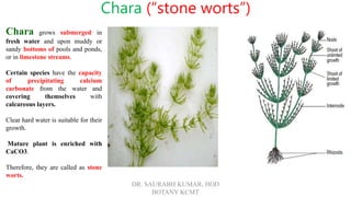
Chara grows submerged in
fresh water and upon muddy or
sandy bottoms of pools and ponds,
or in limestone streams.
Certain species have the capacity
of precipitating calcium
carbonate from the water and
covering themselves with
calcareous layers.
Clear hard water is suitable for their
growth.
Mature plant is enriched with
CaCO3.
Therefore, they are called as stone
worts.
Chara (“stone worts”)
2. DR. SAURABH KUMAR, HOD BOTANY KCMT
Structure of Chara
Plant body or thallus of chara is Multicellular, Macroscopic,
Filamentous and Branched
The plant attains a height of 20-30cm. It has divided into following
parts.
Rhizoids
Arise from lower end, branched, multicellular, no nodes and
internodes, uniseriate, obliquely septate, arise from rhizoidal
plate attached the substratum by means of rhizoids.
Branches
The thallus has long slender upright branches. The axis is
differentiated into node and internodes. From each node arise the
following four types of appendages,
1. Branchlets:
Each node bears a whorl of branches, which has limited growth.
These are called branchlets or branches of limited growth.
3. DR. SAURABH KUMAR, HOD BOTANY KCMT
2. Long Branches:
There are one or two branches of
unlimited growth may also be
present in each node. They arise
singly at the older nodes of the main
axis. They often called as axillary
branches. The axillary branch
continues the growth of thallus.
3. Stipuloids:
These are unicellular out growth that
arises from the basal node of each
branchlet. They are usually two in
number.
4. Cortex:
In some species, the intermodal cells
is covered by a sheath of vertically
elongated narrow cells, the cortex
Such species are called as corticated.
4. DR. SAURABH KUMAR, HOD BOTANY KCMT
Cell Structure
Young cells are always uni-
nucleated structures without
conspicuous vacuole.
In the mature cells, the vacuole
may be developed and may
become multinucleated due to
amitosis.
There are many small ellipsoidal
chloroplasts arranged
longitudinally in peripheral
portion of cytoplasm.
The Cell wall of the internodal
cell is impregnated with silica
and calcium carbonate.
The reserve food material is
starch and the cytoplasm show
characteristic cyclic movement.
5. DR. SAURABH KUMAR, HOD BOTANY KCMT
Growth of the Thallus
The growth of the thallus is by dome-
shaped apical cell which cuts off
derivatives at its posterior surface
Each derivative undergoes a transverse
division to produce two daughter cells.
The upper cell acts as nodal initial and
the lower as internodal initial. The
internodal initial elongates many times
its original length and matures to form
internode of the axis.
The nodal initial divides by vertical
divisions intersecting each other to
produce two central cells surrounded by
6-20 peripheral cells.
Each peripheral cell cuts off apical cells
of branches of limited growth.
6. DR. SAURABH KUMAR, HOD BOTANY KCMT
Reproduction
The Chara reproduces by vegetative and sexual reproduction.
Asexual reproduction by spore formation is absent.
Vegetative Reproduction
The vegetative reproduction in Chara involves various kinds of
reproductive bodies which on detachment from the parent
plant give rise to a new plant. The common means of vegetative
reproduction are as followings:
Amylum Stars: These are star-shaped groups of cells
developed from the lower nodes. The cells contain amylum
starch. After being detached these develop into new
plants.
Bulbils: These are small, rounded or star-shaped, tuberous
bodies that develop either on rhizoids (Chara aspera) or on
stem nodes (Chara baltica). These develop into new plant on
detachment.
Secondary Protonema: In some cases, protonema-like
outgrowths arise from the node of primary protonema or
from the basal cell of the primary rhizoid. These are called
secondary protonema and develop into new plants like primary
protonema.
7. DR. SAURABH KUMAR, HOD BOTANY KCMT
Sexual Reproduction
The sexual reproduction is an advanced oogamous.
The gametes are produced. In antheridia and oogonia
that are enveloped in multicellular sheaths formed
of cells derived from the cells present below the sex
organs.
These specialist complex structures containing the
sex organs are called globule (male fructification)
and nucule (female fructification).
Some species are dioecious while the others are
monoecious. The monoecious species are
protandrous, i. e., the male sex organs develop first.
8. DR. SAURABH KUMAR, HOD BOTANY KCMT
Globule: The male fructification is called as globule. The mature globule is bright
yellow in color. It consists of following parts.
Pedicel cell
The globule is attached to the plant by a large cell called as pedicel cell. It extends within the
cavity of the globule and join with the primary capitula
Shield cells
Each globule is in the form of ball like structure whose wall consists of large plate like eight
cells called as shield cells. The outer wall of each shield cell has radial out growth therefore it
appears as multicellular structure.
Manubrium
On the inner side of shield-cells in the center is attached an elongated cell called as manubrium.
Primary capitula
The inner ends of manubrial cells are united to form eight isodimetric cells called as primary
capitula
Secondary capitula
Each primary capitula has one or two smaller cells towards the cavity of globule called as
secondary capitula.
Antheridial Filament
Attached to the primary or secondary capitula are several branched uniseriate filament called
as antheridial filament. Its contents are metamorphosed into a single antherozoid.
Antherozoid
Each antherozoid is an elongated somewhat coiled structure. It bears two flagella.
Liberation of Antheridia
When the antherozoids are mature, the shield cells are separated from each other, exposing the
antheridial filament. Antherozoids are than escape through a pore. The liberation of
antherozoids may takes place in the morning and they may swim until the evening.
9. DR. SAURABH KUMAR, HOD BOTANY KCMT
Development of Globule
The development of the globule starts from the adaxial
peripheral cell of the lower node of a branch of limited
growth. It divides by a periclinical division into an outer
globule initial and an inner cell that undergoes another
periclinal division. The lowermost cell act as internodal
cell and the middle one forms the basal node of the
antheridium. The basal nodal cell divides to produce five
peripheral cells, the uppermost cell acting as globule
initial.
The globule Initial divides transversely into a basal pedicel
cell and a terminal antheridial mother cell. The pedicel
cell does not divide further rather elongates and protrudes
into the antheridial cavity. The antheridial mother cell
becomes spherical and divides by two vertical divisions at
right angles to each other to form a quadrat. All these cells
divide transversely to produce an octant of eight cells. Each
of these eight cells divide periclinally into an outer and an
inner cell. The cells of outer layer divide periclinally again
and as a result three layers of cells are formed lying one
above the other in the same radius. At this stage, the
antheridium consists of twenty-four cells arranged in eight
diagonal series of three cells each.
10. DR. SAURABH KUMAR, HOD BOTANY KCMT
Nucule
The female fructification is called as nucule. It consists of following
parts.
Pedicel cell
It is present at the base of the nucule on which are present central and
stalk cell.
Oogonium
Upper to the stalk cell is a very much-enlarged structure called as
oogonium. Its contents are transformed into a single large
uninucleate egg.
Tube cells
The oogonium is covered by five elongated spirally twisted cells,
called as tube cells.
Corona
At the top of the oogonium is a crown of small five cells called the
corona.
11. DR. SAURABH KUMAR, HOD BOTANY KCMT
Development of Nucule
The nucule initial divides twice transversely to produce a
filament of three cells.
The lowermost cell elongates and function as pedicel of the
nucule. The middle cell divides by vertical divisions to form
five sheath initials surrounding a central cell. The terminal cell
functions as oogonium mother cell. It elongates and divide
transversely into a lower small stalk cell and an upper
oogonium. The oogonium enlarges and its contents
metamorphose into a single egg.
The sheath initials elongate, grow upward, and divide
transversely to form two tiers of five cells each. The cells of
the upper tier function as corona cells and these forms the
corona of the nucule. The cells of the lower tier act as tube
cells. The tube cells elongate and twist spirally in clockwise
direction around the oogonium.
The egg becomes filled with starch and oil, its nucleus
migrates towards the lower side and a receptive spot develops
at the top of it.
12. DR. SAURABH KUMAR, HOD BOTANY KCMT
Fertilization
When the nucule is Mature, the spirally twisted tube cells
separated from each other just below the corona to form five
small slits.
Antherozoids swim through these slits and enter into the
sheath of nucule. One of the antherozoid enters the egg and
fertilization is completed.
The zygote secretes a thick wall and become oospore.
The zygospore falls to the bottom of the pond and germinates
after a period of rest of few weeks or more.
13. DR. SAURABH KUMAR, HOD BOTANY KCMT
Structure and Germination of
Oospore
The oospore germinates after a period of rest.
Its nucleus divides by meiosis to produce four haploid nuclei.
The oospore divided into two unequal cells by a wall.
The upper cell is small and uninucleate and the lower cell is
larger and contains three nuclei which disintegrate later on.
The oospore wall burst open to expose the upper cell.
It divides by an oblique longitudinal wall into a protonemal
initial and a rhizoidal initial.
The rhizoids develop from the rhizoidal initial and the
protonemal initial develops into an erect primary protonema
which is differentiated into nodes and internodes later on.
A new adult plant develops from the primary protonema.
The life cycle of Chara is Haploid.