Pellia
•
18 likes•15,790 views

This ppt contains all about the genus Pellia of Bryophytes. It contains about the systematic position, habitat, distribution,

Recommended
More Related Content
What's hot
What's hot (20)
Similar to Pellia
Similar to Pellia (20)
More from Sangeeta Das
More from Sangeeta Das (20)
Recently uploaded
Recently uploaded (20)
Pellia
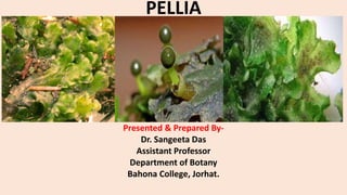
- 1. PELLIA Presented & Prepared By- Dr. Sangeeta Das Assistant Professor Department of Botany Bahona College, Jorhat.
- 2. Systematic Position of Pellia: Division- Bryophyta Class- Hepaticopsida Order- Jungermanniales Family- Pelliaceae Genus- Pellia
- 3. Distribution and Habitat of Pellia: Pellia is a thalloid liverwort spread widely in north temperate regions of the world. In India, it is quite common in Sikkim, Darjeeling and eastern Himalayas and also in western Himalayas and Kumaon regions. Pellia occurs in diverse conditions especially in moist damp soil, more particularly by the sides of moist rocks, ditches, streams and other similar surroundings. It also sometimes grows submerged under flowing water. The form and texture of the thallus varies according to the habitat.
- 4. A. External Morphology: The plant body is thalloid, prostrate, simple, dorsiventral thallus. It is dichotomously branched as in Marchantia but differs in its external form. The thallus is thin, flat, green and lobed. The margin is sinuous and irregularly lobed.
- 5. B. Anatomy: Anatomically, the thallus is very simple with least differentiation of tissues and mainly consists of thin, polyhedral, parenchymatous cells (A-C). The cells are joined together in a honey-comb-like manner. The thallus is several layers of cells thick along the median line or midrib region. In some species (P. epiphylla and P. neesiana) the cells in the midrib region are elongated in the direction of the long axis of the thallus lobes. The cell walls of these elongated cells are thickened by brown or yellow layer of thickening bands forming a kind of network (C). The pores and air chambers characteristic of Marchantia are absent.
- 6. C. REPRODUCTION: Reproduction take place by two methods-vegetative and sexual. 1. Vegetative Reproduction: It takes place by the following methods: (i) Adventitious branches. (ii) Fragmentation, and (iii) Regeneration. 2. Sexual Reproduction: The thallus bears the sex organs-antheridia and archegonia. Some species of Pellia are dioecious e.g., P. neesiana and others monoecious e.g., P. epiphylla. The monoecious species are protandrous (Antheridia are developed first).
- 7. PELLIA An enlarged antheridium V.S. of monoecious thallus showing the position of sex organs
- 8. Sporogonium: At maturity the sporogonium consists of the foot, the seta and the capsule. The foot is distinct and conical in structure and forms the basal, absorbing region of the sporogonium. Its edges project upwards forming a collar-like structure around the base of the seta. The seta is of white in colour, almost transparent at maturity and terminates in a dark green or black nearly globular capsule.
- 9. Structure of capsule: It is globular in outline. The capsule wall is two or more cell layers thick. Within the capsule wall is the spacious spore cavity which contains free elaters intermixed with spores. The spores are large and begin to germinate before they are shed. The mature free elaters are empty considerably long, slender, spindle-shaped cells with 2 or 3 sometimes more up to six, bands of spiral thickening on their walls. They are hygroscopic and thus well known as organs of spore dispersal. Besides the free elaters, there is a prominent basal central core or tuft of 50-100 elaters attached to the centre of the base of capsule. This elateral cluster is often but erroneously termed the elaterophore.
- 10. Life cycle of Pellia sp.