A worldwide community photographing and learning about wildlife

Project Noah Nature School

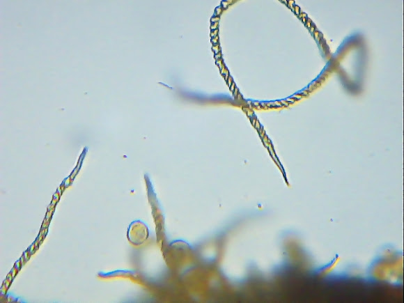
Trichia varia (Pers. ex J.F. Gmel.) Pers. (1794)
Capillitium: Yellow to ochraceous, with long free elaters, 3-5 µm in diameter with2 (3) widely spaced spiral bands. Tips acute, curved lengh twice the diameter of the elaters. Spores : Dull yellow, delicately warted, 12-`14 µm in diameter ( http://hiddenforest.co.nz/slime/index.ht... ), ( http://slimemold.uark.edu/fungi/webforms... varia (Pers. ex J.F. Gmel.) Pers. (1794)&NameKey=CD8A4504-DB5A-46A6-9F9A-C3AC29225FFC&ImageKey=7AA685E0-3587-46DC-B709-E061A79C6CF2 )
------What these three groups have in common is a life cycle that superficially resembles that of the fungi. When conditions become unfavorable, these slime molds form sporangia - clusters of spores, often on the tips of stalks such as in the sporangium of a Physarum shown at right. Spores from the sporangia are dispersed to new habitats, "germinate" into small amoebae, and the life cycle begins again. Similarities in the life cycle do not, however, imply close relationship, especially when one considers that certain bacteria (the myxobacteria) and even an unusual ciliate have very similar life cycles, aggregating to form spores on a sporangium. Slime molds have almost no fossil record, which is not surprising. Not only do slime molds produce few resistant structures (except for spores, which are often overlooked or unidentifiable), but they live in moist terrestrial habitats, such as on decaying wood or fresh cow dung, where their potential for preservation is low. A few fossil slime molds have been found in amber ( http://www.ucmp.berkeley.edu/protista/sl... )
Sporocarps gregarious or crowded, sessile or with a short, stout, black stalk, rarely subplasmodiocarpous. Sporotheca shining yellow, globose, obovoid or somewhat elongate, 0.5-0.9 mm wide. Peridium membranous. Hypothallus broadly expanded, inconspicuous. Capillitium of simple or rarely branched elaters, 3-5 µm diam., bearing 2(-3) irregular spiral bands, these prominent and in places remote, the apices often swollen behind the conical tip. Spore-mass yellow to orange-yellow. Spores dull pale yellow, verruculose, 12-14 µm diam. Plasmodium white ( http://www.discoverlife.org/mp/20q?searc... )----- ( http://slimemold.uark.edu/fungi/WebForms... ), ( http://www.flickr.com/photos/41066614@N0... )= outside ( http://www.projectnoah.org/spottings/935... ) [fopdraadwatje-> dutch]-- while tricha in mind i was looking for some microscope-picture in the internet, not much ( few have posted some - but then good once ), first, not right spirals, then to big,thin,not or too spiny....... this is what i found !

Spotted on Mar 1, 2012
Submitted on Mar 3, 2012


1 Comment
thanks clive,recently i can't put that much time in my hobby ( it is definitly noticeable ), and different, good references,isn't that easy to find (there is you site, very helpfull sometimes).I have much to learn and appreciated your helpfull words! :)