Article

Comparative Morphological Analysis of Two Species of Turtle Leeches Coexisting in North America (Hirudinea: Glossiphoniidae): Embryological Evidence for Character Displacement

Medical Leech Museum, 2 Bryngwili Road, Hendy, Pontarddulais, Swansea SA4 0XT, UK
Taxonomy 2022, 2(2), 160-179; https://doi.org/10.3390/taxonomy2020013
Submission received: 12 January 2022 / Revised: 14 March 2022 / Accepted: 30 March 2022 / Published: 2 April 2022

Abstract

:
The genus Placobdella (Glossiphoniidae) has a centre of species concentration in North America. The type species P. costata is the only representative in the Palaearctic region. American Placobdella which feed on turtles are represented predominantly by two common species, P. parasitica and P. rugosa, which geographically overlap in eastern USA and southern Canada. The latter species is morphologically indistinguishable from P. multilineata of southeast USA. These two tuberculated forms are recognised herein as a clade and treated as a single ‘species’ for comparative purposes. Both P. parasitica and P. rugosa clade commonly coexist and feed on the same turtle species without host preference. This paper addresses morphological differences between them. An unexpected finding is that hatchlings of both species are very difficult to distinguish. A morphologically significant observation is that developmental divergence results in tubercle prominence in P. rugosa clade, but tubercle suppression in P. parasitica, the first example of character displacement in the Hirudinea. Morphological differences are interpreted as reflecting interspecific competition, a phenomenon not found in their Palaearctic counterpart. Why do the two American turtle leech species coexist rather than reduce competition by partitioning their food supply?

1. Introduction

The genus Placobdella Blanchard, 1893, is a group of leeches in the family Glossiphoniidae with a centre of species distribution in North America [1]. For historic reasons the type species is P. costata (Fr. Müller, 1846), the only representative of this genus in the Palaearctic region [2,3,4].
By way of comparison, North American Placobdella are represented predominantly by two geographically overlapping species. One is P. parasitica (Say, 1824) [5] which is distributed throughout most of the eastern half of North America from southern Canada to Florida and Texas [6,7,8].
The other is P. rugosa (Verrill, 1874) [9,10] of the northern half of the United States and adjacent southern Canada [11,12,13]. A recent morphological study has shown that P. rugosa is barely distinguishable from P. multilineata Moore, 1953, of the southeast United States [9,14]. Thus, for the purpose of this paper these closely allied tuberculated forms are considered to represent a clade P. rugosa/ P. multilineata. For didactic and practical purposes this clade is treated herein as a single ‘species’ and simplified as P. rugosa clade.
The one Palaearctic species and the two American species feed almost exclusively on freshwater turtles. P. costata feeds predominantly on the European pond turtle, Emys orbicularis (L., 1758). The two American species also feed on emydine turtles [15] but they feed more commonly on a wide range of other turtle species, without host preference. Furthermore, individuals of P. parasitica are frequently encountered with individuals of the P. rugosa clade throughout their range, evidence for which is presented below.
This study addresses morphological differences between P. parasitica and P. rugosa clade. These differences are interpreted in the context of interspecific competition, and are compared briefly to their Palaearctic counterpart which has no interspecific competition. A remaining evolutionary question from this study is why do the two American leeches coexist over such an extensive range rather than reduce competition by partitioning turtle host species?

2. Objective, Study Area, Material and Methods

2.1. Objective

This study uses comparative methodology to identify morphological differences between two coexisting species of Placobdella. Toward this end, two criteria had to be met: (1) A comprehensive morphological description of each of the two respective leech species; and (2) verification that the specimens used in this study do indeed coexist, i.e., live in precisely the same habitats and feed on the same turtle hosts.

2.2. Study Area

The Outer Banks region of northeast North Carolina and southeast Virginia, USA, is one of the largest freshwater wetlands in the United States. This unique ecosystem lies at the northern tip of a coastal strip warmed by the Gulf Stream and protected by barrier islands known as the Outer Banks. This massive swampland abounds with some of the largest numbers and highest diversity of turtles in the country [16,17,18]. An ongoing study of the Hirudinea of the Outer Banks confirms there are two species of turtle leeches indigenous to this region. Individuals of both species feed on the same turtle hosts and occur in precisely the same habitats. Thus, this region meets both criteria for studying morphological differences between coexisting species of Placobdella.
The more common of the two turtle leeches in this region is provisionally identified as P. multilineata (Figure 1B), whose morphology is the most thoroughly described within the P. rugosa clade [9]. The less common turtle leech here is readily identifiable as P. parasitica [1] (Figure 1A). To further corroborate the identity of this species a morphological comparison is made in this study with published taxonomic accounts of P. parasitica, namely those from Minnesota (type locality), Ontario, Illinois and Massachusetts [7,8,19,20,21]. Finally, a molecular study of specimens of this species, also from the Outer Banks region, confirms its identity is indeed P. parasitica [8].

2.3. Resource Material

2.3.1. P. multilineata

A total of 676 individuals of this species from the Outer Banks have been examined morphologically in a companion study [9]. These specimens were fixed in 5% formalin and deposited along with locality data, hosts, etc, in the Hirudinea collection of the Medical Leech Museum, Swansea, UK. The deposition numbers of this species are prefixed by the rubric PM.

2.3.2. P. parasitica

During the period 2003–2020, individuals of this species were collected at infrequent time intervals and then from only two localities in southern Tyrrell County, NC (both of which also harboured P. multilineata). They were fixed and deposited as for the preceding species. The deposition numbers are prefixed by the rubric PP.
The following individuals of P. parasitica were examined in this study: Tyrrell County, Gum Neck, near FWB Church [35.712480, −76.114413]: PP-1 [26 May 2017, 1 recently fed specimen on snapping turtle, Chelydra serpentina, on land, 36.5 mm]. Tyrrell County, Gum Neck, 460 Grapevine Landing Road [35.719759, −76.107886]: PP-2 [3 May 2018, 1 specimen with some blood in gut, on mud turtle, Kinosternum subrubrum, on land, 22.0 mm. Note this specimen was bitten by its host soon after capture.]; PP-3 [10 October 2013, 1 unfed specimen, on aluminium trap, 12.8 mm]; PP-4a [27 May 2019, 1 brooding adult with numerous hatchlings on venter, on aluminium trap, 39.8 mm]; PP-4b [hatchlings of PP-4a].

2.4. Methodology

The techniques of collection, preservation, dissection, photography and taxonomic terminology are recounted in detail in the companion paper on P. multilineata [9] and need not be repeated here. For P. parasitica all post-hatching individuals collected in this study (PP-1, PP-2, PP-3, PP-4a) were first examined externally. Subsequently, these same individuals were dissected, with the exception of the smallest PP-3 (12.8 mm), with particular attention to variation of dorsal tuberculation and anatomy of the proboscis complex.
An often ignored aspect of morphological variability in leeches is the degree of change an individual undergoes during growth and maturation. In this context special attention is focused on development of key morphological characters defining P. parasitica. The development of P. multilineata had been previously described in a companion paper [9]. Specimens of P. parasitica examined in this study ranged developmentally from recent hatchlings (PP-4b, 6.0 mm) to early juvenile (PP-3, 12.8 mm), and young adult (PP-2, 22.0 mm) to fully mature, brooding adult (PP-4a, 39.8 mm). The hatchlings of both species were scrutinised and photographed under varying lighting and magnification.
The current study is the first comprehensive account of the external and internal morphology of P. parasitica. For the purposes of this comparative study the previous account of P. multilineata serves as a baseline for the entire P. rugosa clade. In lieu of a lengthy description of P. parasitica, the following section focuses on notable differences between the two species under study.

3. Results

3.1. Comparison of External Morphology

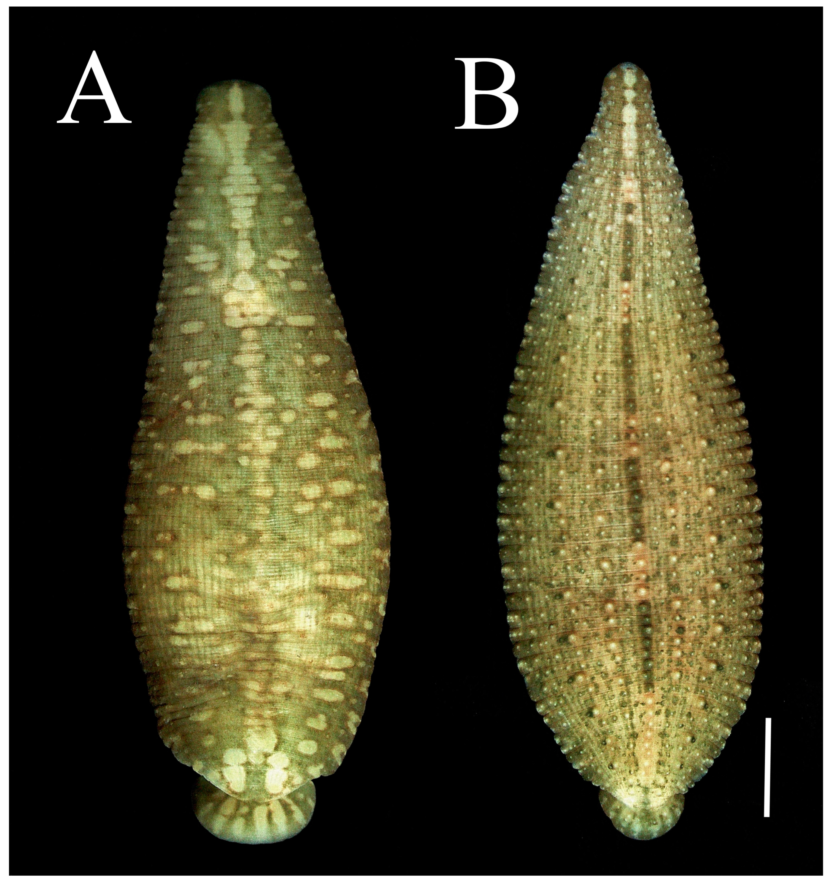
Dorsum. The dorsum of mature P. parasitica encountered in this region is comparatively smoother (PP-4a) (Figure 2), without longitudinal rows of prominent tubercles so diagnostic of P. multilineata from the same region. However, tubercles, specifically those in intermediate position, are detectable in young P. parasitica. As discussed below, the integrity of these structures is lost during maturation.
Note on terminology: The term ‘tubercle’ is used in this paper in preference to ‘papilla’ without comment with regard to cytology or function. Tubercle as used herein refers to pointed or rounded structures well raised from the tegument and potentially on any annulus. In the P. rugosa clade they are typically found in a regular pattern on the dorsum. In this paper ‘smooth dorsum’ refers to the absence of distinct tubercles, or at most bearing only relatively fewer diminutive structures of little overall import. The term sensillum refers to a simple sensory receptor in the epidermis.
Head. The head of P. parasitica is rounded with a broad oral sucker, whereas the head and oral sucker of P. multilineata is noticeably more attenuated. Thus, the width of the oral sucker in relation to body length of relaxed adults is 10.4% (N = 4) in P. parasitica but only 7.5% (N = 3) in P. multilineata. Furthermore, the ratio of length/width of the oral sucker is 58.2% (N = 4) in P. parasitica and 75.7% (N = 4) in P. multilineata.
Annulation. Annuli of the neck segments of P. parasitica are perceptibly foreshortened, i.e., detectably less subdivided, compared to P. multilineata. Thus, in the former species from segment V to as far as segments VII (PP-1), VIII (PP-4a) or IX (PP-2, PP-3), respectively, the first two annuli (a1 + a2) of each respective segment are barely divided, whereas in P. multilineata these serially homologous neck segments are clearly divided. Based on limited data, similar foreshortening may also occur in the caudal region in that segment XXV in P. parasitica appears to be uni-annulate, whereas this segment is consistently bi-annulate in P. multilineata.
The noticeably greater subdivisions in the head and tail segments of P. multilineata are interpreted functionally as manifestation of greater extensibility of P. multilineata compared to the seemingly less mobile P. parasitica.
Caudal sucker. The caudal sucker of P. parasitica is relatively larger and more substantial than that of P. multilineata. Thus, the width of the caudal sucker in relation to body length of relaxed adults is 17.3% (N = 4) in P. parasitica but only 10.8% (N = 18) in P. multilineata. The more substantial caudal sucker in P. parasitica is interpreted functionally as more of a holdfast organ rather than one of locomotion as suggestive in the case of P. multilineata. Interestingly, the caudal sucker of the former species functions amply after death, if pressure is initially applied.
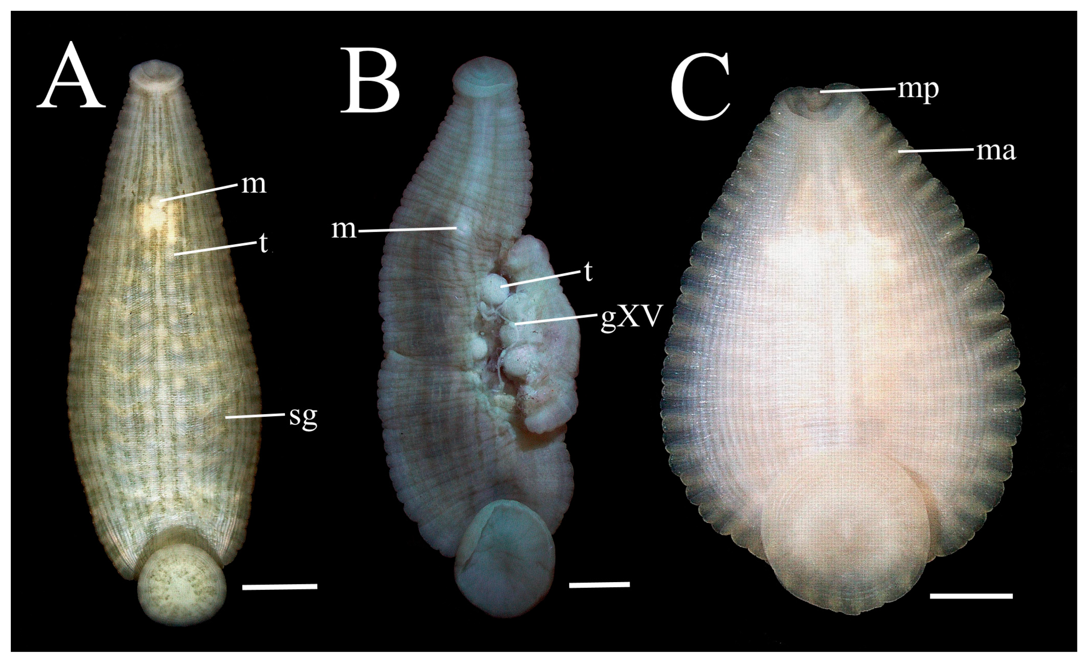
Ventral stripes. The venter of P. parasitica typically bears 8 to 10 prominent longitudinal stripes along its entire length, each comprised of distinctive green chromatophores (Figure 3A, sg). These ventral stripes can be useful for field identification. However, they are not reliable for taxonomic purposes in that they fade or disappear under certain conditions of preservation (PP-1, PP-2, PP-3) (Figure 3B,C), especially in ethanol.
Colour change. P. parasitica is capable of conspicuous colour change. In this study this phenomenon was observed only once in which a recently captured mature individual (PP-4a) changed within minutes from dark brown to brilliant green and cream. The latter colour was retained after fixation and preservation (Figure 1A and Figure 2A). Colour change was not observed to any degree in P. multilineata from the Outer Banks [9].
Placobdellid head pattern. The ‘placobdellid head pattern’ of P. parasitica is basically similar to that which characterises P. multilineata but the two species differ in detail. In P. parasitica this head pattern is comprised of a light-coloured, short rod mid-dorsally in segments IV and V, with an indentation at VIa1 + a2, before expanding widely toward the margins at annulus VIa3 (Figure 2A–C, pp). This medial rod appears to be comprised of homogeneous cellular constituents distinguishable from the cream-coloured patterns posterior to segment VI. In P. multilineata, the pattern typically consists of three consecutive, slightly contiguous cream-coloured patches in IV, V and VI, and as a rule the cream colour of annulus VIa3 does not extend exaggeratedly toward the margins as consistently seen in P. parasitica, at least in this study.
Metameric marginal pattern. The ‘metameric marginal pattern’ along the full length of the body of P. parasitica is basically similar to that so characteristic of P. multilineata. However, that of P. parasitica differs slightly in that the two contiguous light annuli (a3, a1) of each segment at the margins are comprised of numerous small cells which typically extend medially to about the paramarginal position (Figure 2A–C, ma). On the other hand, those of P. multilineata are comprised of fewer ‘colossal’ cells (plus a contiguous unpigmented zone), and are mostly confined to the margins [9] (figs 6A–10A).
Mid-dorsal cream-coloured pattern. Adults of P. parasitica in the Outer Banks display a characteristically variable, cream-coloured pattern at the dorsal mid-line from segment VII to XXIVa3 (Figure 2). This design is derivable from a notional longitudinal stripe in which the cream colour expands or recedes in respective annuli to form a pattern which is more or less unique to the individual. Unlike typical P. multilineata there is no longitudinal brown stripe at the dorsal midline of P. parasitica.
Intermediate row of cream-coloured spots. The dorsum of adult P. parasitica from the Outer Banks displays a longitudinal array of cream-coloured spots in intermediate position, extending from segment VI to XXIV (PP-4a) (Figure 2A). They are most evident in anterior segments VIII–X, and again in caudal segments XXII–XXIV, but lose integrity in mid-body segments in some specimens (PP-2) (Figure 2B). These intermediate spots are on the a2 annulus and accordingly are serially homologous to the intermediate tubercles so diagnostic of P. multilineata. The intermediate spots in adult P. parasitica lack distinct tubercles. However, some spots that have a slightly raised centre comprised of a disorganised concentration of cells.
Interestingly, the dorsal pigmentation of P. parasitica from the Outer Banks is not strictly bilaterally symmetrical, especially evident in mid-body segments where spots may occur irregularly in paramedial or paramarginal positions. This adds to the camouflaged individuality of each specimen. Of developmental interest, much of the constituent pigment is confined strictly to a respective annulus, implying some sort of localised mechanism at work.
Pre-anal segments XXVXXVII. The three pre-anal segments XXV–XXVII of P. parasitica lack prominent paired tubercles so characteristic of P. multilineata [9] (fig. 11A). Instead, in the homologous position corresponding to the pre-anal tubercles in P. multilineata there are three pairs of cream-coloured spots in P. parasitica (Figure 2A). Each bears a slightly raised cellular centre, but no structural tubercles. The cream-colour of each spot is centred on these central cells.

3.2. Comparison of Internal Morphology

Proboscis Complex (Figure 4, Figure 5, Figure 6, Figure 7 and Figure 8).
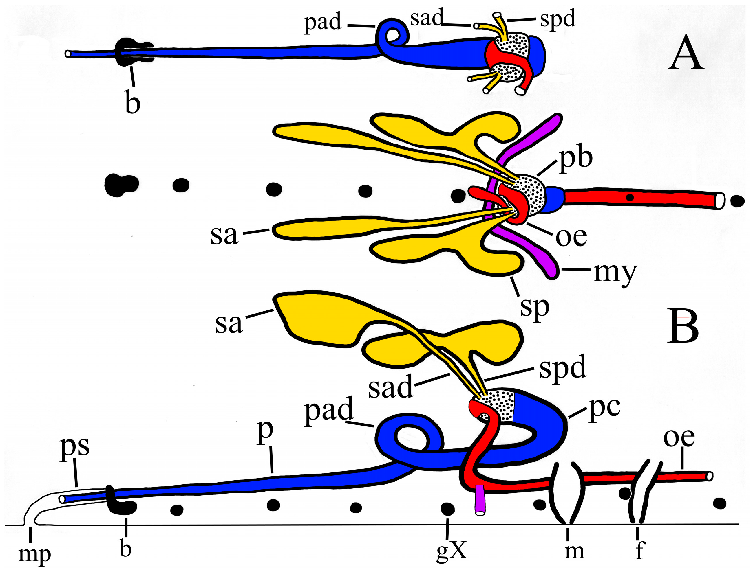
Length and position of proboscis. In P. parasitica the proboscis is long and tapering with its tip positioned in situ anterior to the ‘brain’ in all specimens examined (Figure 8). The position of the posterior end of the proboscis varies among the Outer Banks specimens, from IX/X (PP-1, 36.5 mm), X (PP-2, 22.0 mm) to X/XI (PP-4a, 39.8 mm).
Crook at posterior end of proboscis. In the largest specimen in this study (PP-4a) the posterior end of the proboscis is distinctly crooked along the dorso-ventral axis (Figure 6), whereas in the remaining specimens the proboscis is straight, without a crook (Figure 7, pc). Significantly, in P. multilineata the posterior end of the proboscis is crooked in virtually all specimens, and is diagnostic of that species. Furthermore, the end of proboscis appears to be noticeably further back in P. multilineata [9] (fig. 4, ‘slightly anterior to g. XI’).
Asymmetrical deflection of proboscis. The proboscis of P. parasitica, at least in Outer Banks specimens examined, consistently has a peculiar asymmetrical deflection at segment IX. In the mature specimen (PP-4a) this deflection constitutes an improbable fully formed 360° loop of the proboscis in this segment (Figure 6A, star; Figure 7, pad). In less mature specimens this deflection is less developed, appearing either as a sizeable curve (PP-2) (Figure 6B, star; Figure 8, pad), or simply as a slight deviation (PP-1) (Figure 4, star). In contrast, the proboscis of all P. multilineata examined is straight all along its length, and lacks an asymmetrical deflection.
Compact salivary glands. All dissected specimens of P. parasitica in this paper have two bilateral pairs of compact salivary glands, each with an individual ductule (i.e., no common ductule) which leads into the base of the proboscis.
Note on terminology: Salivary ‘gland’ is used in this paper for convenience. They are not true glands with a lumen typical of vertebrate glands. The leech salivary gland is actually an aggregation of large unicellular exocrine cells each of which has an exceedingly long sub-cellular process leading from the soma into the proboscis, as demonstrated in this species by Moser and Desser (1995) [6]. These individual processes from each gland together form a clearly recognisable bundle. For convenience each multi-cellular bundle is simply called ‘ductule’ in this paper.
Independent salivary ductules. The respective ductules of the anterior and posterior salivary glands of P. parasitica enter the base of the proboscis independently or at most barely abutting (Figure 7, sad, spd), whereas the ductules of P. multilineata join a measurable distance from the proboscis to form a clear and unmistakable common ductule [9] (fig. 4, sb). This taxonomically important distinction alone appears to distinguish P. parasitica from its tuberculated counterpart P. rugosa clade [6,9].
Median annectant lobe. Dissected specimen PP-1 (36.5 mm) reveals a peculiar accumulation of about 25–30 salivary cells at the mid-line at about IX/X. These cells together constitute a delicate median lobe which more or less connects the two posterior salivary glands in this specimen (Figure 4, mac). A similar annectant lobe may also occur in the other two dissected specimens of P. parasitica. However, due to its location at the mid-line it may have been cut inadvertently in order to view the underlying structures. This median annectant lobe appears not to occur in P. multilineata, but this should be re-examined.
Larger and less numerous salivary cells. The salivary glands of P. parasitica contain significantly fewer but larger cells than those of P. multilineata, at least in Outer Banks specimens. Each salivary gland of P. parasitica contains on the order of only 50 cells, typically 175 µm in diameter, but up to 385 µm. Those of P. multilineata are much more numerous but smaller, up to only about 100 µm. The posterior gland of P. parasitica is ‘squatty’ and confined to its segment, extending only to just beyond g. X (Figure 4, Figure 6 and Figure 7, sp), whereas the posterior gland of P. multilineata is typically elongated and extends as far as XI/XII [9] (fig. 4, sp).
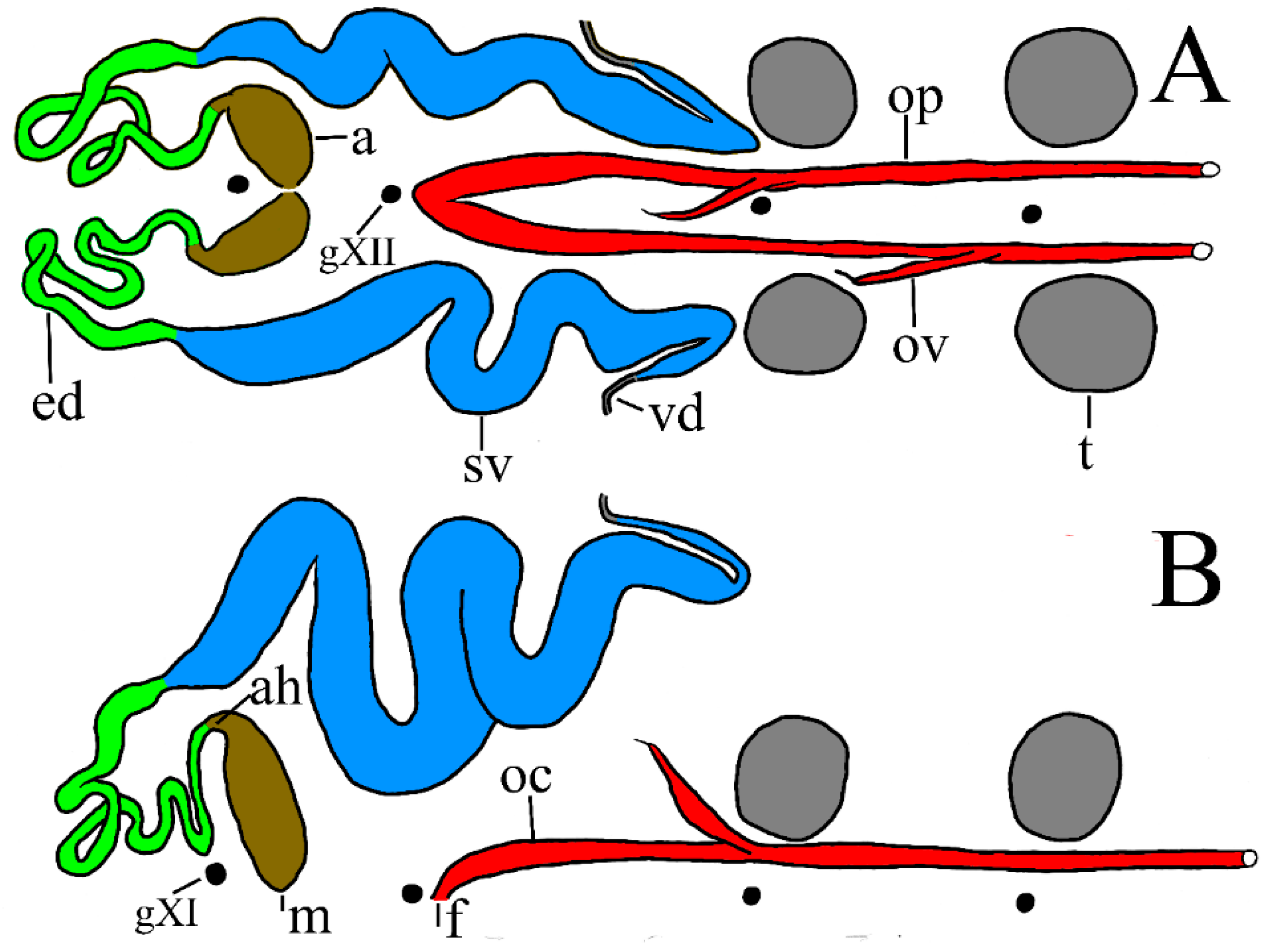
Reproductive systems (Figure 9). Differences between the male anatomy of P. parasitica and P. multilineata are noted in the various dissections, but owing to the physiologically plastic nature of these structures taxonomic significance requires further scrutiny. In P. parasitica the narrow, tubular ejaculatory duct is confined to segment XI and joins the seminal vesicle at about level of the atrium (PP-4a). In one mature specimen (PP-1) the ejaculatory duct extends a little more anterior, to X/XI. In P. multilineata the ejaculatory duct is longer and joins the seminal vesicle at about the level of g. XII [9] (fig. 5, ed). The seminal vesicles of mature P. parasitica are particularly prominent and swollen (Figure 4, sv), and typically observable through the ventral tegument (Figure 3A). The apparent juncture between the ejaculatory duct and seminal vesicle in P. parasitica is at level of the atrium (i.e., behind g. XI), consistently more anterior than the much less swollen seminal vesicles of P. multilineata (i.e., level of g. XII). In both species the seminal vesicle joins the vas deferens just anterior to level of g. XIII. No significant differences between species were noted in the female reproductive system.
Based on limited information, the resting length of gravid/brooding individuals of P. parasitica averaged 38.2 mm (range 36.5–39.8 mm; N = 2), and closely resembled that of P. multilineata in the same region (mean, 36.6 mm; range, 28.0–45.0 mm; N = 23) [9].
Mycetomes. The mycetomes of P parasitica are more substantial than those of P. multilineata. The flattened ductus is relatively wide throughout its length, and typically has a perceptible bulbous ending (Figure 7 and Figure 8, my).
Nephridia. Inexplicably, the nephridia of P parasitica are relatively larger and more conspicuous (Figure 4, n) than those of P multilineata.

3.3. Taxonomic Diagnosis of P. parasitica from Outer Banks

The foregoing section describes external and internal feature of P. parasitica collected from the Outer Banks region, with particular attention to individual variability and differences from the outgroup species P. multilineata from the same locality. This methodology identifies those characters which occur in all adult individuals of P. parasitica, but do not occur in the P. rugosa clade. Taken together these invariant characters constitute a formal diagnosis which defines P. parasitica, at least from the Outer Banks.
Placobdella parasitica (Say, 1824) (Outer Banks).
Diagnosis (post-hatchling). Dorsum is essentially smooth without conspicuous, metameric tubercles. Dorsum displays a bold, irregular and highly variable cream-coloured pigment pattern. The long, tapering proboscis has a peculiar asymmetrical deflection at segment IX. The salivary ductules from the anterior and posterior salivary glands enter the base of the proboscis independently (or at most barely abutting each other). In other words, the respective ductules do not merge to form a clear and unmistakable common ductule a measurable distance from the base of the proboscis as is the case for P. multilineata. In addition, a sometimes useful but taxonomically unreliable character for identifying P. parasitica is the presence of 8–10 broad blue-green ventral stripes.
Hatchlings of P. parasitica are difficult to distinguish externally from P. multilineata in that both possess discrete white pigment patches at intervals, as well as small, but distinct tubercles.

3.4. Morphological Homogeneity of P. parasitica throughout United States and Canada

The current study is the first description of P. parasitica from the southern part of the immense range of this species. In this context, the question is addressed whether specimens from the Outer Banks are anatomically similar to specimens from northern parts of its range. Towards this end, this section critically reviews what is known regarding morphological features of P. parasitica from disparate localities, namely, Minnesota (type locality), Ontario, Illinois and Massachusetts. These four northern localities represent distances of 1000 to 2100 km from the Outer Banks.
P. parasitica from Minnesota (type locality). P. parasitica has been recently redescribed from its type locality at Lily Lake, Waseka County, Minnesota [7]. This redescription was based on a neotype plus nine other mature individuals from Minnesota, one of which was dissected and illustrated. In general, it agrees with the account of this species from the Outer Banks. However, some relatively minor discrepancies, omissions or clarifications are noted below.
P. parasitica from Ontario. In contrast to the scarcity of P. parasitica in the warm swamps of the Outer Banks, this species abounds in the cold lakes of the Algonquin Provincial Park in southeast Ontario [22,23]. It is taxonomically fortuitous that the ultrastructure of the proboscis complex of this species was investigated as part of a morphological study [6]. By way of summary, the proboscis and salivary glands of five specimens of P. parasitica from Ontario were remarkably similar to that of Outer Banks specimens. Of taxonomic relevance for the current study these authors also meticulously described the proboscis complex of the allied tuberculated turtle leech P. rugosa, also from Ontario.
P. parasitica from Illinois. Moore [21] gave the first internal description of this species under the name P. parasitica. He did not give numbers of specimens examined nor specific locations, other than from ‘Illinois’. He specifically commented on the large size of this ‘tortoise’ leech, citing that some individuals could extend to over 100 mm (4 inches). Moore was also the first to compare the external and internal anatomy of P. parasitica and P. rugosa, from Illinois.
P. parasitica from Massachusetts. The first internal description of P. parasitica was based on five specimens from Charles River near Boston, Massachusetts, under the name Clepsine plana [19,20]. The latter name is now recognised as a junior synonym for P. parasitica [7]. Presciently, Whitman was the first to illustrate the taxonomically important proboscis complex of this species [19](fig. 5), [24](fig. 23.4). However, in contrast to P. parasitica presented in this paper, the proboscis of the Boston specimen was clearly illustrated as straight, without a bend or deflection in segment IX, nor with a dorso-ventral crook at its posterior end. These discrepancies may be an error or oversight. However, Whitman was a meticulous researcher, and even made a specific point about his attention to detail, and the accuracy of his reproductions [20] (p. 411).
External features. Specimens from all five localities displayed a dorsum with a bold irregular, cream-coloured or yellowish pigment pattern, and a relatively smooth dorsum (never linear rows of large metameric tubercles, as in the P. rugosa clade). This external uniformity of P. parasitica was further corroborated by exemplary photographs of additional specimens from Minnesota and Connecticut [8] (figs 1 and 2).
Size. The largest specimen of P. parasitica encountered in the Outer Banks (PP-4a, resting length, 39.8 mm) was notably smaller than those found in Minnesota (59.1 mm), Massachusetts (60.0 mm) and Illinois (extended length, 100 mm). This apparent discrepancy may simply be sampling bias.
Ventral stripes. Specimens of P. parasitica from Minnesota [7] (fig. 4), Massachusetts [20] (fig. 2) and the Outer Banks (Figure 3A) displayed 8–10 prominent bluish or greenish longitudinal stripes on the venter. Moore [21] did not specifically mention ventral stripes in specimens from Illinois, but much later he relied on ventral longitudinal stripes in a popular and influential key for identifying this species [24] (p. 550).
By way of caution, while such distinctive stripes are an undeniably useful guide for identifying this species, their presence or absence is not taxonomically sound for diagnosis, especially after preservation (Figure 3B,C). Furthermore, very similar ventral stripes appeared to occur in at least one other nominal species of Placobdella [25] (p. 69). Moreover, interpreting ventral pigmentation can be subjective and misleading. For example, ventral stripes of some individuals of P. multilineata may broadly overlap those found in P. parasitica [5] (fig. 10C).
Pre-anal segments. An apparent discrepancy between Minnesota (type locality) and Outer Banks specimens lies in the pre-anal segments (XXV–XXVII). In some Minnesota specimens three pairs of pre-anal tubercles (‘papillae’) were reported in these segments. However, there is reason to believe the relevant illustration may show instead three pairs of cream-coloured spots in these segments [7] (fig. 3A). Such spots also occurred in Outer Banks specimens, but they were not tubercles in that each spot encompassed slightly disorganised bumps at most. In other words, the presence of distinct pre-anal tubercles is not diagnostic of P. parasitica.

Internal Features

Proboscis complex. Specimens from all five disparate localities reviewed herein possessed two pairs of compact salivary glands and a long slender proboscis whose narrow tip typically extended anterior to the ‘brain’.
Length and position of proboscis. The posterior extension of the proboscis varied somewhat between localities. In most cases, the end of the proboscis was positioned at X (Illinois, Massachusetts, Ontario), whereas in the type locality it was recorded as posterior to X/XI (Minnesota). The position of the posterior end of the proboscis also varied within the Outer Banks population, and accordingly is not considered taxonomically significant.
Crook of posterior end of the proboscis. In the current study, a dorso-ventral crook of the posterior end of the proboscis occurred in only one specimen (PP-4a) encountered in the Outer Banks (Figure 7, pc), the other specimens lack such a crook (Figure 8). Interestingly, a crook also occurred in a specimen from the type locality in Minnesota [7] (fig. 5) and, less demonstrably, from Ontario [6] (fig. 4). No such crook was recorded from Illinois nor from Massachusetts. Owing to its variability, the presence or absence of a dorso-ventral crook is not considered taxonomically significant. On the other hand, such a crook occurred in nearly all specimens of P. multilineata from the Outer Banks [9] (fig. 4).
Asymmetrical bend or deflection of the proboscis at segment IX. This peculiar, possibly unique feature was characteristic of all post-hatching specimens of P. parasitica examined from the Outer Banks. Significantly, it was also found in specimens from the type locality, as illustrated in an excellent dissection from Minnesota in which a veritable loop was present [7] (fig. 5). Similarly, a distinct asymmetrical bend of the proboscis also occurred in specimens from Ontario [6] (fig. 4).
Although such a deflection was not recorded from Illinois nor from Massachusetts, it must be considered that with further data this previously overlooked feature of the proboscis may be diagnostic of post-hatching P. parasitica throughout its range. In this context, it is taxonomically important that this morphological character appeared to be absent in hatchlings of P. parasitica from the Outer Banks (Figure 5C), and significantly in the P. rugosa clade [6] (fig. 3), [9] (fig. 4).
Independent salivary ductules. In all post-hatchling specimens examined from the Outer Banks, the ductules from the anterior and posterior salivary glands, respectively, entered the base of the proboscis independently, or nearly so. This taxonomically important feature may be present in the specimen illustrated from Ontario [6] (fig. 4) but it could not be confirmed unequivocally in specimens from Minnesota, Illinois nor Massachusetts. However, J. P. Moore the doyen of American hirudinology, illustrated what appears to be independent salivary ductules in P parasitica [24] (fig. 23.4).
Conversely, none of the specimens of P. parasitica from any of the localities, including the Outer Banks, have anterior and posterior salivary ductules which join at a measurable distance from the proboscis to form a clear and unmistakable common ductule which enters the base of the proboscis. This latter configuration is characteristic of the P. rugosa clade [9] (fig. 4), [6] (fig. 3, arrowhead, their ornata) and reliably sets these tuberculated forms apart from P. parasitica.
Median annectant salivary lobe. Moore [21] was first to describe a peculiar-overlooked feature of the salivary complex of P. parasitica from Illinois. ‘Very frequently, but apparently not invariably’, a median lobe of salivary tissue links the two posterior salivary glands. A very similar annectant lobe had been illustrated earlier by Whitman [19] (fig. 4) without comment from a specimen from Massachusetts. Finally, at least one specimen from the Outer Banks displayed what appears to be a similar median lobe (Figure 4, mac). This possibly unique annectant lobe is not diagnostic of P. parasitica in that it appears not to be present in all specimens from Illinois nor from the Outer Banks. Of possible taxonomic significance, however, such a median annectant lobe was not present in P. rugosa from Illinois [21] nor in P. multilineata from the Outer Banks [9]. Interestingly, in the current study a similar aggregation of annectant cells occurs in the eyes of P. parasitica (Figure 5D, mac).
Reproductive systems. Unfortunately, details of the reproductive systems from the four northern localities were inadequate for comparison with those from the Outer Banks. The nature of the ejaculatory duct and seminal vesicle from the type locality of P. parasitica could be particularly informative [7] (fig. 6).
By way of summary, morphological features of P. parasitica from these four geographically disparate localities were remarkably similar to those described herein from the Outer Banks, with only minor discrepancies or omissions. Therefore, evidence from comparative anatomy suggests that the smooth turtle leech P. parasitica, is morphologically homogeneous throughout the immense range of this species. This finding concurs well with a parallel study of low molecular diversity of this species [8]. Parenthetically, a molecular phylogenetic study of P rugosa documents a similar low diversity [12,13].

3.5. Similarity of Hatchlings of P. parasitica and P. multilineata

This section focuses on morphological aspects of early development of P. parasitica, with unexpected results. It was found that in terms of tuberculation and pigmentation hatchlings of P. parasitica more closely resemble P. multilineata than adults of their own species (Figure 10). In fact, it is remarkably difficult to distinguish these two species at the hatchling stage. Dorsal tubercles, so characteristic of P. multilineata at all life stages, also occur in hatchlings of P. parasitica, but in this species these structures do not develop and accordingly obfuscate during maturation (Figure 1, Figure 11 and Figure 12). Furthermore, hatchlings of both P. parasitica and P. multilineata display very similar white pigment patches at discrete mid-dorsal intervals, but this pigment pattern loses overall integrity during maturation in P. parasitica (Figure 1 and Figure 10).
Tubercle obfuscation in P. parasitica versus prominence in P. multilineata. Of considerable morphological significance, hatchlings of P. parasitica differ from adults in having small but distinct dorsal tubercles (Figure 11, tu). These are in longitudinal rows in intermediate position on the a2 annulus in segments XIII–XXII, inclusively (Figure 12, tu). Furthermore, these align near the respective tips of the underlying crop caeca, a feature seen in young and adult P. multilineata [9] (fig.15). In contrast, in adult P. parasitica tubercles are substantially obliterated or lacking altogether (Figure 2A,B). In younger, juvenile P. parasitica there are still some discernible tubercle-like protuberances in intermediate position on the a2 annulus in segments IX–XXIV, inclusively (Figure 2C, tu).
Whereas the tubercles of P. parasitic become more indeterminate with age (Figure 1A), those of P. multilineata become more prominent (Figure 1B). This is interpreted as the first example of character displacement in the Hirudinea [26,27]. This reflects a significant taxonomic distinction between smooth and tuberculated turtle leeches throughout much of North America as discussed below.
White pigment patches of hatchling P. parasitica and P. multilineata diverge during development. Hatchlings of P. parasitica invariable display six white pigment patches (Figure 10, wp) not discretely evident in the adult of this species. In hatchlings, these white patches are invariably centred at the midline at segments VIa3 (nuchal), IXa2 (pre-genital), XIIa2 (genital), XV/XVI (mid-crop), XVIII/XIX (pre-intestine) and XXIIa1 (anal), respectively (Figure 11 and Figure 12). During growth and development these same patches in P. parasitica, but not P. multilineata, widen into irregular cream-coloured expanses on either side of the mid-line (Figure 2C). With further maturation these may become broadly confluent anteriorly and posteriorly, but they remain centred at the same ordinal locations as the respective patches of the hatchling. In mature adults this underlying pattern may become obscured or lost entirely, especially in mid-body segments XI–XXII (Figure 2A,B). In other words, the characteristically variable dorsal pigment pattern of adult P. parasitica (39.8 mm) is derivable during development from a more organised discrete condition in the hatchling (6.0 mm). The pigment pattern of the juvenile (Figure 2C, 12.8 mm) is clearly intermediate between that of the hatchling and the adult.
These white pigment patches in hatchling P. parasitica closely resemble in position and number ‘white shields’ described elsewhere in hatchling and post-hatchling P. multilineata [9] (fig. 8), with two notable differences. The position of the last or anal patch appears to differ between the two species. In P. parasitica it is centred at XXIIa1, whereas in P. multilineata it is more posterior at XXIII-XXIV.
In addition, in my earlier description of P. multilineata only five, rather than six, white shields were recounted [9]. In retrospect these actually correspond in P. parasitica to white patches 2 to 6, inclusively. This is because the first white patch in P. multilineata was misinterpreted as part of the placobdellid head pattern. This is an opportunity to correct this discrepancy and bring the two species into closer harmony, i.e., both species have six white patches with the first one at XVI-VII. This discrepancy was anticipated in the P. multilineata study in that the white patch in the nuchal region of VI-VII was observed to be ‘comprised of different and less durable kind of pigmentation’ [9] (p. 14).
In the context of the challenging task of separating the two turtle leech species at an early hatchling stage, there appears to be a morphologically useful difference in detail of white patches 1 and 6, respectively, as discussed briefly above. There may also be a species difference in the nature of the cells which make up the white patches, being diffused and rounded aggregations in P. parasitica (Figure 11C,D), but compact, linear and organised in P. multilineata [9] (fig. 8).

4. Discussion

4.1. Divergent Evolution between Two Coexisting Turtle Leeches in the Outer Banks

The morphology of P. parasitica discussed in detail in this paper is closely compared to that of P. multilineata [9], both of which coexist on turtles of the Outer Banks. Indeed, their biological relationship in the Outer Banks is remarkably close in that they feed on the same turtle hosts, and live in precisely the same habitats. In this context it is proposed here that morphological differences between them largely reflect divergent evolution or character displacement resulting from coexistence in this region [26,27]. The most widely recognised feature distinguishing these species is that P. parasitica is smooth-backed whereas P. multilineata is prominently tuberculated (Figure 1), a distinction not found in early development (Figure 12).

4.2. Divergent Tuberculation between These Coexisting Turtle Leeches Is Widespread in North America

Numerous field studies document that P. parasitica and its tuberculated counterpart P. rugosa clade coexist in local turtle assemblages throughout much, if not most, of eastern United States and southern Canada. They have been recorded as coexisting in such disparate locations as Algonquin Park in southeast Ontario [15,22,23,28], two localities in southwest Ontario [29], southern Quebec [30], Vermont [31], southeast Michigan (George Pond, Livingstone Co) [32], ponds in central and southern Illinois [33], an urban setting in Indianapolis, Indiana [34], west-central Nebraska (Beckius Pond, Keith Co) [35], Mississippi River in northeast Missouri [36], northeast Arkansas [37], southeast Texas [38,39], central North Carolina [40] and northeast North Carolina (this study). In other words, field evidence is overwhelming of a remarkably widespread and close ecological relationship between the same coexisting turtle leeches. In fact, sometimes individuals of both American species occur simultaneously on the same individual turtle [15,28,30]. The two forms may not always exist together, but coexistence appears to be the rule rather than the exception.
In the majority of these field studies P. parasitica is more common than P. rugosa clade. In fact, to date only in the Outer Banks (current study) and in west-central Nebraska [35], P. rugosa clade is more common than P. parasitica. This observation may reflect relative fitness or lifestyle difference of each respective species under different ecological conditions, but this complex question cannot be pursued further here.

4.3. Field Evidence for Competition between Coexisting Turtle Leeches

Numerous field studies of freshwater turtles and their leeches (Placobdella) contribute to our rich understanding of the complex dynamics of the turtle-leech relationship in North America. However, none of these studies focuses specifically on potential competition between coexisting species. In this context a number of these studies confirm that both P. parasitica and P. rugosa clade feed on a remarkable diversity of turtle species without clear overall host preference. In other words, there is no evidence that these two species minimise competition by partitioning their food supply anywhere in their largely sympatric ranges. Inexplicably, however, these two leeches differ in the same anatomical detail of the respective proboscis complexes throughout their range.

4.4. Lack of Interspecific Competition in Palaearctic Placobdella

Molecular analyses of both American species in this study and the one Palaearctic species concur that all three species have undergone a recent post-glacial recolonisation [4,8,13]. Arguably, in not being subject to interspecific competition P. costata has changed little morphologically over this post-glacial time period in possible contrast to the coexistent American species. In fact, putative competition may be a primary factor in the morphological differences reported in this study. In this context it would be desirable to compare the morphology of P. costata methodically with each of its American counterparts. To my knowledge, however, a comprehensive morphological analysis of P. costata is currently lacking.
Taxonomic note: The traditional view is that the Palaearctic region is represented by a single ‘morphological species’ [2,4]. However, a recent molecular analysis of specimens from widely distributed Palaearctic localities presents convincing evidence that, based on COI distances, there are up to five ‘molecular species’ [3], none of which appear to coexist. In this context, unpublished incongruities between morphological and molecular species within the genus Placobdella need to be addressed. These species concepts are not the same and use entirely different criteria [41], each with benefits, such as rate of genetic diversity as indicated above, and drawbacks, such as documenting character displacement. In this paper the morphological species concept is employed for the purpose of meaningful analysis of comparative morphology.

5. Conclusions

From the foregoing study a working hypothesis can be formulated, namely that turtle assemblages throughout the eastern third of the United States and southern Canada are infected predominately by two coexisting species of leeches of the genus Placobdella, namely the smooth-backed P. parasitica and its tuberculated counterpart P. rugosa clade.
Evolutionary advantages of the morphological differences between them invite tentative explanation. For example, the presence or absence of dorsal tubercles may be their way of distinguishing one species from the other, i.e., a reproductive isolating mechanism. However, it must be emphasised there is no evidence for this. Second, differences in dorsal pigment patterns, one being variegated and capable of colour change, and the other reminiscent of a waterlogged leaf, might reflect camouflage against differing predatory pressures, for example on and off the turtle. However, internal differences, particularly in the morphology of the proboscis complex, are more difficult to explain. In any case the nature of the interactions between these coexisting species for food, reproduction, predation, shelter and/or transportation on turtles is complex but nonetheless rich for future study.

Funding

This research received no external funding.

Institutional Review Board Statement

Not applicable.

Informed Consent Statement

Not applicable.

Data Availability Statement

Not applicable.

Acknowledgments

I gratefully acknowledge Dan R. Sawyer of Elizabeth City, NC, and A. Christopher Meekins of Raleigh, NC, for their tangible assistance in field collecting in the Outer Banks region; Charlie Cooper, Gum Neck, Tyrrell County, NC, for collecting leeches on local turtles over many years; and Carl Peters-Bond of Biopharm (UK) Ltd., Wales, for invaluable logistical support and technical advice. Constructive input from three anonymous reviewers contributed significantly to the much improved final version of this paper.

Conflicts of Interest

The author declares no conflict of interest.

References

  1. Moser, W.E.; Govedich, F.R.; Oceguera-Figueroa, A.; Richardson, D.J.; Phillips, A.J. Chapter 12. Phylum Annelida. Subclass Hirudinida. In Thorp and Covich’s Freshwater Invertebrates: Key to Nearctic Fauna, 4th ed.; Thorp, J.H., Rogers, D.C., Eds.; Elsevier: New York, NY, USA, 2016; Volume II, pp. 244–259. [Google Scholar]
  2. Vecchioni, L.; Marrone, F.; Liuzzo, M.; Seglie, D.; Cavalcante, R.; Bazan, G.; Arizza, V.; Arculeo, M. New Molecular Data Attest to the Absence of Cospeciation Patterns between Placobdella costata (Fr. Müller, 1846) (Hirudinea) and Freshwater Turtles (Emys spp.) in Italy. Eur. Zool. J. 2021, 2021, 959–965. [Google Scholar] [CrossRef]
  3. Kvist, S.; Utevsky, S.; Marrone, F.; Ahmed, R.B.; Gajda, Ł.; Grosser, C.; Huseynov, M.; Jueg, U.; Khomenko, A.; Oceguera-Figueroa, A.; et al. Extensive Sampling Sheds Light on Species-Level Diversity in Palearctic Placobdella (Annelida: Clitellata: Glossiphoniiformes). Hydrobiologia 2022, 849, 1239–1259. [Google Scholar] [CrossRef]
  4. Bielecki, A.; Cichocka, J.M.; Jablońsli, A.; Jeleń, I.; Ropelewska, E.; Biedunkiewicz, A.; Terlecki, J.; Nowakowski, J.J.; Szlachciak, J. Coexistence of Placobdella costata (Fr. Müller, 1846) (Hirudinida: Glossiphoniidae) and mud turtle Emys orbicularis. Biologia 2012, 67, 731–738. [Google Scholar] [CrossRef]
  5. Keating, W.H. Class Vermes. In Narrative of an Expedition to the Source of St. Peter’s River, Lake Winnepeek, Lake of the Woods; Carey, H.C., Lea, I., Eds.; GB Whittaker: Philadelphia, PA, USA, 1824; Volume 2, pp. 266–268. [Google Scholar]
  6. Moser, W.E.; Desser, S.S. Morphological, Histochemical, and Ultrastructural Characterization of the Salivary Glands and Proboscises of Three Species of Glossiphoniid Leeches (Hirudinea: Rhynchobdellida). J. Morphol. 1995, 225, 1–18. [Google Scholar] [CrossRef]
  7. Moser, W.E.; Richardson, D.J.; Hammond, C.I.; Lazo-Wasem, E.A. Redescription of Placobdella parasitica (Say, 1824) Moore, 1901 (Hirudinida: Glossiphoniidae). Bull. Peabody Mus. Nat. Hist. 2013, 54, 255–262. [Google Scholar] [CrossRef]
  8. Richardson, D.J.; Hammond, C.I.; Moser, W.; Phillips, A.J.; Lazo-Wasem, E.A.; Barger, M. Molecular Variation and Biogeography of the Common North American Turtle Leech, Placobdella parasitica. Bull. Peabody Mus. Nat. Hist. 2020, 61, 83–95. [Google Scholar] [CrossRef]
  9. Sawyer, R.T. Description and Taxonomic Analysis of a Tuberculated Turtle Leech from the Outer Banks, North Carolina, USA, Provisionally Identified as Placobdella multilineata Moore, 1953 (Hirudinea: Glossiphoniidae). Zootaxa 2021, 4991, 1–35. [Google Scholar] [CrossRef]
  10. Verrill, A.E. Synopsis of North American Freshwater Leeches. In United States Fisheries Commission Report for 1872–1873; United States Government: Washington, DC, USA, 1874; Pt. 2; pp. 666–689. [Google Scholar]
  11. Moser, W.E.; Richardson, D.J.; Hammond, C.I.; Govedich, F.R.; Lazo-Wasem, E. Resurrection and Redescription of Placobdella rugosa (Verrill, 1874) (Hirudinida: Glossiphoniidae). Bull. Peabody Mus. Nat. Hist. 2012, 53, 375–381. [Google Scholar] [CrossRef]
  12. de Carle, D.; Oceguera-Figueroa, A.; Tessler, M.; Siddall, M.E.; Kvist, S. Phylogenetic Analysis of Placobdella (Hirudinea: Rhynchobdellida: Glossiphoniidae) with Consideration of COI Variation. Mol. Phylogenet. Evol. 2017, 114, 234–248. [Google Scholar] [CrossRef]
  13. Mack, J.; de Carle, D.; Kvist, S. Prey, Populations, and the Pleistocene: Evidence for Low COI Variation in a Widespread North American Leech. Mitochondrial DNA A: DNA Mapp. Seq. Anal. 2019, 30, 749–763. [Google Scholar] [CrossRef]
  14. Moore, J.P. Three Undescribed North American Leeches (Hirudinea). Not. Nat. Acad. Nat. Sci. Philadelphia 1953, 250, 1–13. [Google Scholar]
  15. Saumure, R.A. Emydoidea blandingi (Blanding’s Turtle). Parasites. Herpetol. Rev. 1990, 21, 60. [Google Scholar]
  16. Palmer, W.M.; Braswell, A.L. Reptiles of North Carolina; University of North Carolina Press: Chapel Hill, NC, USA, 2013; pp. 1–444. [Google Scholar]
  17. Meyers, J.M.; Pike, D.A. Herpetofaunal Diversity of Alligator River National Wildlife Refuge, North Carolina. Southeast. Nat. 2006, 5, 235–252. [Google Scholar] [CrossRef]
  18. Sawyer, R.T. America’s Wetland. An Environmental and Cultural History of Tidewater Virginia and North Carolina; University of Virginia Press: Charlottesville, VA, USA, 2010; pp. 1–260. [Google Scholar]
  19. Whitman, C.O. Spermatophores as a Means of Hypodermic Impregnation. J. Morphol. 1891, 4, 361–405. [Google Scholar] [CrossRef] [Green Version]
  20. Whitman, C.O. Description of Clepsine plana. J. Morphol. 1891, 4, 407–418. [Google Scholar] [CrossRef]
  21. Moore, J.P. The Hirudinea of Illinois. Bull. Ill. State Lab. Nat. Hist. 1901, 5, 479–546. [Google Scholar] [CrossRef]
  22. Brooks, R.J.; Galbraith, D.A.; Layfield, J.A. Occurrence of Placobdella parasitica (Hirudinea) on Snapping Turtles, Chelydra serpentina, in Southeastern Ontario. J. Parasitol. 1990, 76, 190–195. [Google Scholar] [CrossRef]
  23. Brown, G.P.; Brooks, R.J.; Siddall, M.E.; Desser, S.S. Parasites and Reproductive Output in the Snapping Turtle, Chelydra serpentina. Copeia 1994, 1994, 228–231. [Google Scholar] [CrossRef]
  24. Moore, J.P. Hirudinea. In Fresh-Water Biology, 2nd ed.; Edmondson, W.T., Ed.; John Wiley: New York, NY, USA, 1959; pp. 542–557. [Google Scholar]
  25. Sawyer, R.T.; Shelley, R.M. New Records and Species of Leeches (Annelida: Hirudinea) from North and South Carolina. J. Nat. Hist. 1976, 10, 65–97. [Google Scholar] [CrossRef]
  26. Brown, W.L., Jr.; Wilson, E.O. Character Displacement. Syst. Zool. 1956, 5, 49–64. [Google Scholar] [CrossRef]
  27. Davies, T.J.; Meiri, S.; Barraclough, T.G.; Gittleman, J.L. Species Co-existence and Character Divergence across Carnivores. Ecol. Lett. 2007, 10, 146–152. [Google Scholar] [CrossRef] [PubMed]
  28. Saumure, R.A.; Livingston, P.J. Graptemys geographica (Common Map Turtle). Parasites. Herpetol. Rev. 1994, 25, 121. [Google Scholar]
  29. Davy, C.M.; Shim, K.C.; Coombes, S.M. Leech (Annelida: Hirudinea) Infestations on Canadian Turtles, Including the First Canadian Record of Helobdella modesta from Freshwater Turtles. Can. Field-Nat. 2009, 123, 44–47. [Google Scholar] [CrossRef] [Green Version]
  30. Saumure, R.A.; Bider, J.R. Clemmys insculpta (Wood Turtle). Ectoparasites. Herpetol. Rev. 1996, 27, 197–198. [Google Scholar]
  31. Graham, T.E.; Saumure, R.A.; Ericson, B. Map Turtle Winter Leech Loads. J. Parasitol. 1997, 83, 1185–1186. [Google Scholar] [CrossRef]
  32. Sawyer, R.T. Notes on the Natural History of the Leeches on the George Reserve, Michigan. Ohio J. Sci. 1968, 68, 226–228. [Google Scholar]
  33. Readel, A.M.; Phillips, C.A.; Wetzel, M.J. Leech Parasitism in a Turtle Asemblage: Effects of Host and Environmental Characteristics. Copeia 2008, 2008, 227–233. [Google Scholar] [CrossRef]
  34. Ryan, T.J.; Lambert, A. Prevalence and Colonisation of Placobdella on Two Species of Freshwater Turtles (Graptemys geographica and Sternotherus odoratus). J. Herpet. 2005, 39, 284–287. [Google Scholar] [CrossRef]
  35. Moser, W.E. Leeches (Annelida: Hirudinea) in Central and Western Nebraska. Trans. Nebr. Acad. Sci. Affil. Soc. 1991, 18, 87–91. [Google Scholar]
  36. Ziglar, C.L.; Anderson, R.V. Epizoic Organisms on Turtles in Pool 20 of the Upper Mississippi River. J. Freshw. Ecol. 2005, 20, 389–396. [Google Scholar] [CrossRef]
  37. Cochran, B.G.; Harp, G.L. Aquatic Macroinvertebrates of the St. Francis Sunken Lands in Northeast Arkansas. J. Ark. Acad. Sci. 1990, 44, 23–27. [Google Scholar]
  38. Neely, W.J. A Study of the Parasite Diversity and Community Ecology of Three Species of Texas Freshwater Turtles. Master’s Thesis, Texas A&M University, College Station, TX, USA, 2018. [Google Scholar]
  39. Neely, W.J.; Kelsey, L.G.; Dronen, N.O. Distribution and Diversity of Glossiphoniid Leeches on Three Common Species of Freshwater Turtles from Texas, U.S.A. Comp. Parasitol. 2020, 87, 49–55. [Google Scholar] [CrossRef]
  40. Saumure, R.A.; Beane, J.C. Clemmys muhlenbergh (Bog Turtle). Ectoparasites. Herpetol. Rev. 2001, 32, 38. [Google Scholar]
  41. Rubinoff, D.; Camera, S.; Will, K. A Genomic Perspective on the Shortcomings of Mitochondrial DNA for ‘Barcoding’ Identification. J. Hered. 2006, 97, 581–594. [Google Scholar] [CrossRef] [PubMed] [Green Version]
Figure 1. Smooth and tuberculated turtle leeches coexist on the same turtle assemblages in the Outer Banks, North Carolina. (A) Placobdella parasitica (PP-4a, 39.8 mm). (B) P. multilineata (PM-15a, 39.0 mm). Scale bar: 5.0 mm.
Taxonomy 02 00013 g001
Figure 2. The dorsal pigment pattern of P. parasitica is the most useful character for identifying this species. The pattern is most complex in the adult but is developmentally derivable from a simpler pattern which is ultimately reminiscent of that found also in the P. rugosa clade. Shown are three fixed individuals of decreasing size from the same locality in Outer Banks. (A) PP-4a, 39.8 mm. (B) PP-2, 22.0 mm. This individual was bitten by its turtle host Kinosternon subrubrum shortly after capture. (C) PP-3, 12.8 mm. Note the changes with maturation in the six mid-dorsal white patches (for earlier stages see Figures 10–12). Abbreviations: ma, metameric marginal pattern; pp, placobdellid head pattern; tu, tubercle (diminutive); wp1–wp6, white patch 1 to white patch 6. Scale bars: (A) 5.0 mm; (B) 2.5 mm; (C) 2.0 mm.
Taxonomy 02 00013 g002
Figure 3. Ventral views of P. parasitica from Outer Banks. Shown are the same three individuals as Figure 2. Note the green longitudinal stripes so evident in (A) are not reliable taxonomic characters in (B,C). Abbreviations: gXV, ganglion 15; m, male gonopore; ma, metameric marginal pattern; mp, mouth pore; sg, green stripe; t testisac. Same scale bars as Figure 2.
Taxonomy 02 00013 g003
Figure 4. Dissection of a mature individual of P. parasitica from Outer Banks showing major organs in situ (PP-1, 36.5 mm). Abbreviations: a, atrium; ah, atrial horn; b, brain; cc, crop caecum with blood; ed, ejaculatory duct; mac, median annectant salivary cells; n, nephridia; oc, common ovisac; oe, oesophagus; p, proboscis (withdrawn from sheath); pb, base of proboscis; pt, tip of proboscis; ps, proboscis sheath; sa, anterior salivary gland; sad, ductule of anterior salivary gland; sp, posterior salivary gland (characteristically bilobed); sv, seminal vesicle; t, testisac. Six-sided star: asymmetrical deflection of proboscis at segment IX, characteristic of this species. Scale bar: 2.0 mm.
Taxonomy 02 00013 g004
Figure 5. The anterior proboscis and eyes of adult and recent hatchling of P. parasitica are remarkably similar. (A,B) PP-4a, 39.8 mm. (C,D) P-4b, 6.0 mm, hatchling of foregoing. The proboscis of both adults and hatchlings tapers to a remarkably narrow tip which, when retracted, extends anterior to the brain. Abbreviations: b, brain; e, location of eye in situ; mac, median annectant eye cells; p, proboscis (dissected from sheath in A); pp, placobdellid head pattern (precursor cells); ps, proboscis sheath (anterior to brain); pt, tip of proboscis. Scale bars: (A) 0.5 mm; (B) 100 µm; (C) 10 µm; (D) 3.0 µm.
Taxonomy 02 00013 g005
Figure 6. Taxonomic features of the proboscis complex of P. parasitica during growth and development. (A) PP-4a, 39.8 mm. (B) PP-2, 22.0 mm. (C) PP-4b, 6.0 mm, hatchling offspring of (A). Note the long tapering proboscis extends through and anterior to the brain, and the relative size, structure and positions of the salivary glands remain constant at all developmental stages. On the other hand, the proboscis at segment IX varies (A) from a fully coiled loop at maturity (see also Figure 7, pad), (B) to an asymmetrical deflection at sub-adult (see also Figure 8, pad), (C) to straight in the recent hatchling. Abbreviations: b, brain; p, proboscis; ps, proboscis sheath (portion anterior to brain); sa, anterior salivary gland; sp, posterior salivary gland (bilobed). Six-sided stars indicate locations at segment IX of the asymmetrical deflection of the proboscis characteristic of (post-embryonic) P. parasitica from the outer banks. Scale bars: (A,B) 1.0 mm; (C) 0.25 mm.
Taxonomy 02 00013 g006
Figure 7. Scheme of the proboscis complex of P. parasitica, based on dissection of a mature specimen (PP-4a, 39.8 mm) from Tyrrell County, NC. (A) Dorsal view, with proboscis inset separately for clarity. (B) Lateral view. Note particularly the loop of the proboscis in segment IX (pad). Abbreviations: b, brain; f, female gonopore; gX, ganglion X; m, male gonopore; mp, mouth pore; my, mycetome; oe, oesophagus; p, proboscis; pad, asymmetrical deflection of proboscis at segment IX; pb, base of proboscis; pc, proboscis crook; ps, proboscis sheath; sa, anterior salivary gland; sad ductule of anterior salivary gland; sp, posterior salivary gland (bilobed); spd, ductule of posterior salivary gland.
Taxonomy 02 00013 g007
Figure 8. Scheme of the proboscis complex of P. parasitica, based on dissection of a post-juvenile specimen (PP-2, 22.0 mm) from Tyrrell County, NC. (A) Dorsal view. (B) Lateral view. Note in this younger specimen the proboscis does not assume a full loop in segment IX, nor is there a crook of the base of the proboscis, but the other morphologic features remain constant, most notably the independent insertion of the salivary ductules. Abbreviations: pm, protractor muscle; others same as Figure 7.
Taxonomy 02 00013 g008
Figure 9. Scheme of the reproductive systems of P. parasitica, based on dissection of a mature specimen (PP-1, 36.5 mm) from Tyrrell County, NC. (A) Dorsal view. (B) Lateral view. Abbreviations: a, atrium; ah, atrial horn; ed, ejaculatory duct; f, female gonopore; gXI, ganglion XI; gXII, ganglion XII; m, male gonopore; oc, common ovisac; op, posterior arm of ovisac; ov, vertical arm of ovisac; sv, seminal vesicle; vd, vas deferens; t, testisac.
Taxonomy 02 00013 g009
Figure 10. Recent hatchlings of P. parasitica from the Outer Banks more closely resemble adults of P. multilineata [9] (fig. 6A) than adults of their own species (Figure 1A). Shown are numerous hatchlings (PP-4b) brooded on the venter of the parent (PP-4a). Note each hatchling displays six discrete mid-dorsal white patches (see also Figure 11 and Figure 12) barely recognisable in the adult (but see Figure 2C). Abbreviations: e, eye of hatchling; wp, white patch of hatchling. Scale bar: 1.0 mm.
Taxonomy 02 00013 g010
Figure 11. Cellular basis of white pigment patches manifest in hatchling P. parasitica but not in the adult. Shown are photomicrographs of hatchlings (PP-4b, 6.0 mm) of varying magnifications in transmitted LED light (A,B) and transmitted UV light (C,D). These aggregations of precursor cells are remarkably similar to those in early hatchlings of P. multilineata [9] (fig. 8). This suggests a close but developmentally divergent affinity between these two syntopic turtle leech species. Abbreviations: e, eye; ma, metameric marginal pattern (precursor); pp, placobdellid head pattern (precursor); tu, tubercle (precursor); wp1–wp6, white patch 1 to white patch 6 (constituent precursor cells). Scale bars: (A,B) 0.5 mm; (C,D) 0.2 mm.
Taxonomy 02 00013 g011
Figure 12. Six white pigment patches (wp) and metameric dorsal tubercles (tu) are clearly manifest in the hatchling of P. parasitica, but are barely perceptible in the adult. Shown are (A) photomicrograph of a typical hatchling offspring of PP-4a (dorsal view); (B) composite scheme based on several hatchlings, dorsal view; and (C) composite scheme, ventral view. Abbreviations: a, atrium; an, annuli a1, a2 and a3 of segment XIV; b, brain; cc, crop caecum; e, eye; gVII, ganglion VII; gXX, ganglion XX; m, male gonopore; p, proboscis; pc, proboscis crook; pcc, posterior crop caecum; r, rectum; sa, anterior salivary gland; sp, posterior salivary gland; tu, tubercle; wp1–wp6, white patch 1 to white patch 6. Scale bar: 0.5 mm.
Taxonomy 02 00013 g012
Publisher’s Note: MDPI stays neutral with regard to jurisdictional claims in published maps and institutional affiliations.

