-
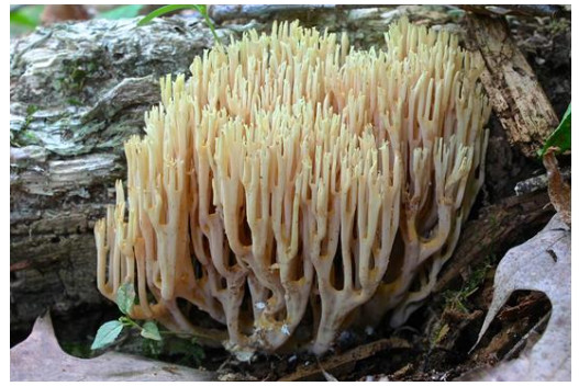
Figure 1. Fruitbody of Ramaria stricta in the field. The photograph was taken at the Sewickley Heights Borough Park, PA, USA by Fluffberger (https://www.inaturalist.org/observations/3596638). The photo was used under the CC BY-NC 4.0 non-commercial use license. Scale bar = 10 cm.
-
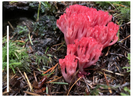
Figure 2. Fruitbody of Ramaria araiospora in the field. The photograph was taken at the Millersylvania State Park, Olympia, WA, USA by Yay4john (https://www.inaturalist.org/observations/62355314). The photo was used under the CC BY-NC 4.0 non-commercial use license. Scale bar = 10 cm.
-
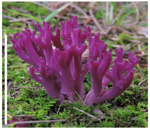
Figure 3. Fruitbody of Clavaria zollingeri in the field. The photograph was taken at Ohio, USA by Rcurtis (https://www.inaturalist.org/observations/1693040). The photo was used under the CC BY-NC 4.0 non-commercial use license. Scale bar = 10 cm.
-
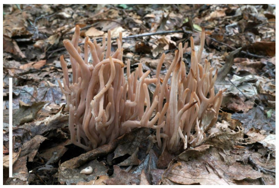
Figure 4. Fruitbodies of Clavaria rubicundula in the field. The photograph was taken at Hewitt, West Milford, NJ 07421, USA by Tombigelow (https://www.inaturalist.org/observations/16044756). The photo was used under the CC BY-NC 4.0 non-commercial use license. Scale bar = 10 cm.
-
Ramaria species Tested extract Employed methods Tested bacteria Tested fungi References Gram-positive Gram-negative R. formosa Ethyl acetate, methanol, and water Percentage of inhibition B. subtili, S. aureus E. coli, K. pneumonia, Proteus vulgaris, P. aeruginosa Not tested Pala et al. 2019 R. formosa Methanol Inhibition zone assay S. aureus P. aeruginosa Candida albicans Ramesh & Pattar 2010 R. zippellii Ethanol, water 96-well microplate bioassay S. aureus E. coli No activity Bala et al. 2011 R. aurea Ethanol Inhibition zone assay S. aureus E. coli, P. aeruginosa, P. vulgari Candida albicans Rai et al. 2013 R. flava Ethanol Inhibition zone assay B. subtili, S. aureus E. coli Fusarium auenaceum, F. graminearum and Cercosporella albomaculans Liu et al. 2013 R. flava Ethanol Agar-well diffusion method S. aureus, Micrococcus luteus, M. flavus, B. subtilis, B. cereus Salmonella enteritidis, Yersinia enterecolitica and K. pneumoniae No activity Gezer et al. 2006 R. botrytis Ethanol Methanol Acetone Ethyl acetate Inhibition zone diameter S. aureus P. aeruginosa, E. coli, Enterobacter cloacae No activity Han et al. 2016 Table 1. Antimicrobial activities of various Ramaria species
-
Ramaria species Place of collection Methods used Activity (EC 50 % of inhibition) References R. flava Turkey DPPH scavenging assay 94.78% at 12 mg/mL Gursoy et al. 2010 β-carotene linoleic acid assay 95.02% at 20 mg/mL Reducing power 95.02% at 20 mg/mL Metal chelating effect 96.75 at 2 mg/mL R. flava Turkey DPPH scavenging assay 276 µg/mL Gezer et al. 2006 β-carotene linoleic acid assay 94.7% at 160 µg/mL R. formosa India DPPH radical scavenging activity 5.8 mg/ml Ramesh & Pattar 2010 R. patagonica Argentina DPPH scavenging assay 770 µg/mL Toledo et al. 2016 β-carotene linoleic acid assay 610 µg/mL Reducing power 170 µg/mL TBARS inhibition activity 60 µg/mL R. Formosa Korea DPPH scavenging assay 117 AsA/mg/mL at 500 µg/mL Kim et al. 2016 Reducing power 36% copper ion inhibition at 20 µg/mL concentration Peroxyl radical scavenging activity 7.8 µM trolox equivalent at 20 µg/mL R. stricta Ukraine Total antioxidant status 4.223±0.054 mmol/l Krupodorova & Sevindik 2020 Oxidative stress index 0.194±0.001 Total oxidant status 8.201 ± 0.095 μmol/l Table 2. Antioxidant activities of various Ramaria species isolated from different places
Figures
(4)
Tables
(2)