by Pat Jaquith
pat@westvalleynaturalists.org

Worldwide, there are about 100 species of Peltigera in the Foliose group of lichens; there are 23 species in the Rocky Mountains. But here in the West Valley, the number is much more manageable! In the following article, I will give some pointers I found helpful as my exploration has evolved.
Lichens are organisms composed of fungi that give them structure and a photobiont of algae or photosynthesizing bacteria. Pelt lichens (The word peltigera comes from Latin, meaning “shield-bearing”) have lobes of different shapes and sizes. When they are dry and dormant, most of them are grayish-colored and a bit difficult to tell apart. When they are moist, the gray-colored outer cover becomes transparent, the color of the photobiont becomes visible, and identification becomes much easier. You can tell which photobiont they have by their color: if green, it’s green algae; if bluish or dark colored, it’s blue-green cyanobacteria. Simply put, the photobiont contains chlorophyll and is the food-producing part. The fungal contributions are absorbing water, giving it structure, and attaching it to its substrate. In some pelt lichens, there is a third component, the cyanobacteria nostoc which ‘fixes’ atmospheric nitrogen, making it available to the organism, and enriching the soil with nitrogen when the lichen dies.
TIP: Many of the key features of lichens are TINY; a hand lens will open windows on some microscopic features that are fascinating, beautiful, and often key to separating one species from another. I have one that consists of 3- 10x lenses that can be used separately or in combination. It is about the size of a walnut; I attached it to a piece of elastic tethered to my backpack with a small carabiner. I can use the hand lens while attached to my bag, lessening the chances of leaving it behind.

Peltigera canina (dry) 
Peltigera canina lobe(dry) 
Peltigera canina wet
These are all Dog Pelt Lichens. These are “terricolous” lichens which means they grow on the ground. Many lichen species may occupy the same small location, and the features are often hard to separate, especially when tree needles, leaves, moss, and grass all compete for space! Enlarged, through the cracks in the dry lobe, the little white threads called “rhizines” are visible. These structures help absorb water and attach it to the soil and/or plants it’s growing on. The photo at the bottom demonstrates how completely different it looks in a wet environment! The lobe’s texture has changed from paperboard to flexible; the cyanobacterial photobiont is revealed as the gray covering is now transparent; and in the growing mode the reproductive parts, called apothecia, are large, and thought to resemble teeth or a dog’s upright ears.
In early times, putting pelt lichen in one’s shoes was said to deter biting dogs! Dog pelt lichen was used as treatment for rabies!


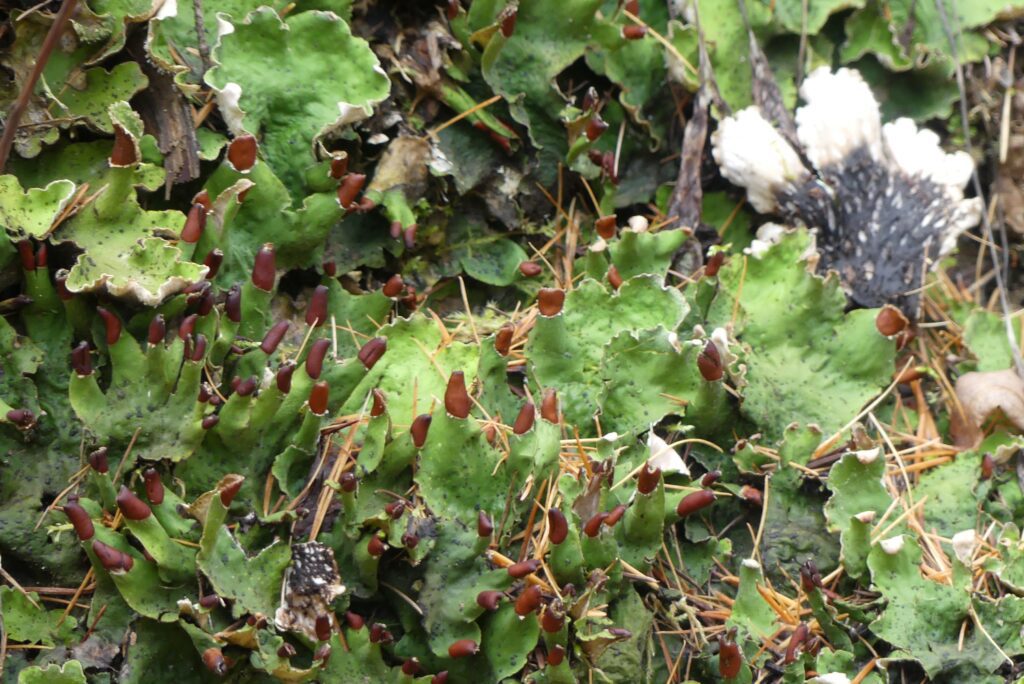
Peltigera leucophlebia (Ruffled Freckle pelt lichen)
These are both Ruffled freckle pelt lichen. There’s another species, Peltigera aphthosa (Studded leather lichen) which is the same bright green color and has black ‘warts’ on the surface. Key differences between them are is the appearance of the lower surface of a lobe and the appearance of the edge of the lobes. As demonstrated in the image on the right, the bottom surface has a pattern of dark brown veins made of fungal material and white tips. The organism on the right is in active growth mode with brown apothecia (reproductive organs) on the tips of some of the ruffles. The black ‘warts’ called cephalodia are collections of cyanobacteria which “fixes” atmospheric nitrogen, making it available to the growing organism. This lichen is an example of a tri-part symbiosis: a fungus (the mycobiont); a photobiont of green algae, and cyanobacteria.
TIP: Don’t go home without making a thorough inspection of the whole organism! The lower surface of many lichens has features that are key to identifying the species. After reading guides, I’ve made return trips many times to look for details I had overlooked!

Apple pelt is a deep blue-green color, thanks to the photobiont of blue-green cyanobacteria. Note that it has no black warts, either!


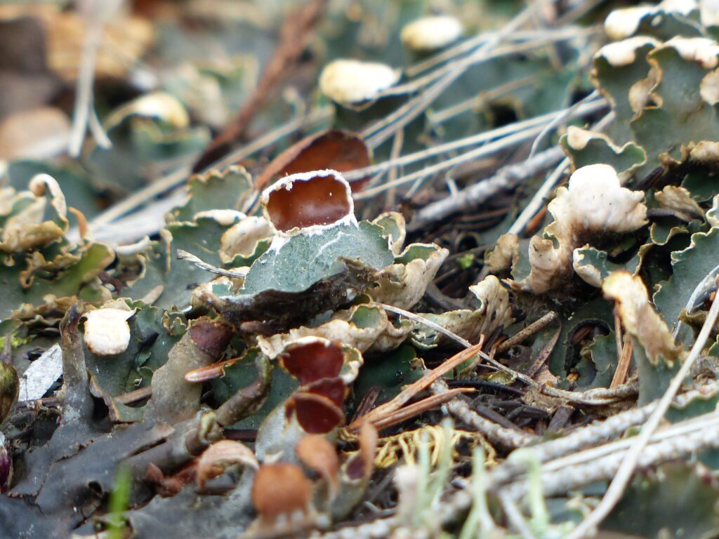
Peltigera venosa
In the upper left image, the little apothecial discs are nearly black. I think that age is a factor.
To confirm the identification, the under surface of Peltigera venosa has a striking pattern of dark fungal veins alternating with white fuzz (tomentum) covering the entire back of the lobes.
The bottom photo shows characteristic habitat of lichens. There are at least 4 lichen species and some mosses covering the soil on a rather steep trailside bank. These lichens stabilize the bank and deter erosion. Peltigera venosa is a very small lichen; note the tiny green chip-shaped bits at the upper left of the reddish discs: they are immature P.venosa organisms. The 1/16th inch reddish-brown apothecia do help point them out, but they are in mature mode only part of their life!
TIP: Give yourself a gift of time to engage your vision in the details of the site! It’s rewarding and refreshing. Let yourself become engaged in the environment. Challenge yourself to look at all those tiny organisms – or limit yourself to inspecting only one with your hand lens. The world of microorganisms is wondrous!
To learn more about the local lichens, check out my post A Bouquet of Lichens here and Skip’s Flora, Fauna, or...at this site.