Grimmia pulvinata (Hedw.) Sm. ex Sm. & Sowerby – Class Bryopsida

Grimmia pulvina spores

Grimmia pulvinata spores (click to enlarge)

Grimmia pulvinata sporophyte in which our spores were taken from. This sample is from the UBC Herbarium at the Beaty Biodiversity Museum.

Grimmia pulvinata sporophyte in which our spores were taken from. This sample is from the UBC Herbarium at the Beaty Biodiversity Museum.

Grimmia pulvinata (Hedw.) Sm. ex Sm. & Sowerby – Bryopsida

Description of spores: Smooth, spherical, a lot of spores from one sporangium
(in the image above, it appears to have grooves and bumps however it is due to the collapse of the spore wall from the vacuum and gold plating)
Size:12 μm in diameter
Habitat: Found frequent in dry climates and on rocks and concrete located in urban areas.
Dispersal Mechanism:  Hygroscopic endostome teeth flick spores out of the sporangia into turbulent air
Predicted spore adaptation: Relatively small in size therefore can be picked up by wind with ease. It is smooth so it decreases drag from the wind which allows it to be dispersed further away. Smooth spore surfaces prevent the spores from sticking to the substrate and allow it to roll away from nearby spores and further disperse.


Acknowledgements

Our project would not have been possible without the following people and organizations!

Professor Shona Ellis of UBC Botany, Instructor for Biology 321

Teaching Assistants David Bell and Erica Hunter

Olivia Lee of the UBC Herbarium

Steve Joya of the UBC Herbarium

Derrick Horne, Senior Technician, of the UBC Bioimaging Facility

References

(January 31, 2013). University and Jepsen Herbaria Portal to California Flora. Retrieved from http://ucjeps.berkeley.edu/

(July 15 2007). Bryophytes: Mosses, part 1.Flora of North America, 27. Retrieved from http://www.efloras.org/volume_page.aspx?volume_id=1027&flora_id=1

Glime, Janice M. 2007.  Bryophyte Ecology. Volume 1. Physiological Ecology. Ebook sponsored by Michigan Technological University and the International Association of Bryologists. accessed on 3/21/2014 at http://www.bryoecol.mtu.edu/ .

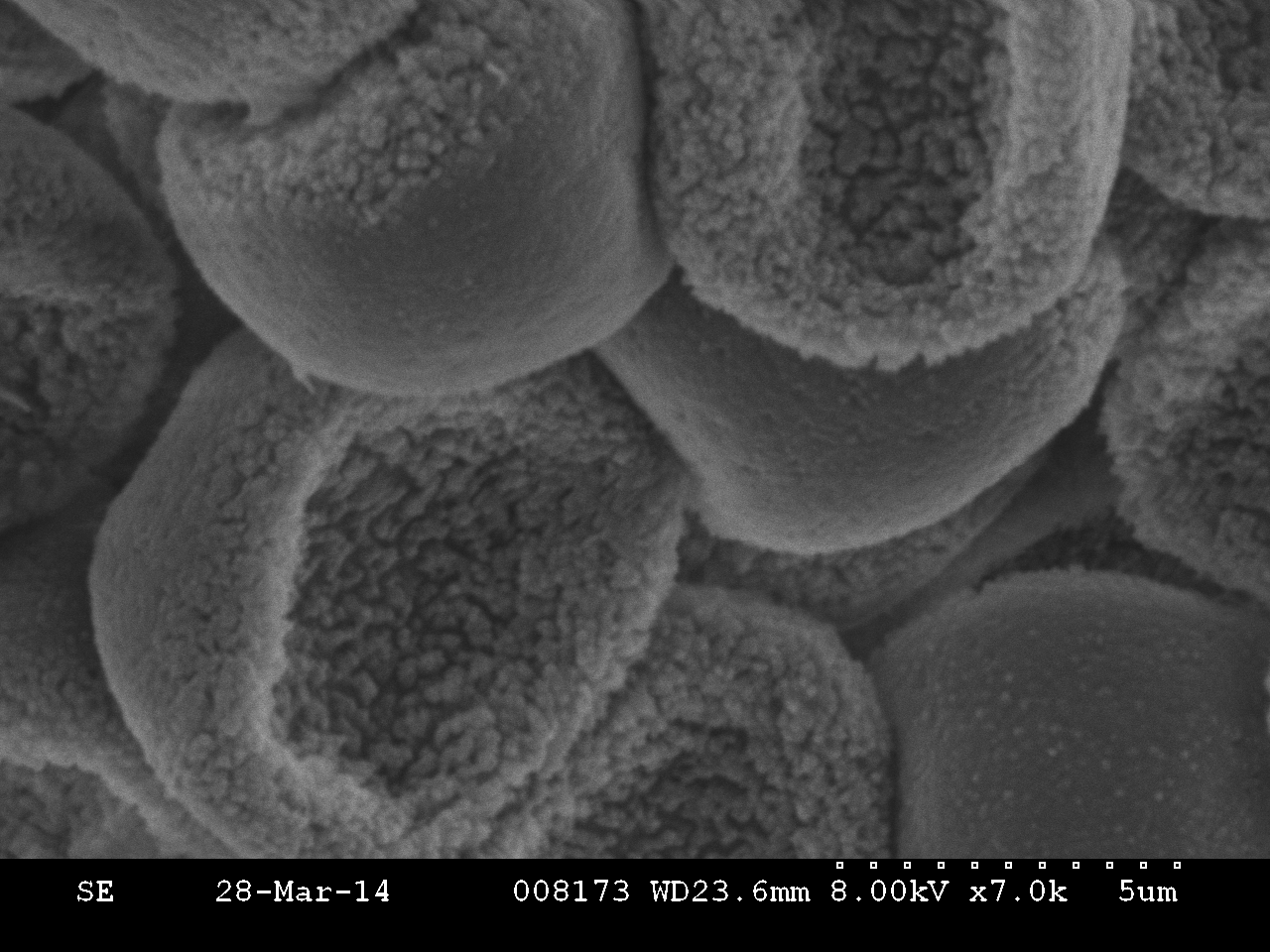
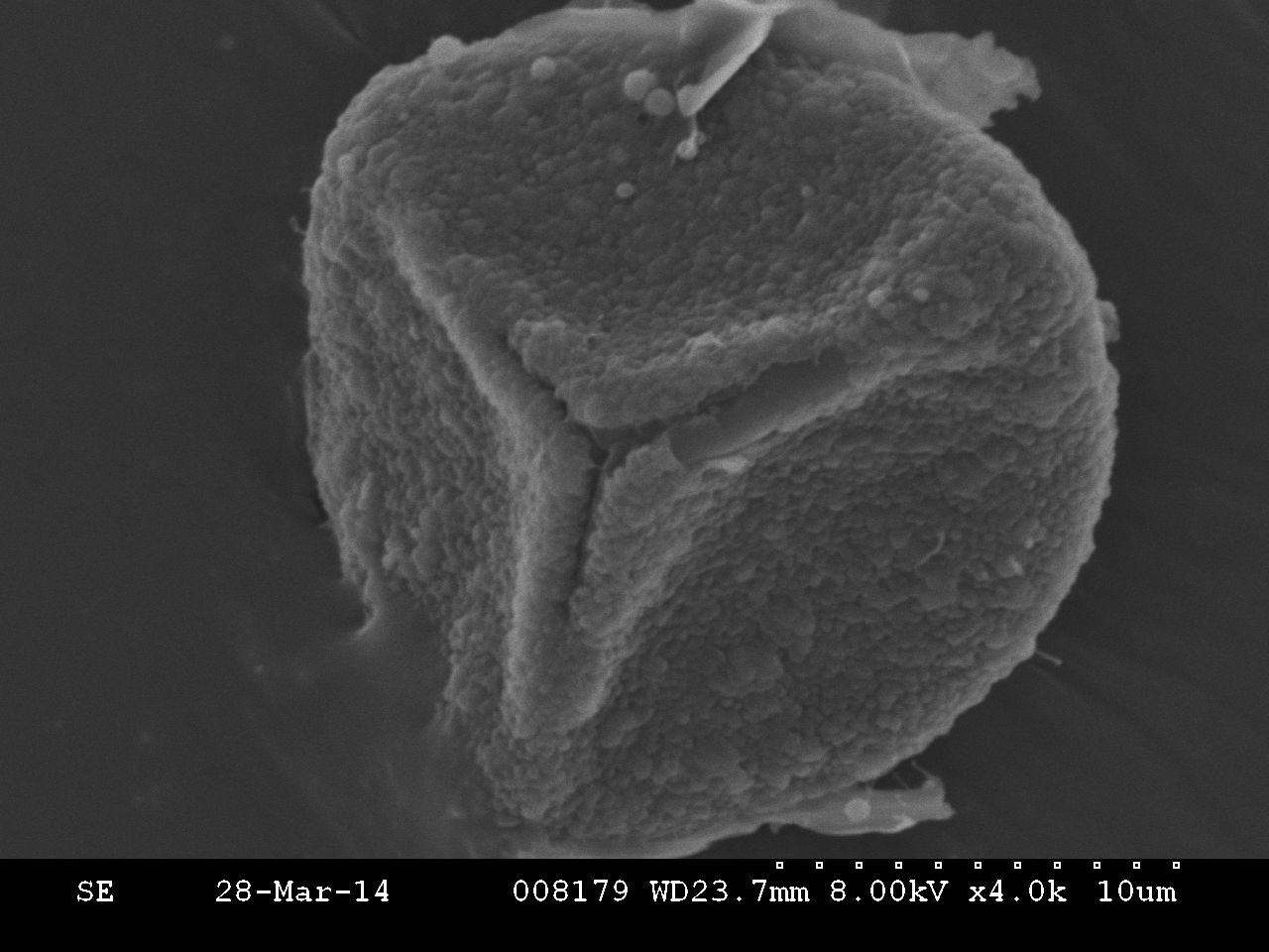
Hafner, Bob. “Scanning Electron Microscopy Primer.” Scanning Electron Microscopy Primer. Chracterization Facility, University of Minnesota – Twin Cities, 16 Apr. 2007. Web. 10 Mar. 2014.

Schofield, Wilf. Funaria hygrometrica Hedw. In Klinkenberg, Brian. (Editor) 2013. E-Flora BC: Electronic Atlas of the Plants of British Columbia [eflora.bc.ca]. Lab for Advanced Spatial Analysis, Department of Geography, University of British Columbia, Vancouver. [Accessed:3/7/2014 8:26:42 PM ]

Schofield, Wilf. Grimmia pulvinata (Hedw.) Sm. ex Sm. & Sowerby. In Klinkenberg, Brian. (Editor) 2013. E-Flora BC: Electronic Atlas of the Plants of British Columbia [eflora.bc.ca]. Lab for Advanced Spatial Analysis, Department of Geography, University of British Columbia, Vancouver. [Accessed:3/7/2014 8:36:10 PM ]

Schofield, Wilf. Hypnum circinale Hook. In Klinkenberg, Brian. (Editor) 2013. E-Flora BC: Electronic Atlas of the Plants of British Columbia [eflora.bc.ca]. Lab for Advanced Spatial Analysis, Department of Geography, University of British Columbia, Vancouver. [Accessed:3/5/2014 6:56:28 PM ]

Schofield, Wilf. Polytrichum commune Hedw. In Klinkenberg, Brian. (Editor) 2013. E-Flora BC: Electronic Atlas of the Plants of British Columbia [eflora.bc.ca]. Lab for Advanced Spatial Analysis, Department of Geography, University of British Columbia, Vancouver. [Accessed:3/5/2014 6:36:15 PM ]

Schofield, Wilf. Sphagnum capillifolium (Ehrh.) Hedw. In Klinkenberg, Brian. (Editor) 2013. E-Flora BC: Electronic Atlas of the Plants of British Columbia [eflora.bc.ca]. Lab for Advanced Spatial Analysis, Department of Geography, University of British Columbia, Vancouver. [Accessed:3/5/2014 6:41:20 PM ]

Hypnum circinale Hook.- Class Bryopsida

Hypnum circinale spores (click to enlarge)

Hypnum circinale spores (click to enlarge)

Sporophyte samples of Hypnum circinale in which our spores were obtained from.

Sporophyte samples of Hypnum circinale in which our spores were obtained from.


Hypnum circinale Hook.- Bryopsida

Description of spores: Smooth to lightly papillae, spherical, papillae are irregularly shaped; a lot of spores from one sporangia
Size: Up to 15 μm
Habitat: Found on tree trunks ,decomposing logs and rocks.
Dispersal Mechanism: Hygroscopic endostome teeth flick spores out of the sporangium into turbulent air. Hygroscopic seta provides some movement to the sporangia to shake out the spores and further increase dispersal distance.
Prediction of spore adaptation: The papillose surface allows for a velcro action to stick to substrates. This can secure its place on the substrate and allow it to germinate without any disruption.


Polytrichum commune Hedw.- Class Polytrichopsida

Polytrichum commune spores (click to enlarge)

Polytrichum commune spores (click to enlarge)

This Polytrichum commune sporophyte was obtained from the UBC Herbarium at the UBC Beaty Biodiversity Museum. The spores from our project were obtained from this sample.

This Polytrichum commune sporophyte was obtained from the UBC Herbarium at the UBC Beaty Biodiversity Museum. The spores from our project were obtained from this sample.

Polytrichum commune Hedw.- Polytrichopsida

Description of spores: Small, smooth, spherical, slightly spiny, a lot of spores from one sporangia
Size: Usually 5-8μm can get up to 12 μm
Habitat: Exposed environments with moist soil and sunny areas, such as bogs and swamps
Dispersal Mechanism: Hygroscopic seta twists and provide movement to disperse spores through the openings between the non-hygroscopic teeth and the epiphragm.
Prediction of spore adaptation: The spines provide grip or wedges into crevices in substrates. It can also allow more secure attachment to wet soil and not get blown away or carried by water from where it has already landed.

Sphagnum capillifolium (Ehrh.) Hedw.- Class Sphagnopsida

Sphagnum capillifolium spore (click to enlarge)

Sphagnum capillifolium spore (click to enlarge)

Sphagnum capillifolium (Ehrh.) Hedw.- Sphagnopsida

Description of spores: Smooth to fine papillae on both surfaces; spherical, smooth surface with distinct bifurcated Y-mark. Very little amount of spores
Size : 20-28 μm
Habitat: Found in bogs and thrive better in better-drained sites
Dispersal Mechanism: As the sporangium dries up, pressure builds up in the structure and causes a rupturing of the sporangium with the operculum popping off, resulting in spores ejecting up into the turbulent air.
Prediction of spore adaptation: The red colour of the sphagnum may attract flies, similar to the methods of Splachnum ampullaceum,  therefore the fine papillae could aid in spore dispersal in way that it act as a velcro and gets tangled on the hairs of the flies. The flies can transport these spores over a far range. The spore is also relatively large therefore indicating that the moss invests a lot of energy manufacturing these massive spores which may contain a lot of essential factors to increase longevity of the spore. This is especially beneficial for short-lived species and it provides energy for a good start.

Funaria hygrometrica Hedw. – Class Bryoposida

Funaria hygrometica spores (click to enlarge)

Funaria hygrometica spores (click to enlarge)

A sample of the Funaria hygrometrica sporophyte in which our spores were obtained from. This sporophyte was from the UBC Herbarium at the Beaty Biodiversity Museum.

A sample of the Funaria hygrometrica sporophyte in which our spores were obtained from. This sporophyte was from the UBC Herbarium at the Beaty Biodiversity Museum.

Funaria hygrometrica Hedw.- Bryoposida

Description of spore: Light green,  finely papillose-smooth, with spherical papillae, many spores in the sporangia
Size: 20μm
Habitat: Found in moist, shaded and damp soil, crevices of rocks and moist walls
Dispersal mechanism: Hygroscopic endostome teeth flicks spores out of the sporangia into turbulent air
Prediction of spore adaptation: The papillose surface allows for a velcro action to occur,  however it is not too papillose to allow for a bit of falling of the spores from higher elevations to reach the soil where it can germinate.


What Are Spores?

Spores are unicellular, haploid diaspores produced via meiosis by the sporophyte used for reproduction in plants. Disapores are structures that are capable of growing into plants, these include vegetative propagules such as fragments and gemmae.  In “higher” up plants, such as angiosperms, there are two types of spores – microspores and megaspores-  which are highly reduced and the gametophyte actually develops within that spore (endosporic is the term used).

In mosses, only one type of spore is produced in the sporangia. When a spore lands on a substrate it can stay dormant until environmental conditions are suitable and this is when germination of the spore occurs. Germination of the spore first results in a thread-like structure, the protonema, which is basically a string of cells,  in which gametophores eventually grow off of to form the dominant gametophyte structure that we see in bryophytes.

Spore Wall Composition

Until recently, fossils of plants have been of spores or pollen, mostly due to the toughness of the spore and their walls. Spore deposits dating from the Silurian and Devonian demonstrate to us that they can very well resist decay and remain numerous.

The outer wall of spores (exine) has a very important component, a waxy compound known as sporopollenin, which is also found in the wall of pollen in angiosperms. This compound is simply one innovation that allows for plants to thrive on land and to disperse to various habitats. Sporopollenin allows for great resistance against environmental stresses, allowing spores to disperse in the air without drying out and to stay dormant until conditions are favourable for germination. The chemical composition of sporopollenin is not yet known.

Along with sporopollenin, the exine is also composed of polysaccharide deposits which sporopollenin are impregnated within. A callose deposit before the exine forms is obtained by each spore. The intine, the inner wall of the spore, is formed after exine formation and is composed of cellulose and other polysaccharides.

The ornamentation on the surface of the spores of some mosses is due to a layer known as perine. This layer is basically composed of left overs from the walls of the spore mother cell and the columella.

There is currently not a lot of research done to link ornamentation with potential spore dispersal mechanism as previous studies have not been to successful. However, we will be including our predictions on how these ornamentations on the spore wall help various species disperse and germinate more efficiently.