|
||
|
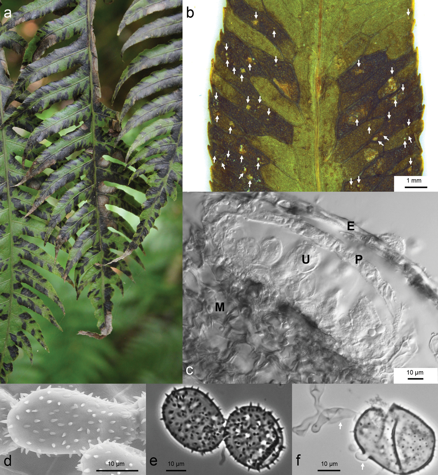
Spore morphology and symptoms on fern fronds of Milesina woodwardiana sp. nov. a Fronds of the host Woodwardia radicans at the collection site in La Palma. Dark spots indicate areas where sori are formed on the underside (La Palma, Cubo de la Galga, ca. 1.2 km SW of parking lot W San Bartolomé, 11 Aug 2017) b Host leaf with uredinia. Sori (arrows) are restricted to areas between leaf veins (KR-M-0048787, dissecting microscope) c Transverse section of uredinium E=epidermis, P=peridial cells, U=urediniospore, M=mesophyll of host plant (KR-M-0048787, LM, interference contrast) d Urediniospores with long echinulae (KR-M-0049036, paratype, SEM) e Urediniospores, cracked, without plasma, germ pores scattered (KR-M-0049033, paratype; LM, phase contrast) f Germinating urediniospores, arrows point to germ tubes (KR-M-0049033, paratype, LM, phase contrast). |