Biology LibreTexts

2.1: Cyanobacteria

  • Page ID
    37188
  • \( \newcommand{\vecs}[1]{\overset { \scriptstyle \rightharpoonup} {\mathbf{#1}} } \) \( \newcommand{\vecd}[1]{\overset{-\!-\!\rightharpoonup}{\vphantom{a}\smash {#1}}} \)\(\newcommand{\id}{\mathrm{id}}\) \( \newcommand{\Span}{\mathrm{span}}\) \( \newcommand{\kernel}{\mathrm{null}\,}\) \( \newcommand{\range}{\mathrm{range}\,}\) \( \newcommand{\RealPart}{\mathrm{Re}}\) \( \newcommand{\ImaginaryPart}{\mathrm{Im}}\) \( \newcommand{\Argument}{\mathrm{Arg}}\) \( \newcommand{\norm}[1]{\| #1 \|}\) \( \newcommand{\inner}[2]{\langle #1, #2 \rangle}\) \( \newcommand{\Span}{\mathrm{span}}\) \(\newcommand{\id}{\mathrm{id}}\) \( \newcommand{\Span}{\mathrm{span}}\) \( \newcommand{\kernel}{\mathrm{null}\,}\) \( \newcommand{\range}{\mathrm{range}\,}\) \( \newcommand{\RealPart}{\mathrm{Re}}\) \( \newcommand{\ImaginaryPart}{\mathrm{Im}}\) \( \newcommand{\Argument}{\mathrm{Arg}}\) \( \newcommand{\norm}[1]{\| #1 \|}\) \( \newcommand{\inner}[2]{\langle #1, #2 \rangle}\) \( \newcommand{\Span}{\mathrm{span}}\)\(\newcommand{\AA}{\unicode[.8,0]{x212B}}\)

    Stromatolites

    The earliest fossils are interpreted to be unicellular organisms similar to modern day cyanobacteria, sometimes referred to as blue green algae. The most widely accepted of these fossils dates back to 3.4 billion years ago from the Strelley Pool Formation in Western Australia. These particular fossils are called stromatolites and are composed of alternating layers of fossilized cells and calcium carbonate. We can use evidence from modern day stromatolite formation in Western Australia to infer that these fossilized cells were doing a process called photosynthesis, using dissolved CO2 in the water to form sugar molecules. This causes calcium to precipitate out of the seawater, forming hardened layers of calcium carbonate on top of the colony of organisms. Because they need access to light to continue photosynthesizing, living cells begin forming a new layer on top of the calcium carbonate. This process continues, making a ringed pattern as the formation grows, much like we see in trees and corals.

    Rocky lumps emerge from the water along the beach. Each lump is a stromatolite.
    A stromatolite fossil showing many concentric rings of growth radiating outward
    Figure \(\PageIndex{1}\): The images above show stromatolites. The first image shows present day stromatolites formed on the coast of Western Australia. The second image shows a fossil stromatolite with the rings of growth formed by successive layers of Cyanobacteria. First photo by AndyGo, CC-BY-NC. Second image: Stromatolite Fossil from Wyoming by James St. John via Wikimedia Commons. CC-BY 2.0.

    Modern Cyanobacteria

    Free-Living

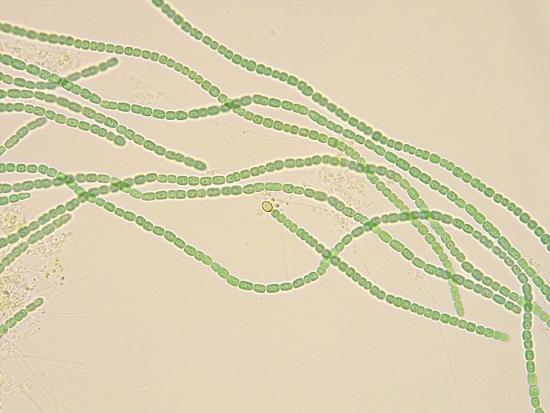
    Cyanobacteria can be found in a vast diversity of places, from floating in the ocean to living in cryptobiotic crusts in the desert. Nostoc is a type of cyanobacteria that can often be found living in gelatinous colonies in wet, terrestrial environments. The colony secretes a mucilaginous sheath that provides a protective barrier and allows for the exchange of materials between cells in the colony.

    Many glassy-looking, brownish green orbs sit in someone's hand. Each orb contains a colony of Nostoc, not visible to the naked eye
    Figure \(\PageIndex{2}\): Each of these glassy orbs contains a colony of Nostoc pruniforme surrounded by a mucilaginous sheath. The bacteria are not visible to the naked eye, so appear as a single organism. Photo by René Stalder, CC-BY-NC.
    A single Nostoc colony viewed through the microscope. A transparent orb filled with chains of green cells tangled into a ball.
    A closer view of a colony. Individual chains are easier to distinguish, still a tangled mess. Some larger, yellow cells present.
    Figure \(\PageIndex{3}\): A view of a Nostoc colony through a microscope. The colony consists of chains of individuals, wrapped together into a ball. This ball is coated by a transparent mucilaginous sheath (visible in the first photo). The green color of the ball is due to the photosynthetic pigments in the Cyanobacteria. In the photo on the right, a colony is shown at a higher magnification. Individuals can be distinguished, including some larger, yellow cells. These latter individuals have converted into heterocysts. First photo by Ivan, CC-BY. Second photo by Mindy Morales, CC-BY-NC.
    A collage of images showing multiple views of the cyanobacterium Rivularia.
    Figure \(\PageIndex{4}\): Multiple views of a gelatinous colony formed by Rivularia polyotis. The thick mucilaginous sheath is visible in the two images in the upper left corner, as well as the central image on the left. Chains of individuals can be distinguished, looking like hairy projections. At lower magnification, colonies are not easily distinguished, surrounded by a thick gelatinous layer. As images reach higher magnification, individual chains can be distinguished, composed of flat, disc-like cells. Photos by Luigi, CC-BY-NC.

    Mutualists

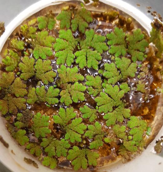
    Many cyanobacteria that you'll see in botany will be in mutualistic relationships. Anabaena is a genus of colonial cyanobacteria that can be found within the leaves of the water fern Azolla, fixing nitrogen in the fern's relatively nutrient-poor aquatic environment. Nostoc is another genus of colonial cyanobacteria capable of fixing nitrogen. It can be found free-living in gelatinous colonies shown above or, as you are likely to see it in your botany course, in compartments of a hornwort thallus. Cyanobacteria can also be found in a mutualistic relationship with fungi in cyanolichens (Figure \(\PageIndex{5}\)).

    A dark bluish grey lichen growing flat against some moss on the ground
    A dark blackish brown lichen growing appressed to a tree trunk
    Figure \(\PageIndex{5}\): Darkly colored lichens, such as this Peltigera (first) and Nephroma (second), often have a cyanobacterial partner. Photos by Maria Morrow CC-BY-NC.

    Looking at Cyanobacteria in Water Ferns

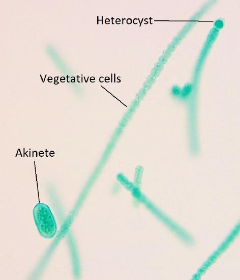
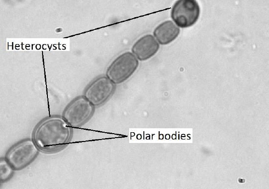
    If you were to chop up a sample of the water fern Azolla and look at it under the microscope, you'd see what looked like strings of green beads. Each bead is an individual cyanobacterium of the genus Anabaena. However, even though each one is an individual, some cells will specialize to provide a service for the colony, as a whole.

    • Heterocysts are thick-walled, chlorophyll-free cells that are fixing atmospheric nitrogen into bioavailable forms using the enzyme nitrogenase. Heterocysts cannot do photosynthesis, as that process produces oxygen and nitrogenase cannot function in the presence of oxygen.
    • Akinetes are individuals that still perform photosynthesis, but also function as a sort of failsafe. Akinetes store large amounts of lipids and carbohydrates so that they have enough energy to begin a new colony if conditions become too cold or too dry for survival. Their formation is triggered by these conditions (dry or cold), so you may not see them from a fresh water fern leaf, as this is a relatively stable, comfortable environment.
    Many water ferns from the genus Azolla in a dish
    Strings of cyanobacterial cells from within the water fern leaves
    Figure \(\PageIndex{6}\) : In the first image, many Azolla plants can be seen floating in a dish of shallow water. Each fern is about the size of a dime (approx. 1 cm across). In the second image, individual Anabaena cells are strung together in colonies. These cells were once inside the leaves of the water fern. Photos by Melissa Ha, CC BY-NC.
    A colony of Anabaena showing all three cell types
    Figure \(\PageIndex{7}\): Three different cell types are visible in this Anabaena colony. There is one large, thick-walled akinete in the lower left. In the upper right, there is a smaller, round heterocyst that is about the size of two vegetative cells. The rest of the cells distinguishable in the image are "normal" vegetative cells. Photo credit: Melissa Ha, CC BY-NC.
    A black and white image of Anabaena showing two heterocysts and their polar bodies
    Figure \(\PageIndex{8}\): Heterocysts usually appear yellow and are more round than the vegetative cells. However, beginners often confuse the larger ones for akinetes (particularly when no akinetes are present. Something that makes heterocysts easier to distinguish from akinetes is the presence of polar bodies. Heterocysts have two distinct regions on either side called polar bodies. Akinetes don't have these and appear more granular. Photo by Maria Morrow.1

    This page titled 2.1: Cyanobacteria is shared under a CC BY-NC 4.0 license and was authored, remixed, and/or curated by Maria Morrow (ASCCC Open Educational Resources Initiative) .

    • Was this article helpful?