Entries by Tristram Brelstaff (3025)

Monday
Dec242012

Springtails

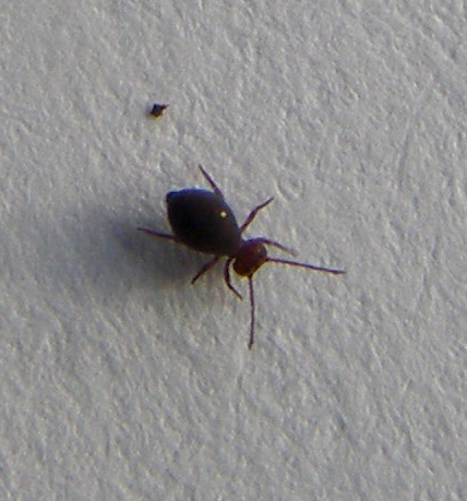
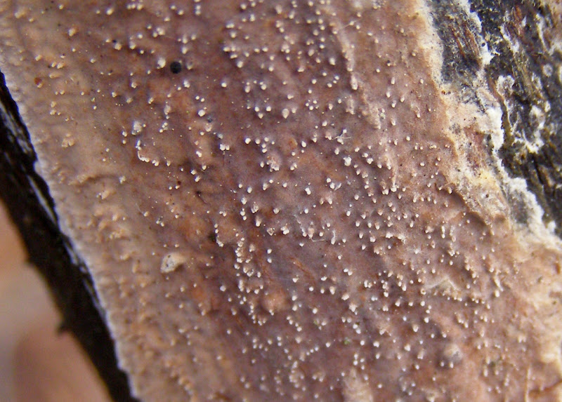
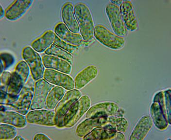
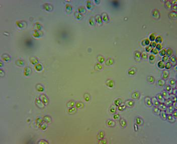
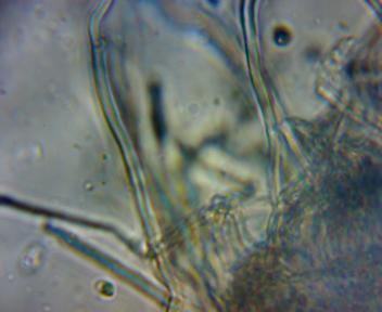
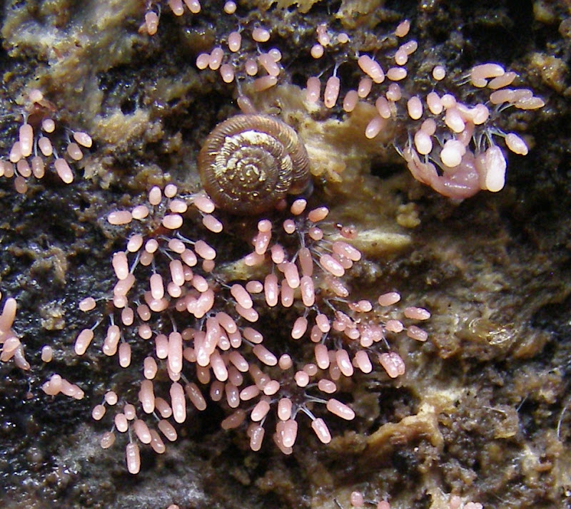
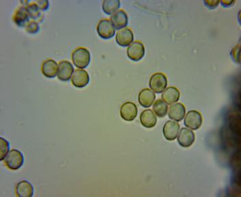
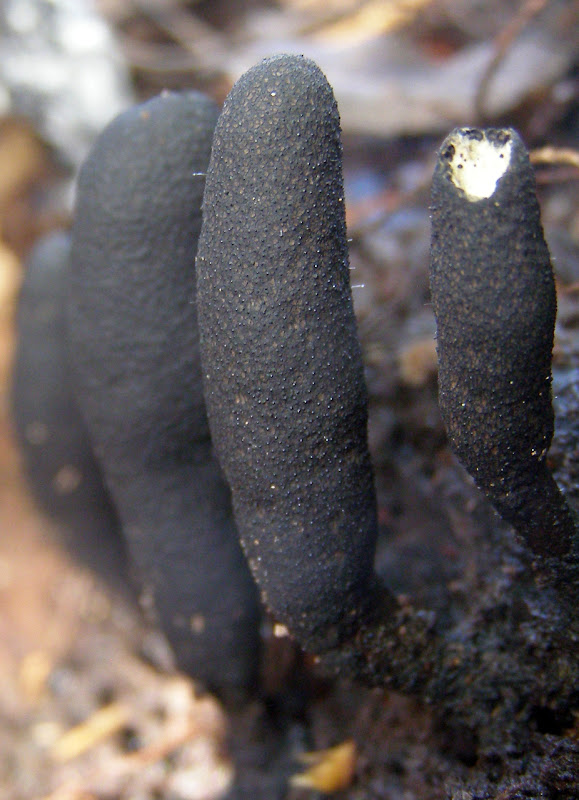
A few weeks back I found some small animals on some fungi I had collected.  They appear to be three different species of springtail. (Collembola). 

[Note added 2012-12-25: They were actually on the Xylaria longipes shown here.  I have since also found them on Xylaria hypoxylon.  Maybe there is an association between Collembola and Xylaria spp or with the places that Xylaria spp grow?]

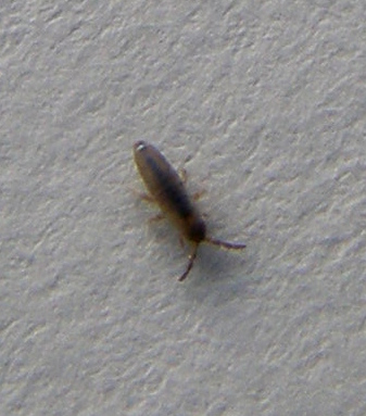
Springtails are arthropods but are not classed as insects.

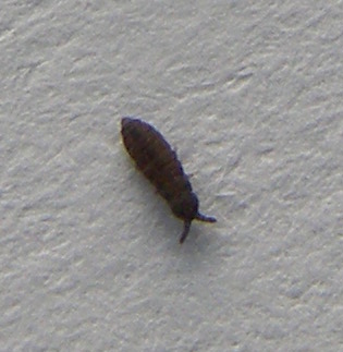
Specimens collected in the Wilderness, Whiteknights Park, Reading, UK on 2012-12-09.

Sunday
Dec232012

Reddish Crust Fungus

A reddish crust fungus, probably Eichleriella deglubans, growing on a horse-chestnut tree (Aesculus hippocastanum).

The spores (prepared by the method given here) look like this at x600:

From the image width of 86um, the spores are about 16 x 6um which agrees well with the size range 12-18 x 6-7um for the European form given here at MycoBank.

First photo taken in the Wilderness, Whiteknights Park, Reading, UK, on 2012-12-18.

Saturday
Dec222012

White Crust Fungus

A white porecrust fungus, probably Schizopora paradoxa, growing on a dead branch of an oak tree (Quercus sp).  The above section was on the side of the branch and shows the pores side-on, while the following section was on the underside and shows the pores end-on:

This is the spores at x600 under the microscope (prepared in the way given here):

The width of the image at x600 is about 86um (determined by photographing a millimetre ruler at x60) so the spores are about 6 x 3um which is consistent with the '4–6 x 3–3.5um' given at Roger's Mushrooms.

The hyphae show clamp connections, see one at the top left of the following image (x600):

First two photos taken in Whiteknights Park, Reading, UK, on 2012-12-18.

Friday
Dec212012

Slime Mould

A slime mould with pale pink cylindrical sporangia on stalks, probably Stemonitopsis typhina (=Comatrichia typhoides).  The sporangia of this species start out as white but rapidly (within only a day or two) turn pink, then brown and black. For some of the same species that I saw last year, see  here.

This is what the spores looked like at x600 under the microscope:

First photo taken in the Wilderness, Whiteknights Park, Reading, UK, on 2012-05-11.

Thursday
Dec202012

Fungus

Dead moll's fingers fungus, Xylaria longipes.  The one on the right has been nibbled, probably by a mouse or a squirrel (would a slug climb all the way to the top before starting to nibble?).

I prepared a microscope slide of the spores using the method given here and this is the result (at x600):

These spores are roughly 1.5 times the length of the spores of Peniophora quercina shown here.  This, and the shape shown above, is consistent with the description given for X .longipes which includes 'Spores 13-15 x 5-7 um; smooth; fusiform' (fusiform = spindle-shaped, ie: pointed at both ends).

At first I assumed this fungus to be Xylaria polymorpha but that has 20-31 x 5-10um spores, which would be 2 to 3 times as long as those of P. quercina.  This is clearly not the case with the spores in the above image.

First photo taken in the Wilderness, Whiteknights Park, Reading, UK, on 2012-12-15.